预约演示
更新于:2026-09-12
AP-5258
更新于:2026-09-12
概要
基本信息
原研机构 |
在研机构- |
非在研机构 |
权益机构- |
最高研发阶段无进展药物发现 |
首次获批日期- |
最高研发阶段(中国)- |
特殊审评- |

关联
100 项与 AP-5258 相关的临床结果
登录后查看更多信息
100 项与 AP-5258 相关的转化医学
登录后查看更多信息
100 项与 AP-5258 相关的专利(医药)
登录后查看更多信息
2
项与 AP-5258 相关的文献(医药)2026-02-26·CURRENT MEDICINAL CHEMISTRY
Unveiling the Small Molecules Binding Site of CD36 Cell Surface Receptor Through Docking and Molecular Dynamics Simulations
Article
作者: Méndez-Luna, David ; Montes-Mondragón, Naomi ; Correa-Basurto, José ; Bello, Martiniano ; Sandoval-Herrera, Vicente ; García-Sánchez, José-Rubén ; Zamudio-Hernández, Sergio-Roberto ; Salgado-Brito, Rosa
Introduction::
CD36 is a transmembrane glycoprotein involved in lipid uptake
and signal transduction, playing a crucial role in various physiological and pathological
processes. Structurally, it is composed primarily of an ectodomain (residues
30-439), which is essential for ligand binding and facilitating Long-Chain Fatty Acid (LCFA)
uptake. Two lysine residues (K164 and K166) have been proposed to contribute
significantly to LCFA internalization. Elucidating the ligand-binding cavities within
CD36 can provide structural insights that may serve as a foundation for drug design.
materials and methods::
Search for probable CD36 ligands
A rational search was conducted using scientific databases including PubMed (https://pubmed.ncbi.nlm.nih.gov/), Scopus (https://www.scopus.com/search/form.uri?display=basic#basic), SciELO (https://scielo.org/es/), and Google Academics (https://scholar.google.es/schhp?hl=es). The keywords used were: CD36 activity, CD36 ligands, docking, and MD simulations. From the search results, only studies that did not involve in silico simulations (which could introduce bias into our analysis) were selected. Instead, preference was given to sources containing robust experimental biological data supporting CD36 modulation. This approach identified three derivatives from N'-(2-hydroxy-4,6-dimethoxybenzylidene)acetohydrazide scaffold (AP5055, AP5156, and AP5258), two polyphenolic compounds (Puerarin and Salvianolic acid), and two well-known inhibitors of this receptor (SSO and MTN) as potential ligands for CD36.
Binding pocket and LCFA cavity detection
The CD36 structure (PDB entry: 5LGD) was obtained from RCSB-PDB (https://www.rcsb.org/). After detailed visualization, all non-essential molecules unrelated to the receptor’s inherent activity (such as water molecules, cofactors, metals, and sugars) were removed. Particularly, the two co-crystallized PLM molecules were eliminated to avoid interference with the analysis, as well as the CIDRa domain from MCvar1 PfEMP1. For this purpose, the protein structure was used without further processing. Detection of the probable LCFA (long-chain fatty acids) acceptor cavities and small-molecules acceptor cavity in CD36 was carried out using two specialized web servers, CavityPlus (http://www.pkumdl.cn:8000/cavityplus/index.php#/) and POCASA (https://g6altair.sci.hokudai.ac.jp/g6/service/pocasa/). CavityPlus is a geometry-based method that uses a probe-sphere model to scan the protein surface, mimicking the kinetic behavior of small molecules attempting to bind the protein. Hence, all the search parameters were kept at their default settings. Briefly, the algorithm first creates 3D grid points encompassing the entire protein, with a spacing of 0.5 Å in each grid point. A 10 Å radius probe sphere then identifies grid points it contacts, labeling them as "outside grid points," while the remaining points are marked as "vacant grid points," representing putative cavity spaces. Refinement of these cavities is performed using the “layer and depth” concepts combined with a “shrink-and-expand” algorithm37. The POCASA algorithm was applied according to our previous methodology38.
Ligand preparation
Ligand structures were initially generated in 3D conformations using ChemSketch software (https://www.acdlabs.com/resources/free-chemistry-software-apps/chemsketch-freeware/). These structures were then converted to Z-matrix format using GaussView 5.039 to delimit a geometrical and energetic minimization and correct proton assignment, using Gaussian 0940, under a semi-empirical AM1 method. The resulting output files were converted to .pdb format for subsequent docking calculations in Autodock (v4.2.6)41. For docking simulations with MOE42, ligands were prepared using the same protocol but employed in .mol2 format.
Molecular docking protocols
Two methodological strategies were employed to validate and strengthen our analysis. Specifically, MOE and AutoDock (v4.2.6) programs were used to evaluate and characterize the potential binding interactions between the tested ligands and CD36. For both software programs, the docking simulations were designed to model two receptor states: CD36-unoccupied (receptor without bound PLM molecules) and CD36-occupied (receptor bound to two PLM molecules), based on the crystal structure of CD36 available in the RCSB-PDB (PDB entry: 5LGD). MOE program was selected as the initial docking tool, considering the Triangle Matcher algorithm for initial ligand placement and the Rigid Receptor for refinement. The scoring functions used were the Affinity dG for initial pose generation (computing 30 poses), and GBVI/WSA dG for final refinement (computing 5 poses). As a complementary approach, the AutoDock (v4.2.6) program was employed, using parameters previously reported by our research group38, with a minor modification: the grid box was set at 126 Å3 across the X, Y, and Z dimensions (blind docking procedure), thereby encompassing nearly the entire ectodomain of the protein. Ligand-receptor interactions were visualized and analyzed using the Ligand interactions tool in MOE (for 2D representations, data not shown) and Pymol v0.9943 (for 3D structural interpretation).
Molecular dynamics simulation with MMGBSA approach
Receptor-ligand complexes were prepared using antechamber and tleap modules of the AMBER22 software package (Case et al., 2005). Systems were solvated using the TIP3P water model and neutralized with counterions by submerging them in a truncated octahedral water at 12 Å. The protein, water, and ions were defined by the ff14SB AMBER force field44. The General Amber force field (GAFF) was taken into consideration when defining the force field characteristics of ligands using AM1-BCC atomic charges45. Four thousand conjugate gradient cycles and five thousand steepest descent cycles made up minimization. Under an NVT ensemble and considering a Berendsen thermostat, the temperature was raised for 200 ps from 0 to 310 K46. A Langevin thermostat47-48 and a Berendsen barostat46 were used to equilibrate the density for 200 ps under an NTP ensemble. Using a Langevin thermostat and a Berendsen barostat, 100 ns long molecular dynamics (MD) simulations with a time step of 2 fs were run at 310 K under an NPT ensemble. The Particle Mesh Ewald approach was used to define the electrostatic term, while a cut-off of 10 Å was taken into consideration for the non-bonded interactions49. The SHAKE algorithm50 was used to limit bond lengths at specific equilibrium. The cpptraj function in Amber22 software was used to analyze the root means squared deviation (RMSD), radius of gyration (Rg), root means squared fluctuations (RMSF), and clustering analysis. To find the binding free energy (ΔGbind), the Molecular Mechanics Generalized Born Surface Area (MMGBSA) method51 was used. Using implicit solvent models52, a 0.10 M salt concentration, and a total of 2000 snapshots, this analysis was conducted over the final 20 ns of equilibrated simulation time. As previously mentioned, the ΔGbind values were calculated53.
Methods::
In this study, we employed a combination of molecular docking, molecular dynamics
simulations, and cavity-detection algorithms to investigate the structural basis of
CD36 interactions with small molecules and fatty acids.
Results::
We identified three main ligand-binding regions: a primary LCFA-binding cavity,
a secondary LCFA-binding cavity, and a Small-Molecule Acceptor Cavity (SMAC).
We evaluated three derivatives of the N-(2-hydroxy-4,6-dimethoxybenzylidene)acetohydrazide
scaffold (AP5055, AP5156, and AP5258), two polyphenolic compounds
(Puerarin and Salvianolic acid), and two well-known CD36 inhibitors (SSO and MTN).
Discussion::
Our findings implicate residues K334, E335, and R337 in disrupting LCFA
uptake, while residues T195, L200, F201, Y202, P203, T207, A208, D209, Y230, and
K231 delineate the initial segment of the SMAC.
Conclusion::
This study highlights critical residues and structural features involved in ligand
recognition by CD36. The identification of the SMAC and its role in modulating
LCFA uptake provides promising insights for the development of therapeutic agents targeting
CD36-related pathologies, including cancer, diabetes, and metabolic disorders.
2015-07-03·Adipocyte4区 · 生物学
Pathways commonly dysregulated in mouse and human obese adipose tissue: FAT/CD36 modulates differentiation and lipogenesis
4区 · 生物学
ArticleOA
作者: A Géloën ; E Berger ; S Héraud ; A Mojallal ; M Weiss-Gayet ; O Damour ; C Lequeux
Obesity is linked to adipose tissue hypertrophy (increased adipocyte cell size) and hyperplasia (increased cell number). Comparative analyses of gene datasets allowed us to identify 1426 genes which may represent common adipose phenotype in humans and mice. Among them we identified several adipocyte-specific genes dysregulated in obese adipose tissue, involved in either fatty acid storage (acyl CoA synthase ACSL1, hormone-sensitive lipase LIPE, aquaporin 7 AQP7, perilipin PLIN) or cell adhesion (fibronectin FN1, collagens COL1A1, COL1A3, metalloprotein MMP9, or both (scavenger receptor FAT/CD36). Using real-time analysis of cell surface occupancy on xCELLigence system we developed a new method to study lipid uptake and differentiation of mouse 3T3L1 fibroblasts and human adipose stem cells. Both processes are regulated by insulin and fatty acids such as oleic acid. We showed that fatty acid addition to culture media increased the differentiation rate and was required for full differentiation into unilocular adipocytes. Significant activation of lipogenesis, i.e. lipid accumulation, by either insulin or oleic acid was monitored in times ranging from 1 to 24 h, depending on differentiation state, whereas significant effects on adipogenesis, i.e., surperimposed lipid accumulation and gene transcriptional regulations were measured after 3 to 4 d. Combination of selected times for analysis of lipid contents, cell counts, size fractionations, and gene transcriptional regulations showed that FAT/CD36 specific inhibitor AP5258 significantly increased cell survival of oleic acid-treated mouse and human adipocytes, and partially restored the transcriptional response to oleic acid in the presence of insulin through JNK pathway. Taken together, these data open new perspectives to study the molecular mechanisms commonly dysregulated in mouse and human obesity at the level of lipogenesis linked to hypertrophy and adipogenesis linked to hyperplasia.
100 项与 AP-5258 相关的药物交易
登录后查看更多信息
研发状态
10 条进展最快的记录, 后查看更多信息
登录
| 适应症 | 最高研发状态 | 国家/地区 | 公司 | 日期 |
|---|---|---|---|---|
| 脂质代谢紊乱 | 药物发现 | 法国 | - |
登录后查看更多信息
临床结果
临床结果
适应症
分期
评价
查看全部结果
| 研究 | 分期 | 人群特征 | 评价人数 | 分组 | 结果 | 评价 | 发布日期 |
|---|
No Data | |||||||
登录后查看更多信息
转化医学
使用我们的转化医学数据加速您的研究。
登录
或
药物交易
使用我们的药物交易数据加速您的研究。
登录
或
核心专利
使用我们的核心专利数据促进您的研究。
登录
或
临床分析
紧跟全球注册中心的最新临床试验。
登录
或
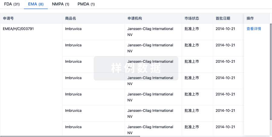
批准
利用最新的监管批准信息加速您的研究。
登录
或
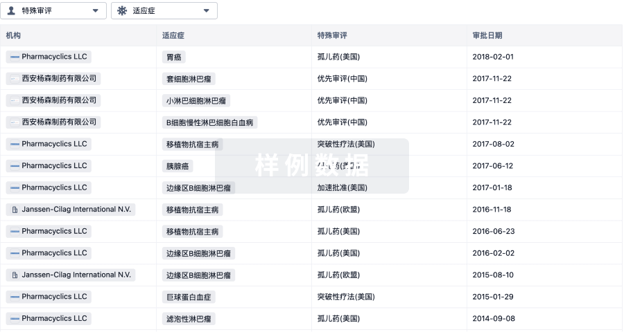
特殊审评
只需点击几下即可了解关键药物信息。
登录
或
提出任何生物医药研究问题
让每一步生物医药决策都精准制胜
基于十亿级生物数据与全球情报的精准 AI,赋能早期药物发现
立即开始免费试用!
智慧芽新药情报库是智慧芽专为生命科学人士构建的基于AI的创新药情报平台,助您全方位提升您的研发与决策效率。
立即接入生物医药数据包
智慧芽新药库数据也通过智慧芽开放平台,以MCP或API服务形式对外开放。立即使用我们的MCP服务定制您的LLM智能体。
生物序列数据库
生物药研发创新
免费使用
化学结构数据库
小分子化药研发创新
免费使用