预约演示
更新于:2025-05-24
FGL
更新于:2025-05-24
概要
基本信息
药物类型 合成多肽 |
别名 FGL、FGLL、NCAM mimetics (Enkam) + [1] |
作用方式 调节剂 |
作用机制 APP调节剂(β-淀粉状蛋白A4调节剂)、FGFRs调节剂(成纤维细胞生长因子受体家族调节剂) |
在研适应症- |
在研机构- |
权益机构- |
最高研发阶段无进展临床1期 |
首次获批日期- |
最高研发阶段(中国)- |
特殊审评- |
登录后查看时间轴
关联
100 项与 FGL 相关的临床结果
登录后查看更多信息
100 项与 FGL 相关的转化医学
登录后查看更多信息
100 项与 FGL 相关的专利(医药)
登录后查看更多信息
53
项与 FGL 相关的文献(医药)2024-08-01·Multiple Sclerosis and Related Disorders
Recurring disease activity in relapsing remitting multiple sclerosis: The multicenter RDA-RMS study
Article
作者: Türkoğlu, Şule Aydın ; Altun, Yaşar ; Bülbül, Nazlı Gamze ; İnanç, Yılmaz ; Yetkin, Mehmet Fatih ; Sıvacı, Ali Özhan ; Güzel, Vildan ; Seferoğlu, Meral ; Tunç, Abdulkadir ; Baydar, Caner ; Sezer, Vasfiye
BACKGROUND:
This study investigates the gap in understanding the dynamics of recurring disease activity (RDA) in RRMS patients after fingolimod (FGL) treatment discontinuation. The aim is to investigate RDA in RRMS patients after stopping FGL, aiming to improve management and comprehension of disease progression post-treatment.
METHODS:
In this multicenter, retrospective study, data from 172 of 944 RRMS patients aged 18-55, across nine centers in Turkey, who discontinued FGL treatment, were analyzed. The collected data included EDSS scores, annualized relapse rates (ARR), lymphocyte counts, and MRI findings, with follow-up assessments conducted at 6 months, 1 year, and up to 2 years.
RESULTS:
RDA was observed in 31.9 % of patients, with incidences of rebound and reactivation at 20.3 % and 11.6 %, respectively. Factors like younger age, longer treatment duration, lower lymphocyte counts, and higher lesion burden increased RDA risk. Notably, 52.9 % of pregnant patients experienced RDA (16.4 % of the overall RDA group), with rebound occurring in six and reactivation in three. Patients with RDA had longer medication-free intervals and increased ARR. Discontinuation reasons varied, with disease progression linked to a lower RDA risk.
CONCLUSION:
Findings highlight the necessity for personalized management and vigilant monitoring after FGL discontinuation in RRMS patients, offering critical insights into RDA risk factors, and the complex interplay between treatment cessation, pregnancy, and disease progression.
2024-05-01·Journal of neurology
Ocrelizumab reduces cortical and deep grey matter loss compared to the S1P-receptor modulator in multiple sclerosis
Article
作者: Castellaro, Marco ; Guandalini, Maddalena ; Pizzini, Francesca B ; Tamanti, Agnese ; Bajrami, Albulena ; Montemezzi, Stefania ; Calderone, Milena ; Peloso, Angela ; Marastoni, Damiano ; Calabrese, Massimiliano ; Ziccardi, Stefano
Abstract:
Introduction:
Ocrelizumab (OCR) and Fingolimod (FGL) are two high-efficacy treatments in multiple sclerosis which, besides their strong anti-inflammatory activity, may limit neurodegeneration.
Aim:
To compare the effect of OCR and FGL on clinical and MRI endpoints.
Methods:
95 relapsing–remitting patients (57 OCR, 38 FGL) clinically followed for 36 months underwent a 3-Tesla MRI at baseline and after 24 months. The annualized relapse rate, EDSS, new cortical/white matter lesions and regional cortical and deep grey matter volume loss were evaluated.
Results:
OCR reduced the relapse rate from 0.48 to 0.04, FGL from 0.32 to 0.05 (both p < 0.001). Compared to FGL, OCR-group experienced fewer new white matter lesions (12% vs 32%, p = 0.005), no differences in new cortical lesions, lower deep grey matter volume loss (− 0.12% vs − 0.66%; p = 0.002, Cohen’s d = 0.54), lower global cortical thickness change (− 0.45% vs − 0.70%; p = 0.036; d = 0.42) and reduced cortical thinning/volume loss in several regions of interests, including those of parietal gyrus (d-range = 0.65–0.71), frontal gyrus (d-range = 0.47–0.60), cingulate (d-range = 0.41–0.72), insula (d = 0.36), cerebellum (cortex d = 0.72, white matter d = 0.44), putamen (d = 0.35) and thalamus (d = 0.31). The effect on some regional thickness changes was confirmed in patients without focal lesions.
Conclusions:
When compared with FGL, patients receiving OCR showed greater suppression of focal MRI lesions accumulation and lower cortical and deep grey matter volume loss.
2024-04-23·Zhurnal nevrologii i psikhiatrii imeni S.S. Korsakova
Experience in the management of pregnant patients with highly active multiple sclerosis in the Moscow region
Article
作者: Yakushin, D.M. ; Yakushina, T.I. ; Shtang, I.O. ; Kotov, S.V.
Objective. To evaluate the effect of discontinuation or prolongation of DMT on the activity of the disease during pregnancy and in the postpartum period in patients with aggressive MS from the Moscow region. Material and methods. The study included female patients with an aggressive course of MS receiving DMT at the time of pregnancy. The patients were followed-up for the period 2016 to February 2024. Results. There were 17 cases of pregnancy during natalizumab (NZ) therapy; discontinuation of therapy in the first trimester of pregnancy provoked a resumption of disease activity in half of the patients. There were no exacerbations in patients whose therapy was prolonged until the 34th week of pregnancy. In 5 patients receiving fingolimod (FGL), therapy was discontinued upon the establishment of pregnancy, which caused the resumption of disease activity in three out of 5 cases. In 3 patients receiving anti-B-cell therapy, pregnancy occurred within a few months after the next infusion, there were no exacerbations during pregnancy. Conclusion. The cancellation of NS therapy in the early stages of pregnancy in most cases leads to the resumption of disease activity during pregnancy. Exacerbations in the postpartum period also correlated with early discontinuation of therapy and with a long period before the restart of infusions. Prolongation of infusions to 30-34 weeks of pregnancy contributed to stabilization of the condition throughout the perinatal period. Discontinuation of FGL therapy at the onset of pregnancy increased the risk of repeated relapses of the disease, up to the development of inflammatory immune restoration syndrome during pregnancy and contributed to the increase in disability in the postpartum period.
100 项与 FGL 相关的药物交易
登录后查看更多信息
研发状态
10 条进展最快的记录, 后查看更多信息
登录
| 适应症 | 最高研发状态 | 国家/地区 | 公司 | 日期 |
|---|---|---|---|---|
| 阿尔茨海默症 | 临床1期 | 丹麦 | - | |
| 脑卒中 | 药物发现 | 丹麦 | - |
登录后查看更多信息
临床结果
临床结果
适应症
分期
评价
查看全部结果
| 研究 | 分期 | 人群特征 | 评价人数 | 分组 | 结果 | 评价 | 发布日期 |
|---|
No Data | |||||||
登录后查看更多信息
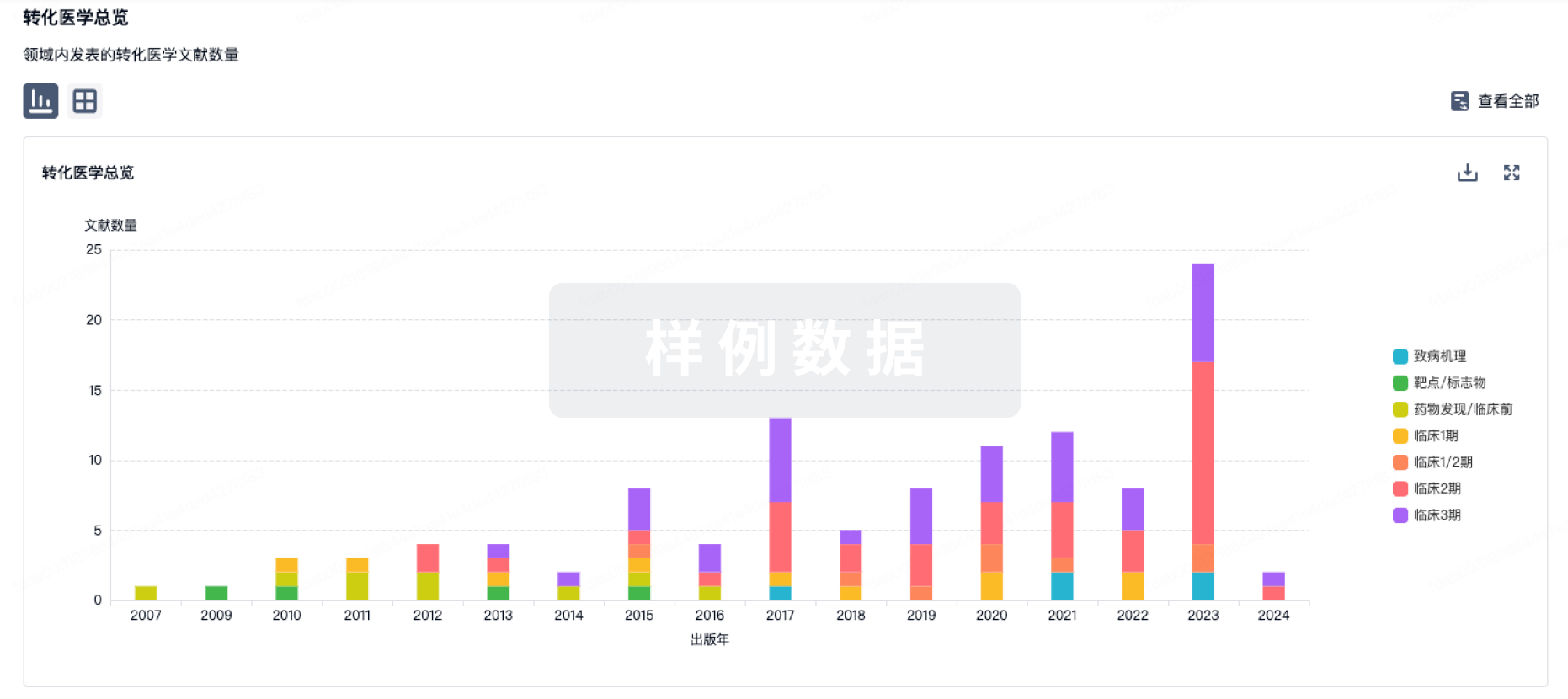
转化医学
使用我们的转化医学数据加速您的研究。
登录
或
药物交易
使用我们的药物交易数据加速您的研究。
登录
或
核心专利
使用我们的核心专利数据促进您的研究。
登录
或
临床分析
紧跟全球注册中心的最新临床试验。
登录
或
批准
利用最新的监管批准信息加速您的研究。
登录
或
生物类似药
生物类似药在不同国家/地区的竞争态势。请注意临床1/2期并入临床2期,临床2/3期并入临床3期
登录
或
特殊审评
只需点击几下即可了解关键药物信息。
登录
或
生物医药百科问答
全新生物医药AI Agent 覆盖科研全链路,让突破性发现快人一步
立即开始免费试用!
智慧芽新药情报库是智慧芽专为生命科学人士构建的基于AI的创新药情报平台,助您全方位提升您的研发与决策效率。
立即开始数据试用!
智慧芽新药库数据也通过智慧芽数据服务平台,以API或者数据包形式对外开放,助您更加充分利用智慧芽新药情报信息。
生物序列数据库
生物药研发创新
免费使用
化学结构数据库
小分子化药研发创新
免费使用