预约演示
更新于:2026-02-27
cRGD-ZW800-1
更新于:2026-02-27
概要
基本信息
非在研机构- |
权益机构- |
最高研发阶段临床2期 |
首次获批日期- |
最高研发阶段(中国)- |
特殊审评孤儿药 (美国)、孤儿药 (欧盟) |
登录后查看时间轴
结构/序列
分子式C78H106N13O16S2 |
InChIKeyYKVVVECWJGQERY-ZVIXCXBOSA-O |
CAS号1239855-63-0 |
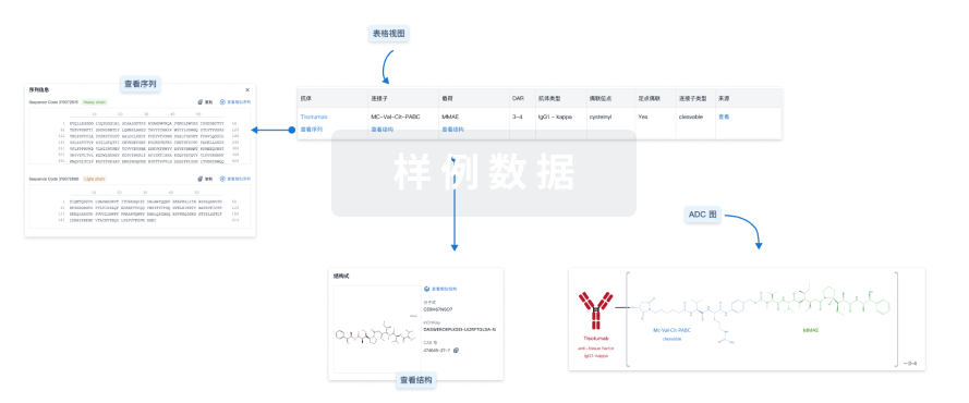
使用我们的ADC技术数据为新药研发加速。
登录
或

Sequence Code 1507071

关联
5
项与 cRGD-ZW800-1 相关的临床试验NCT05752149
The STELLAR Trial: Fluorescence-guided Surgery in Laryngeal- and Hypopharyngeal Cancer: a Feasibility Trial
This is an open-label, single-dose, prospective clinical trial. The study comprises 2 work packages. The main objective of work package I (WP-1) is to assess feasibility of Fluorescence imaing (FLI) during total laryngectomy (TLE) and to assess the optimal dose of the cRGD-ZW800-1. Work package II (WP-II) is designed to assess whether FLI can detect and decrease tumor positive margins after a TLE.
开始日期2025-06-01 |
申办/合作机构 |
NCT05518071
Intraoperative Near-infrared Fluorescence Imaging in Pancreatic- and Extrahepatic Bile Duct Tumors Using cRGD-ZW800-1 and Dedicated Imaging Systems: A Phase II Feasibility Testing, Dose-ranging and Optimal Dose-(Interval) Selection Trial
Pancreas as well as Cholangiocarcinoma have a dismal prognosis at time of diagnosis, due to late onset of clinical symptoms, patients present with advance disease. Complete surgical resection is the only potential curative treatment, however only a small percentage is eligible for upfront total surgical resection due to extension into anatomical related important vascular structures. Neoadjuvant chemo(radio)therapy has become the standard treatment modality for non-primary resectable disease (borderline resectable and locally advanced pancreatic cancer (LAPC)), where subsequent downstaging can make identification of the primary tumor more challenging during surgery. Near-infrared (NIR) fluorescence imaging can aid surgeons by providing real-time visualization of tumors, suspect lymph nodes and vital structures during surgery. Additional intra-operative feedback could possibly reduce the frequency of positive resection margins and increase complete removal of locally spread tumor and involved lymph nodes and could thereby improve patient outcomes as well as overall survival. cRGD-ZW800-1 is a targeted NIR-fluorophore, with specific binding capacity for integrins (αvβ3, αvβ5, αvβ6) which are overexpressed on tumor cells and tumor-associated vascular endothelium associated with neoangiogenesis.
开始日期2022-11-09 |
NCT04191460
Guided by Light: Optimizing Surgical Excision of Oral Cancer Using Real-time Fluorescence Imaging
This is a two-staged clinical trial to investigate the feasibility of intraoperative Fluorescence Imaging (FLI) to adequately assess tumor margins in patients with oral cancer using cRGD-ZW800-1.
开始日期2022-07-12 |
申办/合作机构 |
100 项与 cRGD-ZW800-1 相关的临床结果
登录后查看更多信息
100 项与 cRGD-ZW800-1 相关的转化医学
登录后查看更多信息
100 项与 cRGD-ZW800-1 相关的专利(医药)
登录后查看更多信息
4
项与 cRGD-ZW800-1 相关的文献(医药)2021-09-01·Clinical cancer research : an official journal of the American Association for Cancer Research
NIR Fluorescence Imaging of Colon Cancer With cRGD-ZW800-1—Response
Letter
作者: Vahrmeijer, Alexander L. ; Sier, Cornelis F.M.
We thank Kossatz and Notni for their interest in our article about the use of fluorescent cRGD-ZW800-1 for visualization of tumor tissue in humans during surgery ([1][1]). Their concern seems the specificity of the tracer for integrin αvβ6, which is lower than for αvβ3, and the relation with our
2017-03-28·Oncotarget
Real-time near-infrared fluorescence imaging using cRGD-ZW800-1 for intraoperative visualization of multiple cancer types
Article
作者: Kuil, Joeri ; Vahrmeijer, Alexander L. ; Boonstra, Martin C. ; Sier, Cornelis F.M. ; Valentijn, A. Rob P.M. ; Bordo, Mark W. ; Handgraaf, Henricus J.M. ; Vinkenburg-van Slooten, Maaike L. ; Prevoo, Hendrica A.J.M. ; van de Velde, Cornelis J.H. ; Frangioni, John V. ; Sibinga Mulder, Babs G. ; Boogerd, Leonora S.F. ; Burggraaf, Jacobus
Incomplete resections and damage to critical structures increase morbidity and mortality of patients with cancer. Targeted intraoperative fluorescence imaging aids surgeons by providing real-time visualization of tumors and vital structures. This study evaluated the tumor-targeted zwitterionic near-infrared fluorescent peptide cRGD-ZW800-1 as tracer for intraoperative imaging of multiple cancer types. cRGD-ZW800-1 was validated in vitro on glioblastoma (U-87 MG) and colorectal (HT-29) cell lines. Subsequently, the tracer was tested in orthotopic mouse models with HT-29, breast (MCF-7), pancreatic (BxPC-3), and oral (OSC-19) tumors. Dose-ranging studies, including doses of 0.25, 1.0, 10, and 30 nmol, in xenograft tumor models suggest an optimal dose of 10 nmol, corresponding to a human equivalent dose of 63 μg/kg, and an optimal imaging window between 2 and 24 h post-injection. The mean half-life of cRGD-ZW800-1 in blood was 25 min. Biodistribution at 4 h showed the highest fluorescence signals in tumors and kidneys. In vitro and in vivo competition experiments showed significantly lower fluorescence signals when U-87 MG cells (minus 36%, p = 0.02) or HT-29 tumor bearing mice (TBR at 4 h 3.2 ± 0.5 vs 1.8 ± 0.4, p = 0.03) were simultaneously treated with unlabeled cRGD. cRGD-ZW800-1 visualized in vivo all colorectal, breast, pancreatic, and oral tumor xenografts in mice. Screening for off-target interactions, cRGD-ZW800-1 showed only inhibition of COX-2, likely due to binding of cRGD-ZW800-1 to integrin αVβ3. Due to its recognition of various integrins, which are expressed on malignant and neoangiogenic cells, it is expected that cRGD-ZW800-1 will provide a sensitive and generic tool to visualize cancer during surgery.
International journal of surgery (London, England)
Precise and safe pulmonary segmentectomy enabled by visualizing cancer margins with dual-channel near-infrared fluorescence
Article
作者: G. Kate Park ; Byeong Hyeon Choi ; Hyun Koo Kim ; Haoran Wang ; Chungyeul Kim ; Kai Bao ; Kyungsu Kim ; Hak Soo Choi ; Ok Hwa Jeon ; Shinya Yokomizo ; Jiyun Rho
Background::
Segmentectomy is a type of limited resection surgery indicated for patients with very early-stage lung cancer or compromised function because it can improve quality of life with minimal removal of normal tissue. For segmentectomy, an accurate detection of the tumor with simultaneous identification of the lung intersegment plane is critical. However, it is not easy to identify both during surgery. Here, the authors report dual-channel image-guided lung cancer surgery using renally clearable and physiochemically stable targeted fluorophores to visualize the tumor and intersegmental plane distinctly with different colors; cRGD-ZW800 (800 nm channel) targets tumors specifically, and ZW700 (700 nm channel) simultaneously helps discriminate segmental planes.
Methods::
The near-infrared (NIR) fluorophores with 700 nm and with 800 nm channels were developed and evaluated the feasibility of dual-channel fluorescence imaging of lung tumors and intersegmental lines simultaneously in mouse, rabbit, and canine animal models. Expression levels of integrin αvβ3, which is targeted by cRGD-ZW800-PEG, were retrospectively studied in the lung tissue of 61 patients who underwent lung cancer surgery.
Results::
cRGD-ZW800-PEG has clinically useful optical properties and outperforms the FDA-approved NIR fluorophore indocyanine green and serum unstable cRGD-ZW800-1 in multiple animal models of lung cancer. Combined with the blood-pooling agent ZW700-1C, cRGD-ZW800-PEG permits dual-channel NIR fluorescence imaging for intraoperative identification of lung segment lines and tumor margins with different colors simultaneously and accurately.
Conclusion::
This dual-channel image-guided surgery enables complete tumor resection with adequate negative margins that can reduce the recurrence rate and increase the survival rate of lung cancer patients.
1
项与 cRGD-ZW800-1 相关的新闻(医药)2026-01-07
BONITA SPRINGS, Fla.--(BUSINESS WIRE)--Curadel Pharma, a pioneer in zwitterionicity and innovator in advanced radiotherapies and imaging drugs, announced today that CPI-008 (cRGD-ZW800-1), a novel integrin-targeted, zwitterionic imaging drug for margin detection of pancreatic cancer during surgery, has been granted Orphan Drug Designation (ODD) by the U.S. Food and Drug Administration (FDA) and the European Medicines Agency (EMA).
“Receiving orphan designation from both the FDA and the EMA is a profoundly important milestone for Curadel, granting us valuable incentives to fuel our development efforts,” said John V. Frangioni, MD, PhD, Curadel founder and CEO. “As a pioneering company working to introduce significant advances in surgical imaging, the efficiencies of fee exemptions, credits, along with the potential for market exclusivity are vital tools to help us smartly deploy our resources and focus on delivering value to the surgical community.”
In the U.S., FDA grants ODD to therapeutic candidates for conditions affecting fewer than 200,000 people in the U.S. This designation provides incentives to advance clinical development including exemption from user fees, tax credits for qualified clinical trials, and potential for up to seven years of U.S. market exclusivity if the product is approved for its designated indication. Similarly, EMA's designation includes incentives including protocol assistance, reduced regulatory fees, and potential for early access conditional approvals, as well as market exclusivity up to 10 years if approved.
Pancreatic cancer remains one of the most challenging cancers to treat, and current imaging tools are not fully effective in helping to identify the full extent of cancerous cells to allow for full removal during surgical procedures. By enhancing surgeons’ ability to accurately visualize cancerous cells, Curadel’s technology could become an important asset in the surgical suite to optimize outcomes not only for pancreatic cancer patients, but potentially for other tumor types as well.
CPI-008 has demonstrated strong imaging capabilities in investigator-initiated Phase 2 studies in multiple cancers including pancreatic cancer, head and neck cancer, and colorectal cancer. As part of its strategic pipeline curation, Curadel is evaluating out-licensing opportunities for the program, offering a highly differentiated technology that potential partners can leverage to expand leadership in the competitive imaging market.
About Curadel Pharma
Curadel Pharma, based in Bonita Springs, FL, is an audacious innovator aiming to prevent tumor resistance via disruptive zwitterionic radiopharma technology. Its novel, pan-tumor platform prevents tumor regrowth and resistance while eliminating collateral damage to healthy tissues. Curadel’s lead radiopharmaceutical candidate, CPI-003, represents a potential best-in-class zwitterionic targeted alpha therapy (TAT) initially focused on the treatment of challenging rare cancers. The company is also developing image-guided surgical drugs as well as zwitterionic MRI contrast agents. Its late-stage drug for ureter imaging is the subject of an exclusive distribution agreement with a Tier 1 medical device company and is currently under evaluation in a pivotal trial. For more information, visit www.curadelpharma.com or follow us on LinkedIn.
引进/卖出孤儿药临床2期
100 项与 cRGD-ZW800-1 相关的药物交易
登录后查看更多信息
研发状态
10 条进展最快的记录, 后查看更多信息
登录
| 适应症 | 最高研发状态 | 国家/地区 | 公司 | 日期 |
|---|---|---|---|---|
| 胰腺腺瘤 | 临床2期 | 荷兰 | 2022-11-09 | |
| 胆管癌 | 临床2期 | 荷兰 | 2022-11-09 | |
| 头颈部鳞状细胞癌 | 临床2期 | 荷兰 | 2022-07-12 | |
| 口腔鳞状细胞癌 | 临床2期 | 荷兰 | 2022-07-12 | |
| 头颈部肿瘤 | 临床2期 | 荷兰 | - | |
| 胰腺癌 | 临床2期 | 荷兰 | - |
登录后查看更多信息
临床结果
临床结果
适应症
分期
评价
查看全部结果
| 研究 | 分期 | 人群特征 | 评价人数 | 分组 | 结果 | 评价 | 发布日期 |
|---|
No Data | |||||||
登录后查看更多信息
转化医学
使用我们的转化医学数据加速您的研究。
登录
或

药物交易
使用我们的药物交易数据加速您的研究。
登录
或

核心专利
使用我们的核心专利数据促进您的研究。
登录
或

临床分析
紧跟全球注册中心的最新临床试验。
登录
或

批准
利用最新的监管批准信息加速您的研究。
登录
或

特殊审评
只需点击几下即可了解关键药物信息。
登录
或

生物医药百科问答
全新生物医药AI Agent 覆盖科研全链路,让突破性发现快人一步
立即开始免费试用!
智慧芽新药情报库是智慧芽专为生命科学人士构建的基于AI的创新药情报平台,助您全方位提升您的研发与决策效率。
立即开始数据试用!
智慧芽新药库数据也通过智慧芽数据服务平台,以API或者数据包形式对外开放,助您更加充分利用智慧芽新药情报信息。
生物序列数据库
生物药研发创新
免费使用
化学结构数据库
小分子化药研发创新
免费使用

