预约演示
更新于:2025-06-04
MF-03
更新于:2025-06-04
概要
基本信息
在研机构- |
非在研机构 |
权益机构- |
最高研发阶段终止药物发现 |
首次获批日期- |
最高研发阶段(中国)- |
特殊审评- |

关联
100 项与 MF-03 相关的临床结果
登录后查看更多信息
100 项与 MF-03 相关的转化医学
登录后查看更多信息
100 项与 MF-03 相关的专利(医药)
登录后查看更多信息
8
项与 MF-03 相关的文献(医药)2018-01-11·Pharmaceutical nanotechnology
Microsponge Embedded Tablets for Sustained Delivery of Nifedipine
Article
作者: Tekade, Muktika ; Tekade, Rakesh K. ; Hansbro, Philp M. ; Maheshwari, Rahul ; Sharma, Piyoosh ; Atneriya, Umesh ; Dua, Kamal
BACKGROUND:
Nifedipine is a potential therapeutic agent for the treatment of cardiovascular disturbances, although it suffers from short half-life (t1/2, 2 hr).
OBJECTIVE:
To address the problem, we first prepared nifedipine loaded sustained release microsponges and then formulated tablets for effective clinical application and patient compliance.
METHOD:
Preparations of microsponges were carried out using different compositions of nifedipine and polymer (1:1, 1:2 and 1:3 % molar ratio) using emulsion solvent diffusion technique.
RESULTS:
The microsponges with molar ratio 1:3 (formulation code: MF-3) found optimized as revealed by analyzing surface morphology, better powder flow properties (angle of repose; 28.80 ± 0.9, Hausner ratio 1.15 ± 0.2, % compressibility 15.28 ± 0.5% and higher % drug content (80 ± 1.9 %). Different batches of tablets were then formulated incorporating MF-3 microsponges and different proportions (10-50 %) of microcrystalline cellulose and starch as additives. Among tablet formulations, batch composed of 48% of MF-3, 30% of MCC, 20 % of starch and 2 % of talc (TF-33), showed 92.73 ± 2.19 % drug release during 24 hr in vitro release study in comparison to other batches including commercial formulation which was found to be released completely in 20 hr. Further, stability analysis revealed good drug retention of loaded nifedipine as well as consistent in vitro release pattern over a period of 90 days at 40°C and 75% RH.
CONCLUSION:
The microsponge tablet delivery system was found to be superior concerning the therapeutic advantage as well as manufacturing feasibility of nifedipine.
2013-11-01·Leukemia & lymphoma
Prognostic implications and clinical characteristics associated with bone marrow fibrosis in patients with myelofibrosis
Letter
作者: Nazha, Aziz ; Estrov, Zeev ; Verstovsek, Srdan ; Bueso-Ramos, Carlos E. ; Cortes, Jorge ; Kantarjian, Hagop
To the editor:
Myelofibrosis (MF) is a heterogeneous, hematopoietic stem cell malignancy characterized by abnormal proliferation of myeloid cells of variable maturity and function [1]. The most common clinical manifestations of the disease are anemia, leukocytosis, leukopenia, thrombocytosis, constitutional symptoms, and marked splenomegaly with extramedullary hematopoiesis [1]. Bone marrow fibrosis (BMF), which results from abnormal deposition of reticulin and collagen fibers in the bone marrow plays a major role in the pathophysiology and clinical manifestation of the disease. Various scoring systems have been developed to evaluate the amount of reticulin and collagen fibers in BMF, the most widely accepted is the 4-grade scoring system (MF 0–3) recommended by the European consensus [2]. The prognostic effect of BMF in MF remains controversial; however, it has been suggested that BMF may affect overall survival (OS) in patients with MF [3–5].
We retrospectively analyzed the medical records of 537 patients who were diagnosed with MF according to World Health Organization criteria [6] and were referred to our institution between February 2005 and December 2009. Project was based on a chart review protocol approved by the Institutional Review Board. Paraffin-embedded trephine bone marrow biopsies obtained from patients at their first presentation to MD Anderson were reviewed. Grades from 0 to 3 were documented, with grade 3 representing the most severe grade of fibrosis [2]. Twenty-five patients were excluded from the final analysis because of incomplete documentation of BMF grading in their reviewed biopsies. The OS rate was calculated from the time of first biopsy at MD Anderson to the time of death or last follow-up; the EFS rate was calculated from the time of first biopsy at MD Anderson to time of death, leukemia transformation, or last follow-up. The IPSS and the DIPSS were calculated as described previously [7,8]. Differences among variables were evaluated by the chi-square for categorical variables and by Kruskal-Wallis and Mann-Whitney U tests for continuous variables. A Cox proportional hazard regression module was used to determine the independent predictors of severity of BMF. Time-to-event analyses were performed by the Kaplan-Meier method, and survival curves were compared with the log rank test.
The final analysis included 512 patients (352 patients with primary MF, 88 with post-PV MF, and 72 with post-ET MF). The patient’s demographics and disease characteristics are summarized in Table 1. The median time from diagnosis of MF to the first bone marrow biopsy done at MD Anderson was 4 months (range, 0–388 months), and the median follow-up was 16.5 months (range, 0–79 months). Higher grades of BMF were associated with a lower hemoglobin level (P < 0.001), lower white blood cell count (P = 0.03), and lower platelet count (P = 0.02), higher percentage of blasts in peripheral blood (P = 0.001), as well as, significantly larger spleen (P = 0.01) and liver (P = 0.04). In addition, abnormal cytogenetic findings were associated with higher grades of BMF (P = 0.01); but, neither the presence of the JAK2 V617F mutation nor the allele burden correlated with the severity of BMF (P = 0.28). However, in a multivariate analysis, lower hemoglobin level (P = 0.003), higher percentage of blasts in the peripheral blood (P = 0.015), and larger spleen (P = 0.006) were the only independent prognostic parameters for the severity of BMF. In addition, survival risk assessment of patients using the IPSS and DIPSS scoring systems correlated well with the severity of BMF (Table 1). Vener and colleagues [3] reported the prognostic significance of BMF grading in MF. One hundred thirteen patients were included in their study, 29% had MF-0, 24% MF-1, 23% MF-2, 11% MF-3, and 13% secondary MF. Patients with MF-3 had inferior OS compared to patients with MF-0 (P = 0.001), and MF-1+2 groups (P = 0.003). In addition, Thiele and colleagues [5] had also reported inferior OS among patients with grades MF-2 or MF-3 compared with those with grades MF-0 or MF-1 (statistical significance was not reported) suggesting that BMF grading may play an important role in the prognosis of patients with MF. Recently, Barosi and colleagues [4] compared the clinical features of prefibrotic myelofibrosis (MF-0) to primary myelofibrosis fibrotic type (MF-1,2,and 3) and found that patients with prefibrotic myelofibrosis are predominantly females with younger age, higher hemoglobin, higher platelet count, lower white blood cell count and smaller spleen. Furthermore, with median follow up of 43 months (range, 1–375 months), the median OS for prefibrotic mylofibrosis patients was not reached and was significantly higher than patients with primary myelofibrosis fibrotic type (16.6 years, P <0,001). In our study, although BMF correlated well with DIPSS and IPSS scoring systems and no patients with MF-0 died or had an event, the rates of OS, EFS, and leukemia transformation free survival were not statistically significant between patients with different BMF grades (Table 1; Figure 1). These differences in our result could be explained by the higher number of patients with MF-3 and the lower number of patients with MF-0 in our patient population resulted from the referral of patients to our institution during their disease course rather than at diagnosis, as well as by the very short follow-up during the study period compared to other studies. Furthermore, it is interesting that significant number of patients with low/intermediate-1 risk by DIPSS score had high grades of BMF. This suggests that assessments of BMF at diagnosis and during the disease course may be important even in patients with low risk disease. However, given the difficulties of obtaining bone marrow biopsies regularly in clinical practice, regular assessment of BMF grade may not be necessary outside clinical trials and the incorporation of BMF grading in the prognostication systems of MF need to be further evaluated in larger patient population with longer follow up.
In conclusion, severe BMF has significant impact on the clinical manifestation of MF, as it correlates with lower hemoglobin, higher percentage of blasts in the peripheral blood, and larger spleen as well as the IPSS and DIPSS risk assessments. However, the OS, EFS and transformation to acute leukemia were similar in our experience among patients with various degrees of BMF, possibly due to limited number of patients with no or low grade BMF, and short follow up. Further clinical and biological studies and longer follow up are needed to fully understand the significant impact of BMF in patients with MF.
2012-12-01·Fibrogenesis & tissue repair
Replacement of hematopoietic system by allogeneic stem cell transplantation in myelofibrosis patients induces rapid regression of bone marrow fibrosis
Article
作者: Kröger, Nicolaus ; Thiele, Jürgen ; Kvasnicka, Michael
Bone marrow fibrosis is a hallmark of primary and post ET/PV myelofibrosis. To investigated the impact of replacement of the hematopoietic system in myelofibrosis patients by allogeneic stem cell transplantation on bone marrow fibrosis, we studied bone marrow fibrosis on bone marrow samples from 24 patients with myelofibrosis before and after dose-reduced conditioning followed by allogeneic stem cell transplantation from related or unrelated donor. Using the European Consensus on Grading Bone Marrow Fibrosis, before allografting all patients had advanced fibrosis MF-2 (n = 13) or MF-3 (n = 11). After transplantation, a complete (MF-0) or nearly complete (MF-1) regression of bone marrow fibrosis was seen in 59 % at day +100, in 90 % at day +180, and in 100 % at day +360. No correlation between occurrence of acute graft-versus-host disease, and fibrosis regression on day +180 was seen. We conclude that dose-reduced conditioning, followed by allogeneic stem cell transplantation, resulted in a rapid resolution of bone-marrow fibrosis suggesting the bone marrow fibrogenesis is a highly dynamic rather than static process in patients with myelofibrosis.
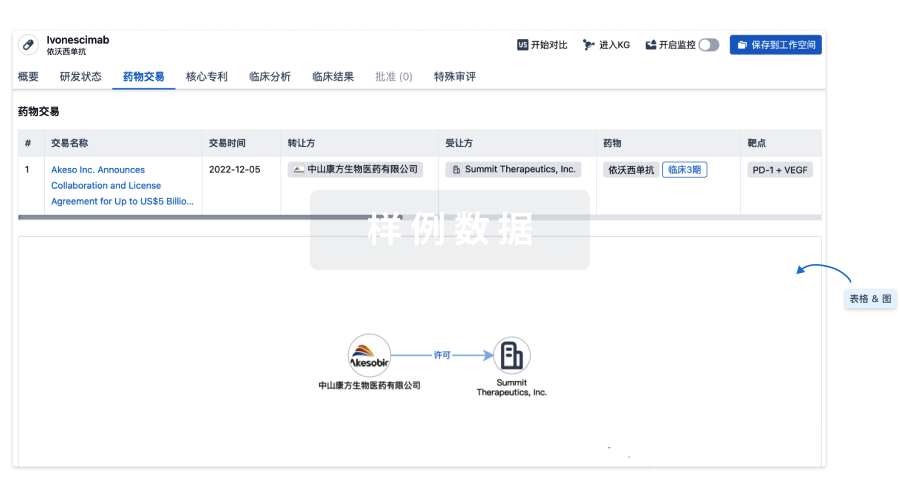
100 项与 MF-03 相关的药物交易
登录后查看更多信息
研发状态
10 条进展最快的记录, 后查看更多信息
登录
| 适应症 | 最高研发状态 | 国家/地区 | 公司 | 日期 |
|---|---|---|---|---|
| 酸中毒 | 药物发现 | 韩国 | 2024-08-26 |
登录后查看更多信息
临床结果
临床结果
适应症
分期
评价
查看全部结果
| 研究 | 分期 | 人群特征 | 评价人数 | 分组 | 结果 | 评价 | 发布日期 |
|---|
No Data | |||||||
登录后查看更多信息
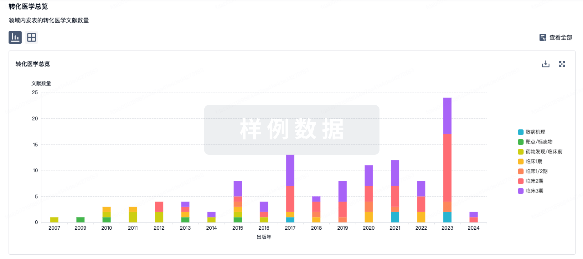
转化医学
使用我们的转化医学数据加速您的研究。
登录
或
药物交易
使用我们的药物交易数据加速您的研究。
登录
或
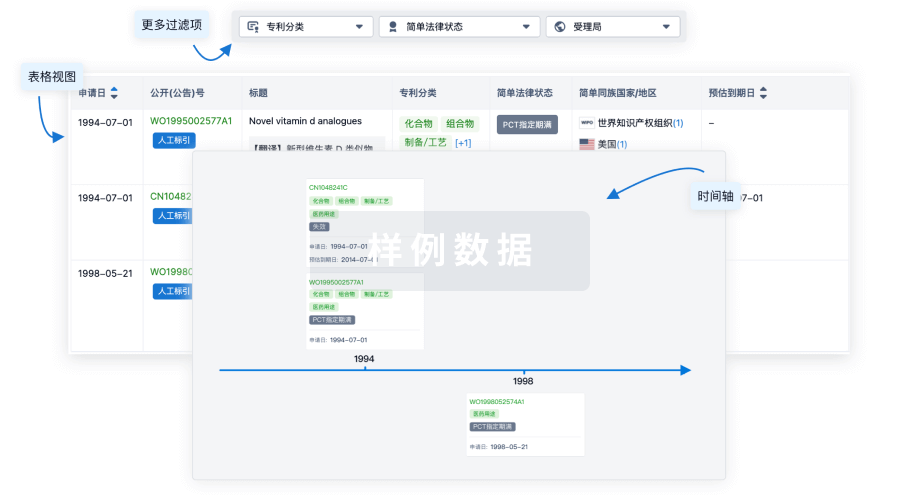
核心专利
使用我们的核心专利数据促进您的研究。
登录
或
临床分析
紧跟全球注册中心的最新临床试验。
登录
或
批准
利用最新的监管批准信息加速您的研究。
登录
或
特殊审评
只需点击几下即可了解关键药物信息。
登录
或
生物医药百科问答
全新生物医药AI Agent 覆盖科研全链路,让突破性发现快人一步
立即开始免费试用!
智慧芽新药情报库是智慧芽专为生命科学人士构建的基于AI的创新药情报平台,助您全方位提升您的研发与决策效率。
立即开始数据试用!
智慧芽新药库数据也通过智慧芽数据服务平台,以API或者数据包形式对外开放,助您更加充分利用智慧芽新药情报信息。
生物序列数据库
生物药研发创新
免费使用
化学结构数据库
小分子化药研发创新
免费使用