预约演示
更新于:2026-07-04
Endomorphin-2
更新于:2026-07-04
概要
基本信息
药物类型 合成多肽 |
别名 EM-2 |
作用方式 激动剂 |
作用机制 Opioid receptors激动剂(阿片样受体激动剂) |
治疗领域- |
在研适应症- |
非在研适应症- |
非在研机构- |
权益机构- |
最高研发阶段临床前 |
首次获批日期- |
最高研发阶段(中国)- |
特殊审评- |
结构/序列
Sequence Code 58736

关联
100 项与 Endomorphin-2 相关的临床结果
登录后查看更多信息
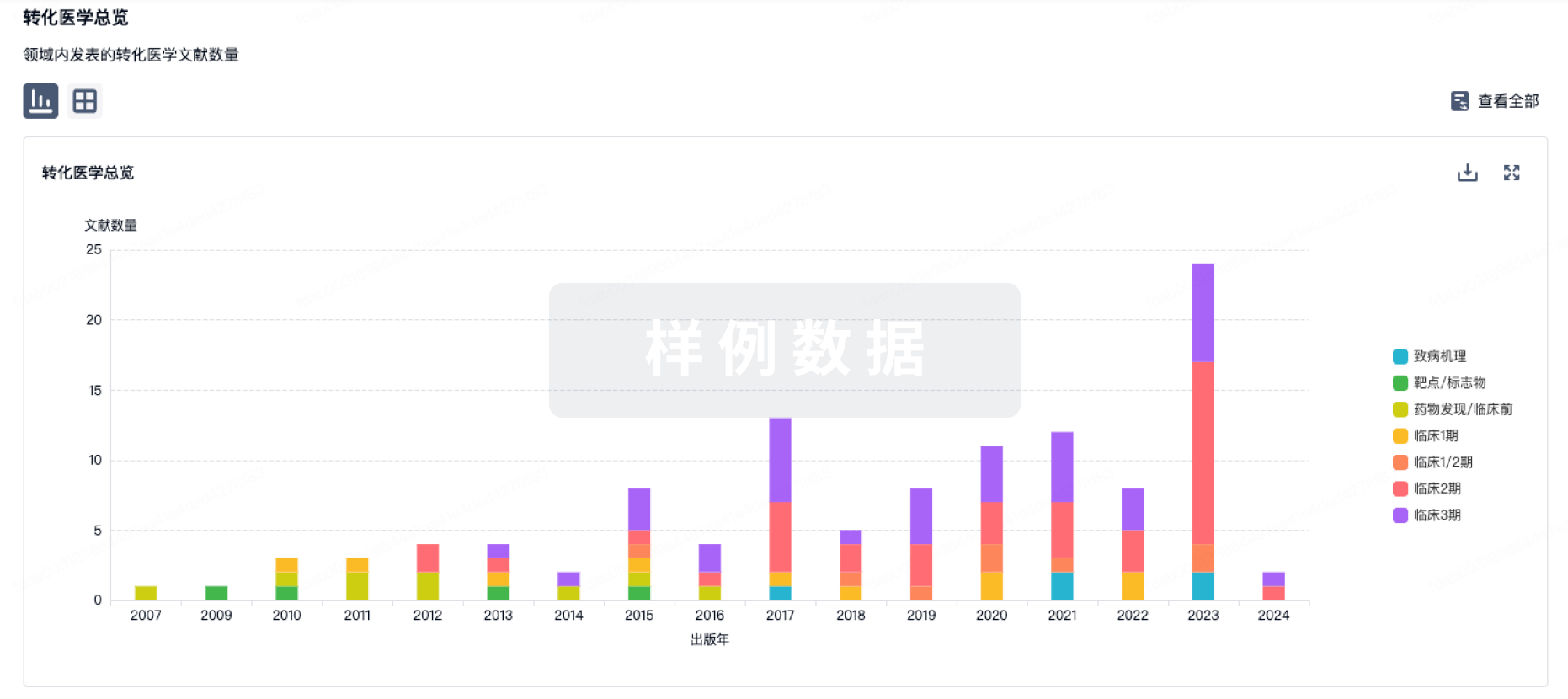
100 项与 Endomorphin-2 相关的转化医学
登录后查看更多信息
100 项与 Endomorphin-2 相关的专利(医药)
登录后查看更多信息
336
项与 Endomorphin-2 相关的文献(医药)2026-03-01·SPECTROCHIMICA ACTA PART A-MOLECULAR AND BIOMOLECULAR SPECTROSCOPY
Non-invasive pharmacological profiling of novel endomorphin-2 analogs by confocal Raman spectroscopy and imaging
Article
作者: Gach-Janczak, Katarzyna ; Piekielna-Ciesielska, Justyna ; Mroczkowska, Julia ; Surmacki, Jakub Maciej
Understanding how biased opioid agonists signal through different intracellular pathways, particularly in the context of pain, is still a major challenge. One reason is the lack of suitable imaging methods that can capture ligand-receptor interactions and their downstream effects in live cells with spatial resolution. In this study, we apply a Raman-based imaging technique that allowed us to observe and compare the intracellular distribution and signaling behavior of endomorphin-2 analogs, JPC-11 and JPC-13, shedding light on the distinct dynamics of G protein versus β-arrestin recruitment by the MOR. The data obtained in this study provide valuable insights into the pharmacological activity of opioid analogs biased toward G protein or β-arrestin 2. We observed changes in the Raman bands at 750, 782, 1003, 1035, 1092, 1126, 1254, 1302, 1310, 1339, 1444, 1583, and 1654 cm-1, which correspond to molecular features of lipids, proteins, carbohydrates and DNA associated with specific cellular organelles. Structural rearrangements can impact the vibrational environment of both ligand- and receptor-associated aromatic residues, leading to measurable differences in the tyrosine-associated Raman bands, particularly in the 800-1100 cm-1 region, with a prominent shift of the band at 1035 cm-1. Here we develop a new method to evaluate and understand the consequences of biased agonism in terms of therapeutic potential and side effects of opioid receptor ligands.
2026-01-31·FASEB JOURNAL
EM2, a Novel
Elephantopus mollis
H.B.K. Monomer, Enhances Radiosensitivity in Cervical Cancer Through Dual Inhibition of AKT and Autophagy
Article
作者: Li, Fengying ; Li, Guiqing ; Zhang, Xiaoying ; Xue, Lujiadai ; Li, Nan ; Jiang, Jianwei ; Zhou, Shimin ; Tang, Jie ; Tang, Lindong ; Wang, Xiaoyu ; Gu, Jianyi
ABSTRACT:
Radiotherapy activates both the PI3K/AKT pathway and autophagy in cervical cancer, contributing to radioresistance. To address this, EM2, a dual AKT/autophagy inhibitor, was investigated for its potential to enhance radiosensitivity. RNA‐Seq, Western blot, qRT‐PCR, and transmission electron microscopy were employed to analyze PI3K/AKT and autophagy pathways following irradiation, while CCK8, clone formation, and flow cytometry assays evaluated proliferation, apoptosis, and cell cycle effects. KEGG and GSEA analyses confirmed irradiation‐induced activation of the PI3K/AKT pathway. Both PI3K and autophagy inhibitors significantly improved efficacy, whereas EM2 suppressed AKT pathway activation and autophagy, synergistically inducing G2/M phase arrest, and increasing apoptosis. In vivo experiments using a nude mouse xenograft model demonstrated that EM2 combined with irradiation effectively suppressed tumor growth, PI3K/AKT activation, and autophagy without significant toxicity. These results underscore EM2 as a promising therapeutic agent to overcome radioresistance by simultaneously targeting the PI3K/AKT pathway and autophagy.
2026-01-01·Advanced Science
EM2, a Natural Product MST1/2 Kinase Activator, Suppresses Non‐Small Cell Lung Cancer via Hippo Pathway Activation
Article
作者: Ruijie Yuan ; Mingyu Pan ; Sijia Li ; Wenlin Wang ; Junxi Liu ; Ziling Tang ; Liujin Zhou ; Yubo Zhang ; Hao Wang ; Huayan Xie ; Zekai Fang ; Guocai Wang ; Siyu Yang ; Qiang Lin ; Jinxuan Su ; Xiaoyong Dai
Abstract:
Lung cancer, 85% of which is non‐small cell lung cancer (NSCLC), is the cancer with the highest incidence and mortality rate worldwide. Despite recent advancements in therapeutic approaches, the efficacy of conventional radiotherapy and chemotherapy remains suboptimal, highlighting the urgent need for more effective treatment strategies. Dysregulation of kinases MST1 and MST2 (MST1/2) is implicated in the progression of NSCLC, positioning MST1/2 as a potential therapeutic target. However, no high‐selectivity and high‐efficacy MST1/2 activator is identified to date. In this study, by using computer‐aided virtual screening combined with cell experiments, EM2 is identified as a promising MST1/2‐binding candidate. Subsequent experimental validation demonstrates that EM2 significantly suppresses the proliferation, migration, and invasion of NSCLC cells by directly targeting MST1/2 and enhancing its kinase activity, thereby activating the Hippo signaling pathway and reducing nuclear translocation of the downstream effector YAP. Both in vivo xenograft models and organoid models demonstrates that EM2 effectively suppresses NSCLC tumor growth. In summary, this study not only reaffirms MST1/2 as a viable therapeutic target for NSCLC but also provides compelling experimental evidence supporting EM2 as a highly effective and promising anti‐cancer agent.
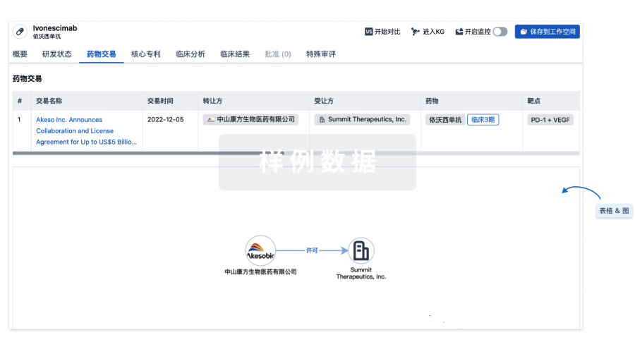
100 项与 Endomorphin-2 相关的药物交易
登录后查看更多信息
研发状态
登录后查看更多信息
临床结果
临床结果
适应症
分期
评价
查看全部结果
| 研究 | 分期 | 人群特征 | 评价人数 | 分组 | 结果 | 评价 | 发布日期 |
|---|
No Data | |||||||
登录后查看更多信息
转化医学
使用我们的转化医学数据加速您的研究。
登录
或
药物交易
使用我们的药物交易数据加速您的研究。
登录
或
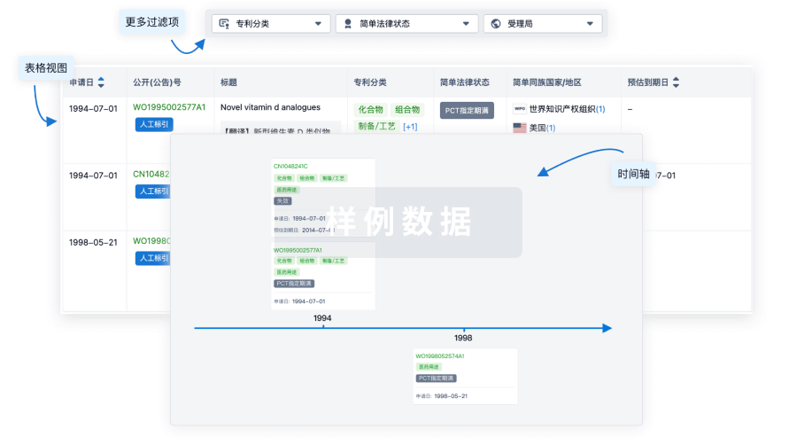
核心专利
使用我们的核心专利数据促进您的研究。
登录
或
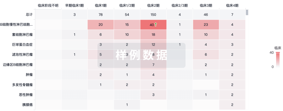
临床分析
紧跟全球注册中心的最新临床试验。
登录
或
批准
利用最新的监管批准信息加速您的研究。
登录
或
生物类似药
生物类似药在不同国家/地区的竞争态势。请注意临床1/2期并入临床2期,临床2/3期并入临床3期
登录
或
特殊审评
只需点击几下即可了解关键药物信息。
登录
或
生物医药百科问答
全新生物医药AI Agent 覆盖科研全链路,让突破性发现快人一步
立即开始免费试用!
智慧芽新药情报库是智慧芽专为生命科学人士构建的基于AI的创新药情报平台,助您全方位提升您的研发与决策效率。
立即开始数据试用!
智慧芽新药库数据也通过智慧芽数据服务平台,以API或者数据包形式对外开放,助您更加充分利用智慧芽新药情报信息。
生物序列数据库
生物药研发创新
免费使用
化学结构数据库
小分子化药研发创新
免费使用