预约演示
更新于:2026-02-07
Octreotide (Imagine Pharma)
更新于:2026-02-07
概要
基本信息
非在研机构- |
权益机构- |
最高研发阶段临床前 |
首次获批日期- |
最高研发阶段(中国)- |
特殊审评- |
关联
100 项与 Octreotide (Imagine Pharma) 相关的临床结果
登录后查看更多信息
100 项与 Octreotide (Imagine Pharma) 相关的转化医学
登录后查看更多信息
100 项与 Octreotide (Imagine Pharma) 相关的专利(医药)
登录后查看更多信息
3,092
项与 Octreotide (Imagine Pharma) 相关的文献(医药)2026-03-01·American journal of ophthalmology case reports
Neuroendocrine tumor of the lung presenting as slowly progressive, painful bilateral choroidal masses
Article
作者: Zhao, Sharon H ; Park, Susanna S
Purpose:
To report a rare case of a metastatic slow growing neuroendocrine tumor of the lung that presented as slow-growing bilateral choroidal masses with progressive vision loss and retrobulbar pain.
Observations:
A 64-year-old male presented with vision loss and retrobulbar pain bilaterally, progressively worsened for four months. He had no significant past medical history but had seen an ophthalmologist in Mexico starting 1.5 years earlier and was informed he may have a mass in the eye when last seen five months earlier. Ophthalmic examination revealed visual acuity of 20/400 bilaterally with large elevated amelanotic choroidal masses in both maculae with overlying pigmentary changes and subretinal fluid. The initial differential diagnosis included choroidal metastasis and infectious and inflammatory etiologies such as posterior scleritis and granuloma. Whole body PET-CT scan revealed a focus of hypermetabolic activity in the lung suspicious for a slow growing malignancy or infectious/inflammatory lesion. Due to insurance issues, there was a delay in referral to medical oncology. The choroidal lesions gradually increased in size over four months. Eventual biopsy of the lung confirmed the diagnosis of a slow-growing neuroendocrine tumor of the lung. Systemic treatment with octreotide, Capecitabine, and Temozolomide was initiated with regression of choroidal tumors, improvement in visual acuity, and resolution of eye pain.
Conclusions and importance:
We describe a rare case of a bilateral choroidal metastasis from a slow growing neuroendocrine tumor of the lung that presented with slowly progressive bilateral vision loss and eye pain. Despite delays in diagnosis and treatment, eye pain resolved, and vision loss improved with systemic therapy alone.
2026-02-01·JOURNAL OF PHARMACEUTICAL SCIENCES
The impact of bile acid sequestrants on octreotide absorption
Article
作者: Houser, Sydney ; Al-Tamimi, Zahraa ; Feng, Mei ; Hageman, Michael J ; Elballa, Waleed
Despite significant advancements in peptide drug development, there is still a challenge in formulating and delivering peptide drugs orally. Current oral peptide drugs have very low bioavailability (<1%), which could be attributed, in part, to enzymatic instability, poor membrane permeability/flux, and the sequestration by intestinal colloids composed of bile acid and phospholipids that form bile acid-phospholipid mixed micelles (BAPMM). In this work, we examined the effect of perturbing the BAPMM with bile acid sequestrants (BAS) on the membrane flux and enzymatic stability of octreotide in vitro, and its potential impact on peptide absorption and bioavailability in vivo. Additionally, we tested the effect of adding cyclic E-cadherin peptide (ECP) permeation enhancers on the bioavailability of octreotide. The results suggest that using BAS decreases the bile acid levels and putatively disrupts the micellar structure, leading to a higher concentration of the free peptide to diffuse across the membrane. In vitro bile acid sequestration enhanced the overall peptide flux rate without compromising the improved enzymatic stability. Our in vivo data suggests that using the BAS, colestipol, did not have a significant impact on peptide absorption though. These results highlight the important role of BAPMM on bioaccessible drug concentration, as well as membrane permeation.
2026-01-23·MEDICINE
Individualized treatment strategies for primary hepatic neuroendocrine carcinoma: Two case reports and literature review (A CARE-compliant case report)
Review
作者: Lan, Xiang ; Chen, Kai ; Xiao, Heng ; Hu, Haiyang
Rationale::
Primary hepatic neuroendocrine carcinoma (PHNET) is an exceptionally rare malignancy with limited standardized treatment options.
Patient concerns::
Two patients presented with incidentally detected hepatic masses and nonspecific gastrointestinal symptoms.
Diagnoses::
Case 1 was diagnosed as primary hepatic neuroendocrine carcinoma (NEC, G3), and Case 2 as primary hepatic large-cell neuroendocrine carcinoma (LCNEC, G3), based on histopathology and immunohistochemistry after excluding extrahepatic origins.
Interventions::
Case 1 received transarterial chemoembolization (TACE), etoposide–cisplatin chemotherapy, hepatic arterial infusion chemotherapy (HAIC), and octreotide. Case 2 underwent 3 cycles of drug-eluting bead TACE (d-TACE), HAIC, and long-acting octreotide for symptomatic control of diarrhea.
Outcomes::
Case 1 experienced progressive disease and died of sepsis. Case 2 achieved significant tumor regression, allowing curative resection. No recurrence was observed at one-month follow-up.
Lessons::
The combination of d-TACE, HAIC, and octreotide may provide a potential downstaging approach for unresectable PHNET, but evidence remains preliminary and hypothesis-generating.
100 项与 Octreotide (Imagine Pharma) 相关的药物交易
登录后查看更多信息
研发状态
10 条进展最快的记录, 后查看更多信息
登录
| 适应症 | 最高研发状态 | 国家/地区 | 公司 | 日期 |
|---|---|---|---|---|
| 类癌 | 临床前 | 美国 | 2024-09-29 |
登录后查看更多信息
临床结果
临床结果
适应症
分期
评价
查看全部结果
| 研究 | 分期 | 人群特征 | 评价人数 | 分组 | 结果 | 评价 | 发布日期 |
|---|
No Data | |||||||
登录后查看更多信息
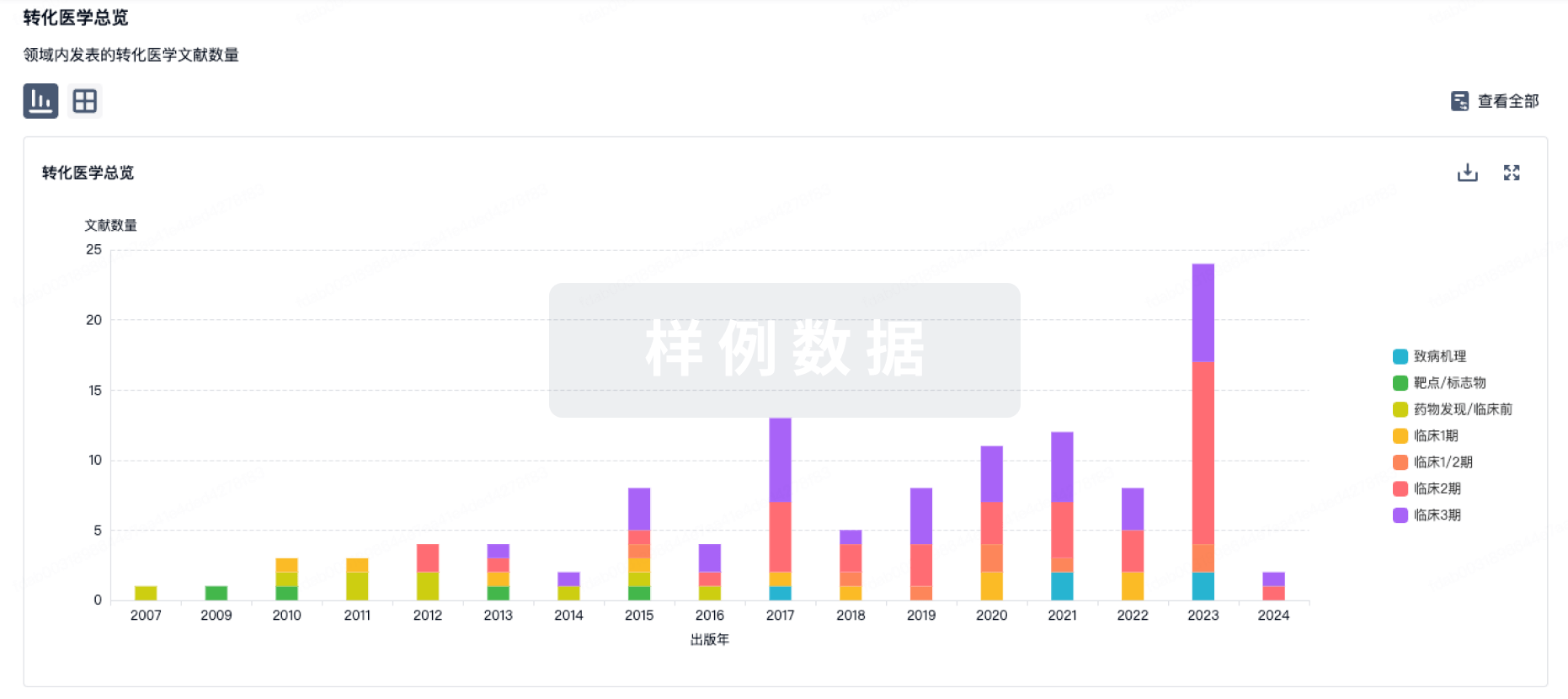
转化医学
使用我们的转化医学数据加速您的研究。
登录
或
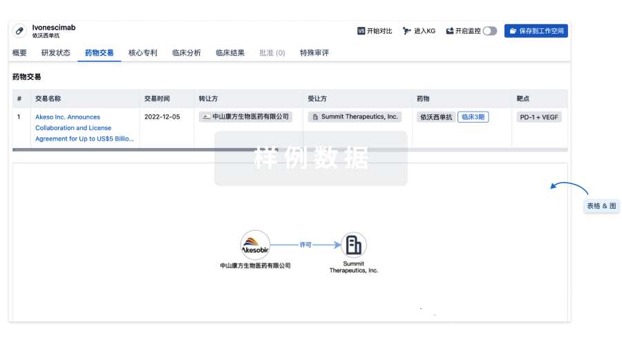
药物交易
使用我们的药物交易数据加速您的研究。
登录
或
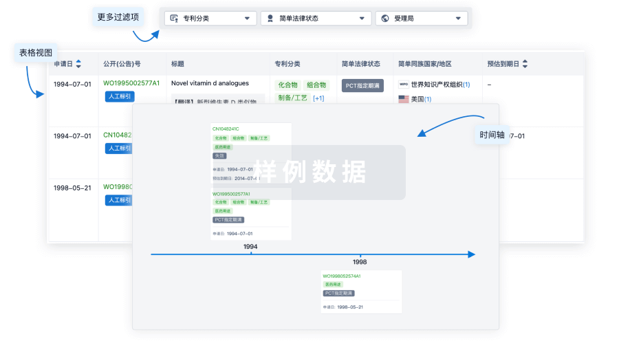
核心专利
使用我们的核心专利数据促进您的研究。
登录
或
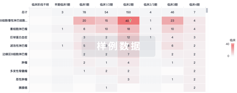
临床分析
紧跟全球注册中心的最新临床试验。
登录
或
批准
利用最新的监管批准信息加速您的研究。
登录
或
生物类似药
生物类似药在不同国家/地区的竞争态势。请注意临床1/2期并入临床2期,临床2/3期并入临床3期
登录
或
特殊审评
只需点击几下即可了解关键药物信息。
登录
或
生物医药百科问答
全新生物医药AI Agent 覆盖科研全链路,让突破性发现快人一步
立即开始免费试用!
智慧芽新药情报库是智慧芽专为生命科学人士构建的基于AI的创新药情报平台,助您全方位提升您的研发与决策效率。
立即开始数据试用!
智慧芽新药库数据也通过智慧芽数据服务平台,以API或者数据包形式对外开放,助您更加充分利用智慧芽新药情报信息。
生物序列数据库
生物药研发创新
免费使用
化学结构数据库
小分子化药研发创新
免费使用