预约演示
更新于:2026-05-25

Riverside Health Care System, Inc.
更新于:2026-05-25
概览
关联
100 项与 Riverside Health Care System, Inc. 相关的临床结果
登录后查看更多信息
0 项与 Riverside Health Care System, Inc. 相关的专利(医药)
登录后查看更多信息
48
项与 Riverside Health Care System, Inc. 相关的文献(医药)2025-11-18·Cureus Journal of Medical Science
Rhabdomyolysis Associated With Excess Pine Bark Extract: A Case Report
作者: Master, Sana ; Kermanshah, Arya ; Kaceli, Tea ; Kalaydjian, Garine ; Bedi, Ashmeet
[This corrects the article DOI: 10.7759/cureus.93956.].
2025-11-05·Cureus Journal of Medical Science
Radiologic Assessment of Malpositioned Indwelling Catheters and Medical Devices: A Retrospective Case Series With Pictorial Review
Article
作者: Pai, Raghav ; Kermanshah, Arya ; Gozlan, Etienne C ; Sobhan, Armaan M ; Castillo, Patricia ; Bedi, Ashmeet ; Padilla, Carolina ; Galan, Daniela C
BACKGROUND:
Indwelling medical devices such as central venous catheters (CVCs), ureteral stents, ventriculoperitoneal (VP) shunts, inferior vena cava (IVC) filters, percutaneous endoscopic gastrostomy (PEG) tubes, and nasogastric (NG) tubes are integral to modern clinical care. Malposition or migration, however, can result in severe complications, including vascular injury, organ perforation, infection, and obstruction. Radiologists are often the first to detect these complications, but interpretation may be challenging due to variable device types, insertion techniques, and patient anatomy.
METHODS:
We conducted a retrospective case-based review of malpositioned indwelling devices encountered in clinical practice at our institution. Each case was analyzed individually with attention to device type, insertion pathway, radiographic landmarks, abnormal positioning patterns, and associated complications. Representative examples were selected to create a pictorial review highlighting imaging pearls and diagnostic pitfalls.
RESULTS:
The cases illustrate a wide spectrum of malposition patterns across commonly placed devices. Vascular access complications included arterial cannulation, aberrant venous placement, and migration into unexpected vascular channels. Ureteral stent malpositions demonstrated distal migration into the urethra and proximal perforation through the renal cortex. VP shunt cases showed distal extrusion into hollow viscera such as the rectum. IVC filter malpositions included tilt, strut perforation, and extension into adjacent organs. PEG tube malpositions resulted in intraperitoneal leakage and peritonitis, while NG tube misplacements involved tracheobronchial insertion and gastric perforation. These examples highlight both typical and rare presentations, underscoring the importance of systematic radiographic evaluation.
CONCLUSION:
This retrospective case series and pictorial review underscore the critical role of radiologists in recognizing malpositioned indwelling devices. Accurate interpretation requires a structured imaging approach, correlation with clinical context, and familiarity with device-specific pitfalls. By presenting illustrative examples across multiple device types, this work provides a practical reference to enhance radiologic vigilance, improve diagnostic accuracy, and ultimately prevent adverse outcomes in patients with indwelling catheters and medical devices.
2025-08-17·Cureus Journal of Medical Science
Atypical STING-Vasculopathy Phenotype: Definite Usual Interstitial Pneumonia (UIP)-Pattern CT With Mixed Fibrotic Histology: A Case Report
Article
作者: Aher, Aman ; Mirzabeigi, Yasamin ; Kermanshah, Arya ; Aher, Pritish ; Kawade, Urvi ; Gerstner Saucedo, Jochen
STING‑associated vasculopathy with onset in infancy (SAVI) is a rare autoinflammatory disorder that causes systemic inflammation, vasculopathy, and progressive interstitial lung disease (ILD). The pulmonary manifestations of SAVI typically resemble a nonspecific interstitial pneumonia (NSIP) pattern both radiologically and histologically. We present a case of a 20‑year‑old male with genetically confirmed SAVI who developed acute hypoxic respiratory failure triggered by respiratory syncytial virus infection, despite appropriate treatment. Imaging revealed extensive subpleural fibrosis, traction bronchiectasis, and honeycombing. These findings were observed in the setting of a CT appearance strongly consistent with a definite usual interstitial pneumonia (UIP) pattern. Ultimately, the patient underwent a lung transplant, and histopathology of the explanted lungs revealed fibrotic changes, including diffuse interstitial thickening, lymphoid aggregates, and honeycombing, without definitive fibroblastic foci, indicating a mixed UIP and NSIP pattern of fibrosis. This case illustrates a rare UIP‑pattern imaging phenotype in SAVI‑associated ILD, despite a mixed UIP‑NSIP histologic pattern of pulmonary fibrosis, and highlights the potential for acute viral infection to precipitate decompensation in patients with interferon‑driven pulmonary fibrosis. Written informed consent was obtained from the patient for publication of this report and accompanying images.
100 项与 Riverside Health Care System, Inc. 相关的药物交易
登录后查看更多信息
100 项与 Riverside Health Care System, Inc. 相关的转化医学
登录后查看更多信息
组织架构
使用我们的机构树数据加速您的研究。
登录
或
管线布局
2026年09月09日管线快照
无数据报导
登录后保持更新
药物交易
使用我们的药物交易数据加速您的研究。
登录
或
转化医学
使用我们的转化医学数据加速您的研究。
登录
或
营收
使用 Synapse 探索超过 36 万个组织的财务状况。
登录
或
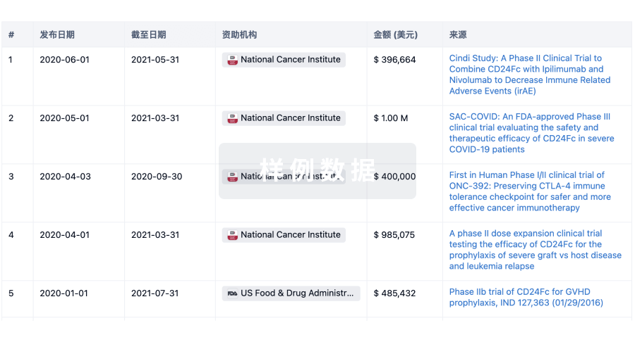
科研基金(NIH)
访问超过 200 万项资助和基金信息,以提升您的研究之旅。
登录
或
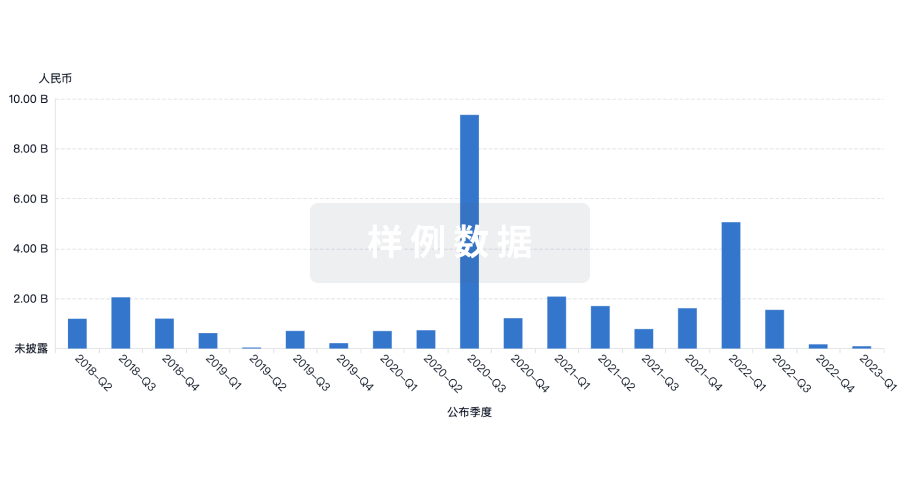
投资
深入了解从初创企业到成熟企业的最新公司投资动态。
登录
或
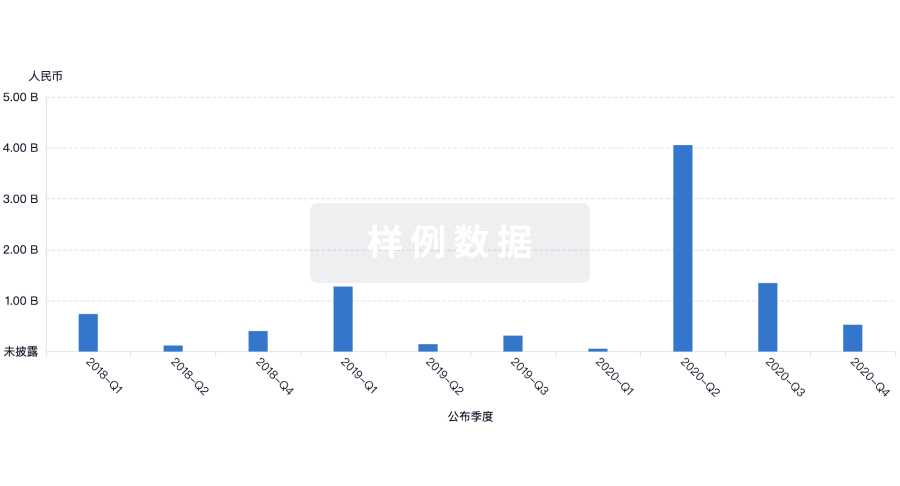
融资
发掘融资趋势以验证和推进您的投资机会。
登录
或
提出任何生物医药研究问题
让每一步生物医药决策都精准制胜
基于十亿级生物数据与全球情报的精准 AI,赋能早期药物发现
立即开始免费试用!
智慧芽新药情报库是智慧芽专为生命科学人士构建的基于AI的创新药情报平台,助您全方位提升您的研发与决策效率。
立即接入生物医药数据包
智慧芽新药库数据也通过智慧芽开放平台,以MCP或API服务形式对外开放。立即使用我们的MCP服务定制您的LLM智能体。
生物序列数据库
生物药研发创新
免费使用
化学结构数据库
小分子化药研发创新
免费使用