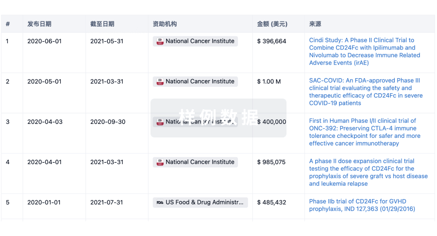
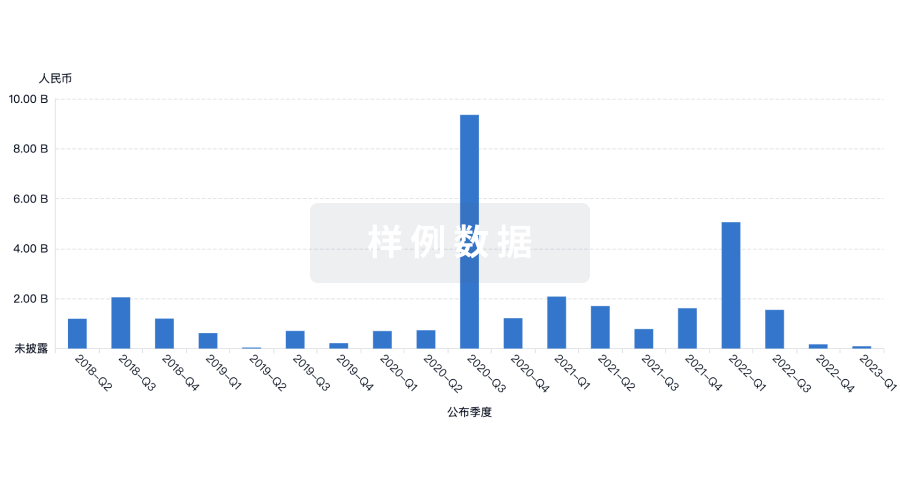
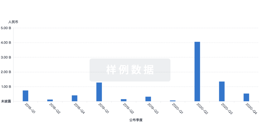
A whole-body visualization model of skin squamous cell carcinoma (SCC) in nude mice was established, and the development of skin cancer by real-time imaging was observed The pEGFP-N1 plasmid was transfected into human skin cancer cell line SCL-1 to obtain pEGFP-SCL-1 cells which could emit fluorescence stably. The pEGFP-SCL-1 cells were then implanted s.c. into BALB/C nude mice to establish the mouse model bearing tumors. The fluorescence emitted from the tumors was quant. detected and imaged with a fluorescence stereo-microscope for real-time visualization of the tumor growth. The transfected skin cancer cells can express EGFP stably and efficiently. Tumor tissues developed after s.c. implantation in nude mice. The result was confirmed by histopathol. The whole-body visualization model bearing the skin cancer provides a reliable method for studying the mechanisms of cancer development, and may be useful for the investigation of novel means for skin cancer treatment.