预约演示
更新于:2026-06-18
Topcon Corp.
更新于:2026-06-18
概览
关联
JPRN-UMIN000059337
Real-world field studies aimed at implementing future ophthalmological examinations in society

NCT07350304
Comparison of Two OCT Device RNFL and GCIPL Measurements in Mild, Moderate, and Severe Glaucoma

JPRN-UMIN000031747
Research on retinal / choroidal structure analysis by novel image analysis technique and machine learning.
100 项与 Topcon Corp. 相关的临床结果
登录后查看更多信息
登录后查看更多信息
2026-06-01AMERICAN JOURNAL OF OPHTHALMOLOGY
Effects of Optic Nerve Head Structures on Retinal Nerve Fiber Layer Defects in Primary Open Angle Glaucoma Eyes: Analyses Using Retinal Optical Texture Analysis
Article
作者: Kikawa, Tsutomu ; Weinreb, Robert N ; Saito, Hitomi ; Leung, Christopher Kai Shun ; Nishigaki, Masashi ; Tomita, Goji ; Aihara, Makoto ; Higashide, Tomomi ; Miki, Atsuya ; Zangwill, Linda M ; Iwase, Aiko ; Murata, Hiroshi ; Sugiyama, Kazuhisa ; Kim, Tae-Woo ; Araie, Makoto ; Ohno-Matsui, Kyoko ; Nakazawa, Toru
PURPOSE:
To elucidate associations between optic nerve head (ONH) structures and the retinal nerve fiber layer optical texture analysis (ROTA)-detected retinal nerve fiber layer defects (RNFLDs) in primary open angle glaucoma (POAG) eyes.
DESIGN:
Prospective cross-sectional observational study.
PARTICIPANTS:
This study enrolled 136 eyes of 109 POAG patients.
METHODS:
All participants underwent comprehensive ophthalmologic examinations including standard automated perimetry and swept-source optical coherence tomography (SS-OCT). Two independent graders assessed ROTA images for the presence, location, and width of RNFLDs. Multivariable linear mixed effects model was used to investigate factors independently associated with ROTA-detected RNFLDs. Explanatory variables were systemic and ocular factors such as age, axial length (AXL), SS-OCT-derived ONH structural parameters such as Bruch membrane opening-centered circumpapillary retinal nerve fiber layer thickness (cpRNFLT) and gamma zone area, and average visual field sensitivity (1/Lambert) (VFSaverage).
MAIN OUTCOME MEASURES:
Width and number of RNFLDs.
RESULTS:
RNFLD detection rates by ROTA were 86.8% overall, 69.0% and 94.7% in the early and moderate stages of POAG eyes, and 86.5% and 87.2% in the highly myopic (AXL >26.0 mm) and non-highly myopic eyes, respectively. Summed width of RNFLD per eye was positively correlated with gamma-zone area (P = .0007) and negatively correlated with age, cpRNFLT, and VFSaverage (P = .0111, .0001, .0124), whereas the number of RNFLDs per eye correlated negatively with age, cpRNFLT, and VFSaverage (P = .0153, .0029, .0007).
CONCLUSIONS:
The ROTA-detected extent of axonal damage was associated with ONH structural change represented by gamma zone area in POAG eyes after adjustment for other possible confounding factors.
2026-04-30International Journal of Retina and Vitreous
Exploratory study on a novel automatic quantification software by artificial intelligence for geographic atrophy associated to age-related macular degeneration.
Article
作者: Akiba, Masahiro ; Mao, Zaixing ; Su, Aiwen ; Ruiz-Moreno, José M ; Ruiz-Medrano, Jorge
2026-03-09Translational Vision Science & Technology
Larger Real-World OCT Reference Database Improves Accuracy of Glaucoma Flagging Using Summary Metrics
Article
作者: Lee, Chris ; Durbin, Mary ; Guzman, Anya ; Hood, Donald C. ; Tsamis, Emmanouil ; Wang, Yujia ; Gebhardt, Tayna ; De Moraes, Carlos Gustavo
Purpose:
To compare the healthy control and glaucoma eyes flagged as outside normal limits (yellow or red) by a commercial reference database (C-RDB) and a larger real-world (RW)-RDB.
Methods:
The C-RDB consisted of 398 eyes/individuals. Based only on optical coherence tomography (OCT) reports, the RW-RDB consisted of 4830 eyes/individuals selected from optometry practices using a reading center method. The fifth and first percentile quantile regression lines (QRLs) versus age were calculated for both RDBs for common OCT metrics, including global circumpapillary retinal nerve fiber (g-cpRNFL) and global ganglion cell layer plus inner plexiform layer (g-GCL+) thickness. The test dataset contained 175 healthy control (H) eyes and 183 eyes with OCT defects consistent with optic neuropathy-glaucoma (ON-G). These eyes were flagged as yellow (red) if they fell below the fifth (first) percentile QRL. The QRLs were also compared to a Gaussian model and Monte Carlo simulations.
Results:
The C-RDB and RW-RDB did not flag an identical set of eyes as red or yellow. In fact, 16% (g-cpRNFL) and 7% (g-GCL+) of the 183 ON-G eyes had a different color flag. The results of the model and simulations support the hypothesis that both RDBs are sampled from essentially the same underlying "normal" population. Thus, the difference between them is largely due to less random error in the larger sample.
Conclusions:
There is a difference in the eyes flagged, and this difference is largely due to the greater size of the RW-RDB.
Translational Relevance:
These findings support the clinical value of expanding reference databases to improve diagnostic accuracy of glaucoma flagging.
100 项与 Topcon Corp. 相关的药物交易
登录后查看更多信息
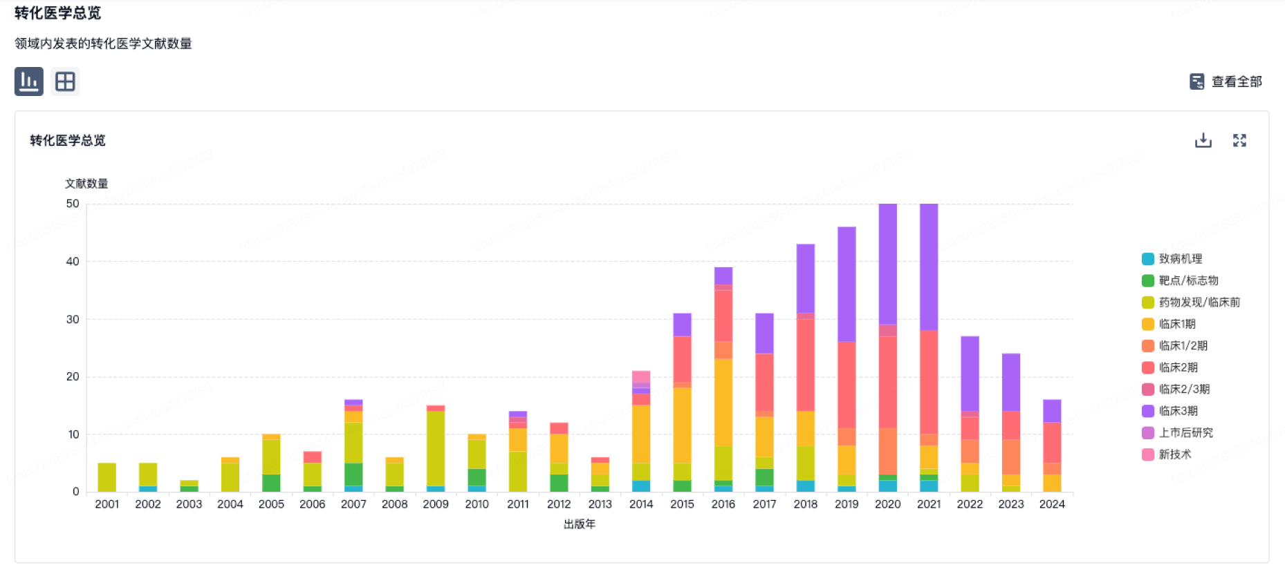
100 项与 Topcon Corp. 相关的转化医学
登录后查看更多信息
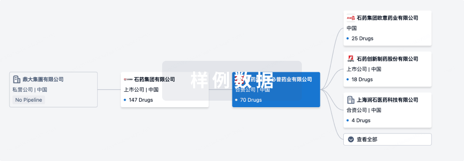
组织架构
使用我们的机构树数据加速您的研究。
登录
或
管线布局
2026年06月19日管线快照
无数据报导
登录后保持更新
药物交易
使用我们的药物交易数据加速您的研究。
登录
或

转化医学
使用我们的转化医学数据加速您的研究。
登录
或
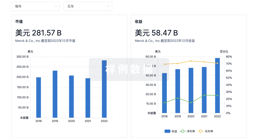
营收
使用 Synapse 探索超过 36 万个组织的财务状况。
登录
或
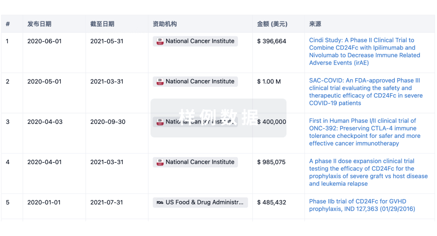
科研基金(NIH)
访问超过 200 万项资助和基金信息,以提升您的研究之旅。
登录
或
投资
深入了解从初创企业到成熟企业的最新公司投资动态。
登录
或
融资
发掘融资趋势以验证和推进您的投资机会。
登录
或
生物医药百科问答
全新生物医药AI Agent 覆盖科研全链路,让突破性发现快人一步
立即开始免费试用!
智慧芽新药情报库是智慧芽专为生命科学人士构建的基于AI的创新药情报平台,助您全方位提升您的研发与决策效率。
立即开始数据试用!
智慧芽新药库数据也通过智慧芽数据服务平台,以API或者数据包形式对外开放,助您更加充分利用智慧芽新药情报信息。
生物序列数据库
生物药研发创新
免费使用
化学结构数据库
小分子化药研发创新
免费使用