预约演示
更新于:2025-05-07

Institut Du Cancer De Montreal
更新于:2025-05-07
概览
关联
100 项与 Institut Du Cancer De Montreal 相关的临床结果
登录后查看更多信息
0 项与 Institut Du Cancer De Montreal 相关的专利(医药)
登录后查看更多信息
233
项与 Institut Du Cancer De Montreal 相关的文献(医药)2025-03-20·Journal of Biomedical Optics
Development and preclinical evaluation of an endonasal Raman spectroscopy probe for transsphenoidal pituitary adenoma surgery
Article
作者: Ember, Katherine ; Leblond, Frédéric ; Marple, Eric ; Sheehy, Guillaume ; Blanquez-Yeste, Victor ; Tran, Trang ; Janelle, Félix ; Dallaire, Frédérick ; Urmey, Kirk ; Labidi, Moujahed
2025-01-24·Journal of Biomedical Optics
Quantitative assessment of the generalizability of a brain tumor Raman spectroscopy machine learning model to various tumor types including astrocytoma and oligodendroglioma
Article
作者: Le Moël, Alice ; Ember, Katherine ; Dudley, Roy ; Sheehy, Guillaume ; Tavera, Hugo ; Blanquez-Yeste, Victor ; Petrecca, Kevin ; Leblond, Frédéric ; Hadjipanayis, Costas ; Guiot, Marie-Christine ; Dallaire, Frédérick ; Tran, Trang ; Weil, Alexander G
2024-09-01·Molecular Therapy: Oncology
Oncolytic vesicular stomatitis virus alone or in combination with JAK inhibitors is effective against ovarian cancer
Article
作者: Geoffroy, Karen ; Leclerc-Desaulniers, Kim ; Mullins-Dansereau, Victor ; Bourgeois-Daigneault, Marie-Claude ; Viens, Mélissa
1
项与 Institut Du Cancer De Montreal 相关的新闻(医药)2013-12-19
Montreal, October 2, 2013. The five Contenders of the first AmorChem KNOCK-OUT Event valiantly climbed into the ring today at BioContact to duke it out against a panel of Heavyweight Champions for a chance to win a coveted $500,000 financing from AmorChem. Earlier in the summer, AmorChem, the innovative Quebec-based venture capital seed fund, launched a province-wide call for proposals to participate in its KNOCK-OUT Event.
All the Contenders displayed remarkable courage and agility in the ring, yet only one could stand victorious.
It is with great pleasure that AmorChem announces the winner of the 2013 KNOCK-OUT Event:
Dr. John Stagg
“Targeting CD73 with small molecules in oncology”
Institut du cancer de Montréal, Centre de recherche du Centre Hospitalier de l’Université de Montréal and Faculty of Pharmacy, Université de Montréal
“This first KNOCK-OUT Event has been quite an adventure and we are very pleased with the outcome of the battle in the ring today. The Heavyweights had a tough job and we are very enthusiastic about their selection,” comments Inès Holzbaur, General Partner at AmorChem. “We would like to thank the Contenders for rolling with the punches, our Heavyweights for bringing their individual expertise into the ring today and Christopher Hall for successfully keeping everyone battling without the rule.”
“Our hopes and objectives have been well surpassed for this Event,” says Elizabeth Douville, General Partner at AmorChem. “We created this Event to increase the Quebec research community’s awareness of AmorChem and reach out to researchers who may not have been aware of the possibility of commercialising aspects of their work. This Event definitely reached those goals.”
The final bell has rung; AmorChem now looks forward to working with Dr Stagg.
The AmorChem KNOCK OUT Event is proudly sponsored by ROBIC, LLP.
ABOUT AMORCHEM
AmorChem L.P. ( ) is a venture capital fund located in Montreal focused on investing in promising life science projects originating from Quebec-based universities and research centres. The principal limited partners of this fund are Investissement-Québec, FIER Partenaires, Fonds de solidarité FTQ and Merck & Co. This fund is the latest addition to the GeneChem portfolio of funds, a fund manager in existence since 1997. AmorChem’s innovative business model involves financing research-stage projects to enable them to reach pre-clinical proof-of-concept (“POC”) in a semi-virtual mode within 18-24 months. The fund seeks to generate returns through a two-pronged exit strategy: sell projects having reached POC to large biotechnology or pharmaceutical companies; or bundle them into new spin-out companies. AmorChem using external resources will manage the projects. To that effect, AmorChem has established a strategic partnership with the Biotechnology Research Institute in order to access its R&D platforms. In addition, to enabling projects requiring small molecules as tools or drug leads, AmorChem has founded NuChem Therapeutics Inc., a medicinal chemistry contract-research company.
Media Contact for AmorChem
Mélanie Larouche
T
418 521 3771
E
melanie.larouche@citoyenoptimum.com
.ABOUT ROBIC
Founded in 1892, ROBIC, LLP is an internationally renowned firm of lawyers, patent and trade-mark agents specialized in Intellectual Property and Business Law. Headquartered in Montreal, ROBIC is a Canadian firm with more than 175 people including 23 partners and 45 professionals. We provide dynamic and entrepreneurial assistance and representation to clients in executing business transactions in addition to managing, protecting, enforcing and commercialising their intellectual capital.
ABOUT BIOCONTACT
BioContact Quebec 2013: The 19th edition will take place on October 2nd and 3rd 2013, at the Château Laurier in Québec City. Representatives from over 24 biopharmaceutical companies from Canada, the United States and Europe will be attending.
Help employers find you! Check out all the jobs and post your resume.
小分子药物合作
100 项与 Institut Du Cancer De Montreal 相关的药物交易
登录后查看更多信息
100 项与 Institut Du Cancer De Montreal 相关的转化医学
登录后查看更多信息
组织架构
使用我们的机构树数据加速您的研究。
登录
或
管线布局
2026年02月08日管线快照
无数据报导
登录后保持更新
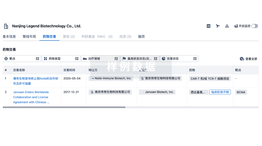
药物交易
使用我们的药物交易数据加速您的研究。
登录
或
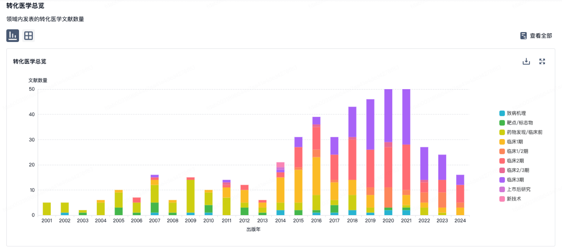
转化医学
使用我们的转化医学数据加速您的研究。
登录
或
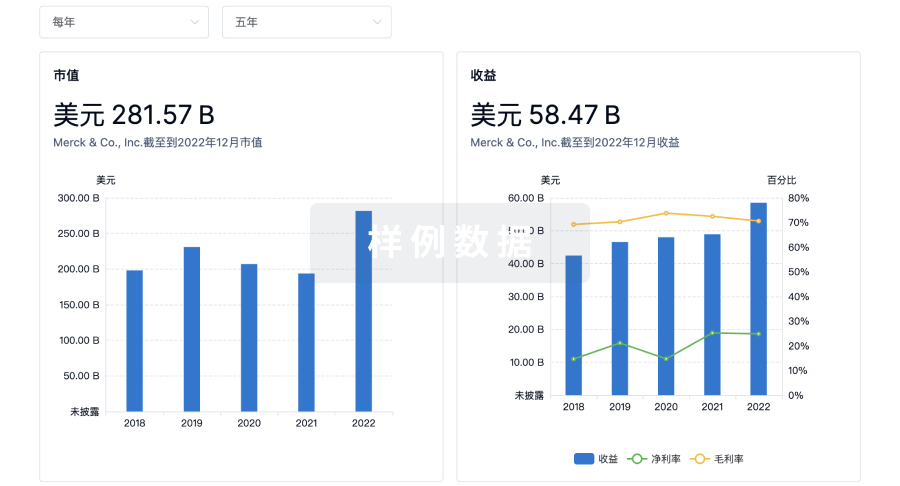
营收
使用 Synapse 探索超过 36 万个组织的财务状况。
登录
或
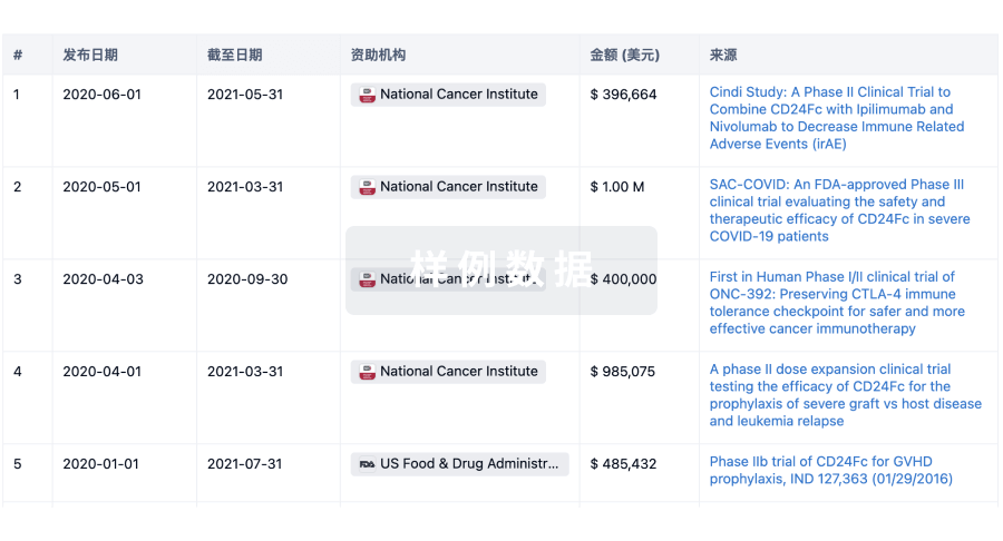
科研基金(NIH)
访问超过 200 万项资助和基金信息,以提升您的研究之旅。
登录
或
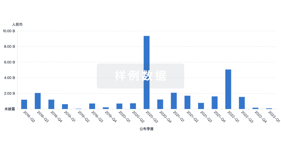
投资
深入了解从初创企业到成熟企业的最新公司投资动态。
登录
或
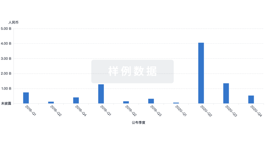
融资
发掘融资趋势以验证和推进您的投资机会。
登录
或
生物医药百科问答
全新生物医药AI Agent 覆盖科研全链路,让突破性发现快人一步
立即开始免费试用!
智慧芽新药情报库是智慧芽专为生命科学人士构建的基于AI的创新药情报平台,助您全方位提升您的研发与决策效率。
立即开始数据试用!
智慧芽新药库数据也通过智慧芽数据服务平台,以API或者数据包形式对外开放,助您更加充分利用智慧芽新药情报信息。
生物序列数据库
生物药研发创新
免费使用
化学结构数据库
小分子化药研发创新
免费使用