预约演示
更新于:2026-03-05
Neusoft Medical Systems Co., Ltd.
更新于:2026-03-05
概览
关联
NCT04241614
Comparison and Analysis of Predictive Performance of CT and Raw Data in Benign and Malignant Classification of Pulmonary Nodules
100 项与 东软医疗系统股份有限公司 相关的临床结果
登录后查看更多信息
登录后查看更多信息
2026-02-01Quantitative Imaging in Medicine and Surgery
Performance of free-breathing contrast-enhanced and unenhanced stack-of-spirals ultrashort echo time MRI for lung follow-up in patients with malignancies at 1.5T
Article
作者: Sun, Peng ; Wu, Jia-Wei ; Benkert, Thomas ; Li, Zi-Kai ; Meng, Xue-Ni ; Lu, Hao-Hao ; Xiao, Ao-Dong ; Sun, Xin ; Fu, Qing ; Yin, Ting ; Li, Qian
Background:
Stack-of-spirals ultrashort echo time (spiral-UTE) has been reported to be feasible for lung imaging; however, the performance of free-breathing contrast-enhanced and unenhanced spiral-UTE (UTEe and UTEu) is still unknown. Therefore, this study aimed to evaluate their performance of lung imaging in patients with malignant tumors.
Methods:
A total of 76 patients with malignancies suspected of pulmonary metastatic nodules were enrolled in free-breathing UTEe, UTEu and routine contrast-enhanced T1-weighted imaging [volumetric interpolated breath-hold examination (VIBE)] for lung follow-up. Two radiologists independently assessed the image quality, and qualitative analysis was scored via a 5-point scale (4, excellent; 0, unreadable) with respect to the visibility of fissures, airways and vessels; signal homogeneity; motion artifacts; lesion conspicuity; and overall image quality. Quantitative analysis included measurements of the apparent contrast-to-noise ratio (CNR) and apparent signal-to-noise ratio (SNR). Pulmonary nodules detected via magnetic resonance (MR) images were compared with those by computed tomography (CT) as the reference standard.
Results:
Both UTEu and UTEe outperformed VIBE in all the qualitative metrics (P<0.001) and there was no significant difference between UTEu and UTEe (P>0.05) in those metrics. UTEe exhibited the best performance in depicting pulmonary vessels, achieving the highest apparent SNR and apparent CNR values. Among the 130 pulmonary nodules identified via CT, spiral-UTE had a sensitivity of 76.9% and a positive predictive value (PPV) of 99.0%, significantly outperforming VIBE (sensitivity of 47.7% and PPV of 95.4%). The detection rates for spiral-UTE were 90.0% for nodules larger than 5 mm, 98.5% for nodules larger than 7 mm and 100.0% for nodules larger than 10 mm.
Conclusions:
Spiral-UTE demonstrated superior image quality and greater sensitivity for pulmonary nodule detection than breath-hold VIBE did. Both unenhanced and enhanced spiral-UTE showed comparable performance in nodule detection, highlighting its potential as a reliable imaging modality for patients with malignant tumors during follow-up imaging.
2025-09-01NEUROIMAGE
Accelerating multi-directional diffusion MRI through patch-based joint reconstruction
Article
作者: Wang, Yaohui ; Chen, Zhifeng ; Liang, Xiaoyun ; Xu, Zhongbiao ; Cheng, Junying ; Guo, Li ; Liu, Feng ; Deng, Guanhua ; Zhang, Rongli ; Huang, Wei ; Chen, Zhaolin
Diffusion magnetic resonance imaging (dMRI) is a valuable technique for studying tissue microstructure and connectivity in the brain. However, acquiring high-resolution dMRI data is time-consuming, limiting its clinical applicability. Traditional parallel imaging techniques can accelerate the acquisition of dMRI, but they are constrained by the geometry factor. In this study, we propose a novel patch-based multiple diffusion directions joint reconstruction method that simultaneously capitalizes on the intra- and inter-image correlation across multiple diffusion directions by grouping similar 3D image patches and then enforces the sparsity of these groups in sensitivity encoding (SENSE) reconstruction, termed PB-SENSE. The simulation and in vivo experiments demonstrated that the proposed method can achieve high-quality images comparable to those obtained from fully sampled data, even with an acceleration of 5. This suggests that the proposed method has the potential to enhance the practical application of high-resolution diffusion imaging.
2025-08-01Quantitative Imaging in Medicine and Surgery
Ultra-low-dose hepatic computed tomography with a novel real-time deep learning-based noise reduction algorithm: a prospective cross-sectional analysis of image quality and lesion detection
Article
作者: Feng, Xiangnan ; Xu, Chensi ; Chen, Yan ; Gao, Jianbo ; Zhou, Zhigang ; Jiang, Yaojun ; Liu, Jie ; Hou, Ping ; Lyu, Peijie ; Wang, Xiaopeng
Background:
Contrast-enhanced computed tomography (CT) is essential for tumor assessment, but the detection of low-contrast liver lesions remains challenging. Reducing the radiation dose increases image noise, compromising image quality and diagnostic accuracy. Iterative reconstruction (IR) algorithms can reduce noise; however, they can also alter image texture and limit lesion detection. Deep-learning image reconstruction (DLIR) represents a promising alternative, but its efficacy in ultra-low-dose (ULD) hepatic CT for detecting small, low-contrast lesions remains underexplored. Thus, this study aimed to evaluate a novel real-time DLIR algorithm in ULD hepatic CT, focusing on image quality and lesion detection.
Methods:
In total, 65 patients with hepatic lesions underwent both standard-dose and ULD abdominal CT scans during the portal venous phase. The standard-dose protocol (group A) used 120 kV with a signal-to-noise ratio (SNR) of 1.0, and the images were reconstructed using 50% IR. The ULD protocol (group B) used 120 kV with an SNR of 0.5, and the images were reconstructed using 50% IR and 50% DLIR (groups B1 and B2, respectively). The quantitative and qualitative image quality parameters were assessed. The lesion detection rates were evaluated by lesion type and size using the metrics of detection rate, sensitivity, specificity, positive predictive value (PPV), and negative predictive value (NPV).
Results:
Group B showed a 73.3% reduction in the radiation dose compared to group A (1.5±0.8 vs. 6.9±2.0 mSv, P<0.001). Image noise differed significantly across the protocols: group B1 had the highest noise [10.05±2.94 Hounsfield units (HU)], followed by groups A (8.29±2.82 HU) and B2 (8.04±2.71 HU; all pairwise P<0.05 except group A vs. group B2: P=0.625). The CT values and contrast-to-noise ratios (CNRs) were comparable between groups B2 and A (all P>0.05), while group B2 had a 29.9-42.2% higher CNR than group B1 (all P<0.001). The qualitative assessments confirmed that the image quality and diagnostic acceptability of groups B2 (100%) and A (all P>0.05) were comparable, while the images of group B1 were diagnostically unacceptable (all scores <3). Overall, lesion detection was comparable in groups B2 (90.5%, 133/147) and A (98.0%, 144/147; P>0.05). However, group B2 had a significantly lower detection rate for small lesions (<0.5 cm: 77.8%, 42/54) compared to group A (P<0.05), but outperformed group B1 (57.4%, 31/54; P<0.05). Group B2 also had a significantly improved lesion detection rate and sensitivity for low-contrast lesions (87.2%, 95/109) compared to group B1 (75.2%, 82/109; P<0.05). The novel DLIR algorithm achieved a reconstruction speed of 60 images per second (ips), which was significantly faster than that of other DLIR approaches, while maintaining comparable performance to the IR algorithm.
Conclusions:
The combination of tube current reduction with a novel real-time DLIR algorithm enabled ULD abdominal CT to achieve a 73.3% reduction in the radiation dose while maintaining image quality and diagnostic performance for detecting hepatic lesions larger than 0.5 cm.
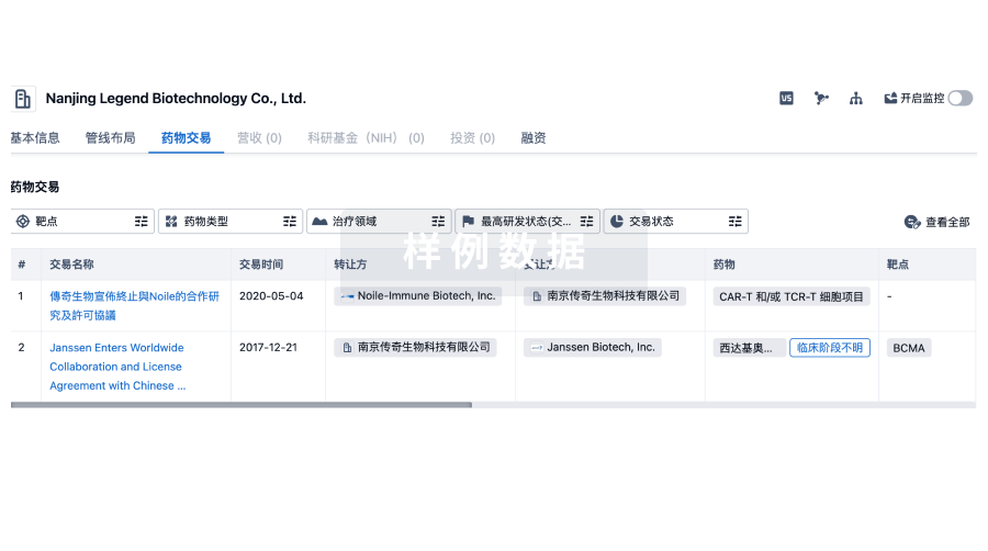
100 项与 东软医疗系统股份有限公司 相关的药物交易
登录后查看更多信息
100 项与 东软医疗系统股份有限公司 相关的转化医学
登录后查看更多信息
组织架构
使用我们的机构树数据加速您的研究。
登录
或
管线布局
2026年04月02日管线快照
无数据报导
登录后保持更新
药物交易
使用我们的药物交易数据加速您的研究。
登录
或
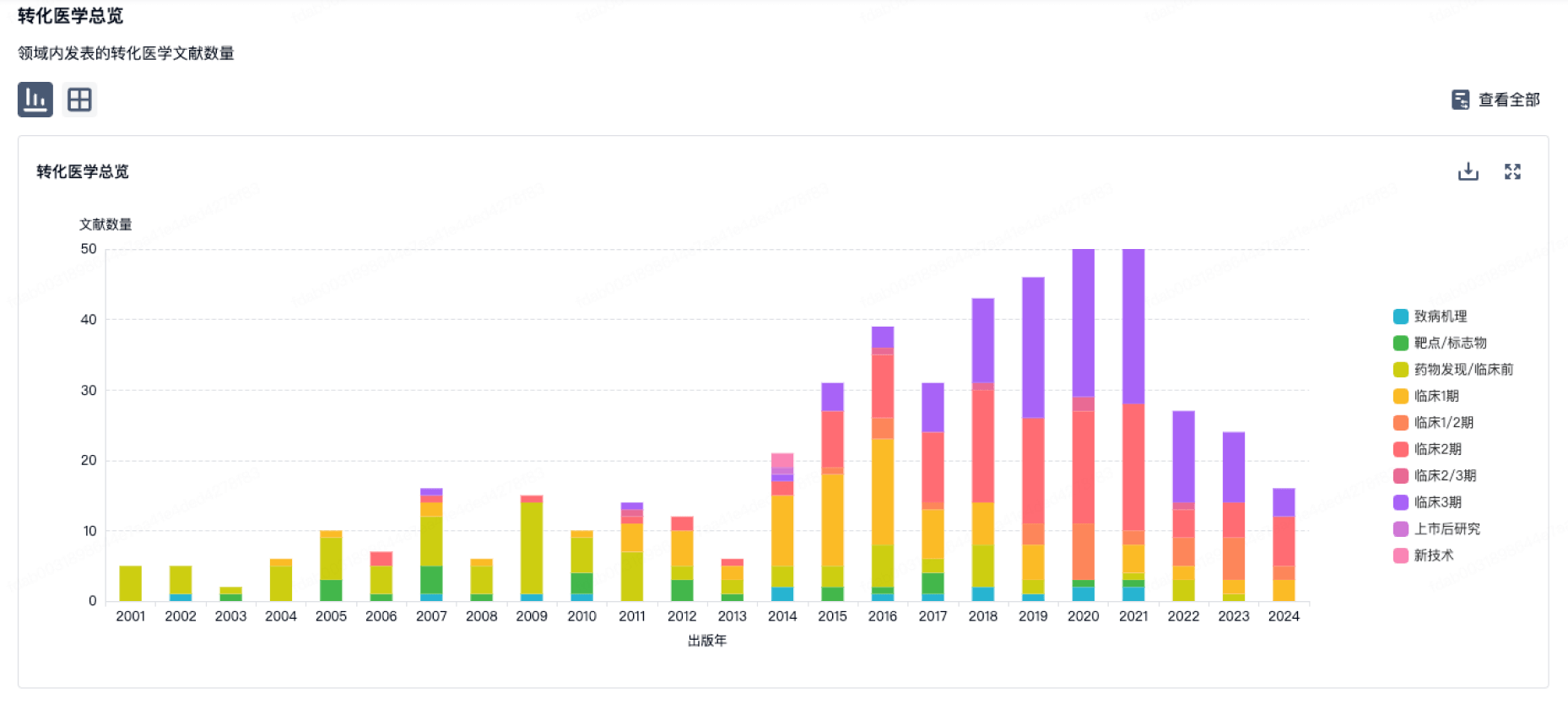
转化医学
使用我们的转化医学数据加速您的研究。
登录
或
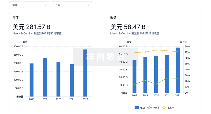
营收
使用 Synapse 探索超过 36 万个组织的财务状况。
登录
或
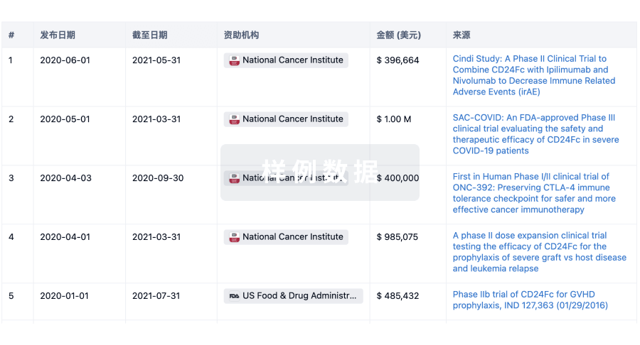
科研基金(NIH)
访问超过 200 万项资助和基金信息,以提升您的研究之旅。
登录
或
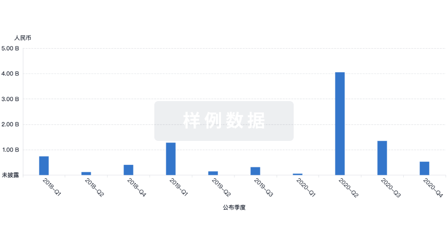
投资
深入了解从初创企业到成熟企业的最新公司投资动态。
登录
或
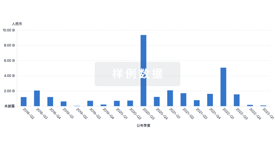
融资
发掘融资趋势以验证和推进您的投资机会。
登录
或
生物医药百科问答
全新生物医药AI Agent 覆盖科研全链路,让突破性发现快人一步
立即开始免费试用!
智慧芽新药情报库是智慧芽专为生命科学人士构建的基于AI的创新药情报平台,助您全方位提升您的研发与决策效率。
立即开始数据试用!
智慧芽新药库数据也通过智慧芽数据服务平台,以API或者数据包形式对外开放,助您更加充分利用智慧芽新药情报信息。
生物序列数据库
生物药研发创新
免费使用
化学结构数据库
小分子化药研发创新
免费使用