预约演示
更新于:2026-05-08

Immuno-Biological Laboratories Co., Ltd.
更新于:2026-05-08
概览
关联
100 项与 Immuno-Biological Laboratories Co., Ltd. 相关的临床结果
登录后查看更多信息
0 项与 Immuno-Biological Laboratories Co., Ltd. 相关的专利(医药)
登录后查看更多信息
65
项与 Immuno-Biological Laboratories Co., Ltd. 相关的文献(医药)2025-12-01·Clinical and Experimental Nephrology
Co-localization of IgG with nephrin in immune-mediated idiopathic nephrotic syndrome
Article
作者: Sakakibara, Nana ; Horinouchi, Tomoko ; Tanaka, Yu ; Kitakado, Hideaki ; Ishimori, Shingo ; Yamamura, Tomohiko ; Nagano, China ; Maruyama, Hironobu ; Fujii, Hideki ; Iijima, Kazumoto ; Aoyama, Shuhei ; Ueda, Chika ; Inoki, Yuta ; Okamoto, Hayaki ; Shima, Yuko ; Ichikawa, Yuta ; Nozu, Kandai ; Kimura, Yuka
Abstract:
Background:
Increased serum anti-nephrin antibody titers and co-localization of nephrin and IgG in kidney tissues have been reported in minimal change disease (MCD) and post-transplant recurrent focal segmental glomerulosclerosis (FSGS). These results indicate an association of anti-nephrin antibodies with nephrotic syndrome (NS); however, the exact relationship remains unclear. Herein, we evaluated nephrin/IgG co-localization in the glomeruli of patients with various kidney diseases, including monogenic NS, to clarify the association between idiopathic nephrotic syndrome (INS) and anti-nephrin antibodies.
Methods:
IgG and nephrin co-localization was investigated in 52 kidney tissue biopsy samples, comprising INS in the active phase (
n
= 26; MCD,
n
= 19; FSGS,
n
= 7) and remission (
n
= 6), monogenic NS (
n
= 3), and other kidney diseases (
n
= 17). Double-immunofluorescence staining for nephrin/IgG was performed in unfixed frozen sections for 2 h at room temperature with Alexa Fluor-labeled nephrin/IgG cocktail antibodies. Nephrin/IgG co-localization was assessed using optical sectioning under a fluorescence microscope.
Results:
Nephrin/IgG co-localization was observed in 81% (21/26, children: 15/17, adults: 6/9) of active INS cases, 84% (16/19) of MCD cases, and 71% (5/7) of FSGS cases. No co-localization was observed in NS with monogenic variants or other kidney diseases.
Conclusion:
Nephrin/IgG co-localization in the kidney tissue is finding observed in active INS
,
strongly indicating an association between anti-nephrin antibodies and INS onset. The nephrin/IgG cocktail antibody is a rapid and effective approach for investigating INS pathogenesis that facilitates the differential diagnosis of immune-mediated NS from other kidney diseases, including monogenic NS.
2025-08-01·JOURNAL OF GASTROENTEROLOGY
Molecular imaging of gastrointestinal stromal tumor using anti-c-KIT antibody and its fragments
Article
作者: Yasushi Sato ; Naoki Muguruma ; Tatsuya Segawa ; Takanori Miyake ; Mamoru Shimizu ; Takanori Kashihara ; Shota Fujimoto ; Tetsuji Takayama ; Koichi Okamoto ; Yutaka Kawano
BACKGROUND:
Gastrointestinal stromal tumors (GISTs) are malignant subepithelial tumors, known for their poor prognosis due to distant metastasis. Because GIST is covered by a normal mucosal layer, effective tissue biopsy under conventional endoscopy is difficult, thereby leading to delayed diagnosis and a dismal prognosis. We performed molecular imaging of GIST targeting c-KIT using fluorescence-labeled anti-c-KIT antibody/fragments and fluorescent endoscopy.
METHODS:
Mouse anti-human c-KIT monoclonal antibody, its F(ab')2 and Fab fragments were labeled with AF680. Two GIST cell lines (GIST-T1, GIST-882M) were used for experiments. Antibodies were intravenously administered to mice xenografted with GIST-T1 or GIST-882M, and each tumor was observed using IVIS Spectrum and self-developed simple fluorescent endoscopy.
RESULTS:
The GIST-T1 cell live imaging revealed strong signals on cell membranes after 1 min incubation, and thereafter, they aggregated and internalized inside the cells within 130 min in all antibody/fragment groups. In vivo mouse experiments, AF680-labeled IgG slowly accumulated in tumors peaking at 24 h after injection. However, AF680-labeled F(ab')2 and Fab rapidly accumulated in tumors peaking at 1-2 h, and completely cleared from the body within 24 h. Fab showed the strongest fluorescence intensity in tumors. Fluorescence endoscopy could clearly detect GIST xenograft tumors 1-2 h after AF680-labeled F(ab')2 and Fab injection.
CONCLUSIONS:
AF680-labeled antibody/fragments showed clear and specific fluorescence signals in GIST xenografts in mice. Particularly, AF680-labeled Fab showed the strongest signal intensity at 1-2 h post-administration and rapid clearance, suggestive of the safety. This approach may enable molecular imaging diagnosis of GIST by endoscopy in outpatient settings in the future.
2023-07-01·The Journal of biological chemistry
Targeting apolipoprotein E and N-terminal amyloid β-protein precursor interaction improves cognition and reduces amyloid pathology in Alzheimer’s mice
Article
作者: Maeda, Masahiro ; Koyama, Naoki ; Fujiwara, Masakazu ; Mori, Takashi ; Sawmiller, Darrell ; Segawa, Tatsuya
Apolipoprotein E (apoE) interaction with amyloid β-protein precursor (APP) has garnered attention as the therapeutic target for Alzheimer's disease (AD). Having discovered the apoE antagonist (6KApoEp) that blocks apoE binding to N-terminal APP, we tested the therapeutic potential of 6KApoEp on AD-relevant phenotypes in amyloid β-protein precursor/presenilin 1 (APP/PS1) mice that express each human apoE isoform of apoE2, apoE3, or apoE4 (designated APP/PS1/E2, APP/PS1/E3, or APP/PS1/E4 mice). At 12 months of age, we intraperitoneally administered 6KApoEp (250 μg/kg) or vehicle once daily for 3 months. At 15 months of age, blockage of apoE and N-terminal APP interaction by 6KApoEp treatment improved cognitive impairment in most tests of learning and memory, including novel object recognition and maze tasks in APP/PS1/E2, APP/PS1/E3, and APP/PS1/E4 mice versus each vehicle-treated mouse line and did not alter behavior in nontransgenic littermates. Moreover, 6KApoEp therapy ameliorated brain parenchymal and cerebral vascular β-amyloid deposits and decreased abundance of amyloid β-protein (Aβ) in APP/PS1/E2, APP/PS1/E3, and APP/PS1/E4 mice versus each vehicle-treated mouse group. Notably, the highest effect in Aβ-lowering by 6KApoEp treatment was observed in APP/PS1/E4 mice versus APP/PS1/E2 or APP/PS1/E3 mice. These effects occured through shifting toward lessened amyloidogenic APP processing due to decreasing APP abundance at the plasma membrane, reducing APP transcription, and inhibiting p44/42 mitogen-activated protein kinase phosphorylation. Our findings provide the preclinical evidence that 6KApoEp therapy aimed at targeting apoE and N-terminal APP interaction is a promising strategy and may be suitable for patients with AD carrying the apoE4 isoform.
3
项与 Immuno-Biological Laboratories Co., Ltd. 相关的新闻(医药)2026-01-19
·搜狐新闻
自身免疫试剂,是指用于自身免疫疾病研究、检测和诊断的化学或生物学试剂,通常包括特异性抗体、抗原、酶标物及信号检测试剂等。这类试剂可用于检测自身抗体水平、识别免疫反应靶点、评估免疫系统异常及辅助临床诊断,广泛应用于实验室研究、临床检测和药物开发中。自身免疫试剂通过高特异性、高灵敏度的设计,能够准确反映免疫系统状态,为自身免疫疾病的早期发现、监测和治疗提供重要工具。自身免疫试剂市场规模及未来增长趋势预测根据问可汇(WENKH)深度研究分析,2025年全球自身免疫试剂的市场规模达到134亿元。自身免疫试剂市场规模持续增长,全球自身免疫性疾病患病群体不断扩大,临床对疾病早期精准诊断的需求逐步释放。检测技术持续迭代升级,提升了检测的效率与准确性,降低了应用门槛。相关医疗政策给予支持,扩大了诊断服务的覆盖范围与可及性,临床研究的深入推进进一步拓展了产品应用场景,共同驱动市场需求稳步上升。未来预计以8.84%的年复合增长率增长至2032年的243亿元。资料来源:问可汇(WENKH)研究整理,2025全球自身免疫试剂市场主要参与者竞争格局根据问可汇(WENKH)深度研究分析,全球自身免疫试剂市场竞争较激烈,主要市场参与者包括EUROIMMUN、HUMAN、Bio-Rad Laboratories、AESKU、Inova、MBL、Orgentec、Trinity、Phadia、江苏浩欧博生物医药股份有限公司、深圳市亚辉龙生物科技股份有限公司、广州市康润生物科技有限公司、Immuno-Biological Laboratories、Biomedical diagnostics、上海科新生物技术股份有限公司等,其中前三大厂商占有大约49%的市场份额。资料来源:问可汇(WENKH)研究整理,2025以下列出的公司是主要参与者,凭借其创新的解决方案和丰富的产品组合推动着全球市场的发展。1.EUROIMMUN总部:德国主营业务:EUROIMMUN核心业务集中在自身免疫性疾病、感染性疾病及过敏性疾病的诊断检测系统,同时提供配套的自动化设备与软件解决方案,其技术涵盖免疫荧光、酶联免疫、免疫印迹等多种检测方法,还设有专业参考实验室,为全球实验室提供诊断技术支持与服务。2.HUMAN总部:德国主营业务:HUMAN(HUMAN Diagnostics Worldwide)核心业务涵盖临床生化、自身免疫、分子诊断等多类产品,同时提供糖尿病管理、床旁检测等相关诊断方案,旗下拥有丰富的试剂与配套设备,产品遵循德国质量标准,通过广泛的全球分销网络服务多个国家的实验室及医疗专业人员。3.Bio-Rad Laboratories总部:美国主营业务:Bio-Rad Laboratories业务专注生命科学研究与临床诊断领域,研发生产相关仪器、试剂、耗材及软件,覆盖蛋白质与核酸分析、细胞研究、药物研发等生命科学应用,同时提供糖尿病监测、自身免疫疾病检测、血型鉴定等临床诊断解决方案。4.AESKU总部:德国主营业务:AESKU专注自身免疫性疾病体外诊断领域,研发生产涵盖免疫荧光、酶联免疫等技术的诊断试剂、自动化设备及相关耗材,同时提供疾病早期检测、诊断与预后相关服务,旗下业务还涉及实验室自动化、自身免疫治疗及国际研究合作,产品与服务覆盖全球多个国家。5.Inova总部:美国主营业务:Inova专注自身免疫疾病诊断领域,研发生产自身免疫抗体检测试剂和相关设备,提供创新诊断方案,产品覆盖多种自身免疫疾病检测需求,通过广泛渠道服务全球多个国家的医疗机构,助力临床精准诊断与检测效率提升。全球自身免疫试剂市场区域分布根据问可汇(WENKH)深度研究分析,自身免疫试剂市场的区域分布呈现差异化格局,2025年北美地区占比30.5%,该区域医疗体系成熟,诊断需求稳定,国际企业如Bio-Rad、Inova在此布局完善的分销网络与本地化服务,同时依托科研资源推动产品迭代,当前处于市场稳步拓展阶段,美国作为核心市场,供应能力覆盖本土及周边国家。欧洲地区占比43.6%,是当前份额最高的区域,EUROIMMUN、AESKU等本土企业在此构建了密集的生产与服务网络,同时吸引其他国际厂商布局,该区域诊断标准统一,临床应用普及率较高,供应体系可辐射欧洲多数国家,处于市场成熟发展阶段。亚太地区占比21.2%,区域内医疗基建持续完善,诊断需求逐步释放,国际企业通过合作或本地化生产的方式布局,核心国家的供应能力逐步提升,当前处于市场快速拓展阶段,产品供应覆盖区域内主要国家。拉美地区占比2.1%,区域内医疗资源逐步整合,国际企业通过区域分销渠道提供产品,供应覆盖部分重点国家,当前处于市场拓展初期阶段。中东和非洲地区占比2.7%,医疗服务能力逐步提升,国际企业通过区域合作方式提供产品支持,供应覆盖区域内部分核心国家,当前处于市场培育阶段。资料来源:问可汇(WENKH)研究整理,2025自身免疫试剂产品细分及特性深度解析根据问可汇(WENKH)深度研究分析,自身免疫试剂主要细分为印迹法、酶联免疫吸附试验法、间接荧光抗体实验法、化学发光免疫分析法。不同类别基于自身技术特性,在临床中形成差异化的应用布局,各类别依据自身优势覆盖相应的检测需求,共同构成自身免疫试剂的产品体系。自身免疫试剂的分类涵盖多种检测方法,印迹法是较为传统的类别,其检测结果具有较高的特异性,在临床中多用于特定抗体的精准鉴别,应用场景相对聚焦。酶联免疫吸附试验法是当前应用最为广泛的类别,操作流程相对简便,适配多数实验室的常规检测需求,覆盖的疾病检测类型较为全面。间接荧光抗体实验法在自身免疫疾病的筛查中应用较多,可实现多靶点的同时检测,适配大规模样本的初步检测场景。化学发光免疫分析法是当前增长速度最快的类别,其检测效率与灵敏度均处于较高水平,适配临床对快速精准诊断的需求,应用场景正逐步拓展。自身免疫试剂下游应用领域市场表现与需求分析根据问可汇(WENKH)深度研究分析,自身免疫试剂应用于医院、第三方检测机构。两类应用领域基于自身的服务定位,形成差异化的试剂需求特征,共同推动自身免疫试剂的市场应用覆盖范围持续延伸。医院是当前应用最多的领域,其作为临床诊断的核心场景,对自身免疫试剂的需求贯穿疾病筛查、诊断及监测全流程,适配不同检测方法的试剂均在此有广泛应用,试剂供应与使用体系较为成熟。第三方检测机构是当前增长速度最快的应用领域,随着医疗服务分工的细化,其承接的检测需求逐步增加,自身免疫试剂的使用规模持续扩大,同时该领域对试剂的检测效率与适配性有较高要求,适配多种检测类别的试剂在此场景的应用逐步拓展。*本文内容皆为问可汇原创,如需转载或引用,务必注明出处。如有违背,我司将保留追究法律责任的权利。以上数据来源于问可汇发布的市场分析报告《全球与中国自身免疫试剂市场规模、竞争格局及产业链研究报告2025》。问可汇精研细分行业研究。在化工、电子和半导体、医疗、机械设备、消费等领域提供市场研究报告、定制化调研、行业白皮书、专项调研、可行性报告等,专注于市场现状及预测、企业竞争分析、市场前景分析、企业定位。以专业数据与深刻洞察助力企业决策,推动合作共赢。返回搜狐,查看更多
诊断试剂
2026-01-19
·今日头条
自身免疫试剂,是指用于自身免疫疾病研究、检测和诊断的化学或生物学试剂,通常包括特异性抗体、抗原、酶标物及信号检测试剂等。这类试剂可用于检测自身抗体水平、识别免疫反应靶点、评估免疫系统异常及辅助临床诊断,广泛应用于实验室研究、临床检测和药物开发中。自身免疫试剂通过高特异性、高灵敏度的设计,能够准确反映免疫系统状态,为自身免疫疾病的早期发现、监测和治疗提供重要工具。
自身免疫试剂市场规模及未来增长趋势预测
根据问可汇(WENKH)深度研究分析,2025年全球自身免疫试剂的市场规模达到134亿元。自身免疫试剂市场规模持续增长,全球自身免疫性疾病患病群体不断扩大,临床对疾病早期精准诊断的需求逐步释放。检测技术持续迭代升级,提升了检测的效率与准确性,降低了应用门槛。相关医疗政策给予支持,扩大了诊断服务的覆盖范围与可及性,临床研究的深入推进进一步拓展了产品应用场景,共同驱动市场需求稳步上升。未来预计以8.84%的年复合增长率增长至2032年的243亿元。
资料来源:问可汇(WENKH)研究整理,2025
全球自身免疫试剂市场主要参与者竞争格局
根据问可汇(WENKH)深度研究分析,全球自身免疫试剂市场竞争较激烈,主要市场参与者包括EUROIMMUN、HUMAN、Bio-Rad Laboratories、AESKU、Inova、MBL、Orgentec、Trinity、Phadia、江苏浩欧博生物医药股份有限公司、深圳市亚辉龙生物科技股份有限公司、广州市康润生物科技有限公司、Immuno-Biological Laboratories、Biomedical diagnostics、上海科新生物技术股份有限公司等,其中前三大厂商占有大约49%的市场份额。
资料来源:问可汇(WENKH)研究整理,2025
以下列出的公司是主要参与者,凭借其创新的解决方案和丰富的产品组合推动着全球市场的发展。
1.
EUROIMMUN
总部:德国
主营业务:
EUROIMMUN核心业务集中在自身免疫性疾病、感染性疾病及过敏性疾病的诊断检测系统,同时提供配套的自动化设备与软件解决方案,其技术涵盖免疫荧光、酶联免疫、免疫印迹等多种检测方法,还设有专业参考实验室,为全球实验室提供诊断技术支持与服务。
2.
HUMAN
总部:德国
主营业务:
HUMAN(HUMAN Diagnostics Worldwide)核心业务涵盖临床生化、自身免疫、分子诊断等多类产品,同时提供糖尿病管理、床旁检测等相关诊断方案,旗下拥有丰富的试剂与配套设备,产品遵循德国质量标准,通过广泛的全球分销网络服务多个国家的实验室及医疗专业人员。
3.
Bio-Rad Laboratories
总部:美国
主营业务:
Bio-Rad Laboratories业务专注生命科学研究与临床诊断领域,研发生产相关仪器、试剂、耗材及软件,覆盖蛋白质与核酸分析、细胞研究、药物研发等生命科学应用,同时提供糖尿病监测、自身免疫疾病检测、血型鉴定等临床诊断解决方案。
4.
AESKU
总部:德国
主营业务:
AESKU专注自身免疫性疾病体外诊断领域,研发生产涵盖免疫荧光、酶联免疫等技术的诊断试剂、自动化设备及相关耗材,同时提供疾病早期检测、诊断与预后相关服务,旗下业务还涉及实验室自动化、自身免疫治疗及国际研究合作,产品与服务覆盖全球多个国家。
5.
Inova
总部:美国
主营业务:
Inova专注自身免疫疾病诊断领域,研发生产自身免疫抗体检测试剂和相关设备,提供创新诊断方案,产品覆盖多种自身免疫疾病检测需求,通过广泛渠道服务全球多个国家的医疗机构,助力临床精准诊断与检测效率提升。
全球自身免疫试剂市场区域分布
根据问可汇(WENKH)深度研究分析,自身免疫试剂市场的区域分布呈现差异化格局,2025年北美地区占比30.5%,该区域医疗体系成熟,诊断需求稳定,国际企业如Bio-Rad、Inova在此布局完善的分销网络与本地化服务,同时依托科研资源推动产品迭代,当前处于市场稳步拓展阶段,美国作为核心市场,供应能力覆盖本土及周边国家。
欧洲地区占比43.6%,是当前份额最高的区域,EUROIMMUN、AESKU等本土企业在此构建了密集的生产与服务网络,同时吸引其他国际厂商布局,该区域诊断标准统一,临床应用普及率较高,供应体系可辐射欧洲多数国家,处于市场成熟发展阶段。
亚太地区占比21.2%,区域内医疗基建持续完善,诊断需求逐步释放,国际企业通过合作或本地化生产的方式布局,核心国家的供应能力逐步提升,当前处于市场快速拓展阶段,产品供应覆盖区域内主要国家。
拉美地区占比2.1%,区域内医疗资源逐步整合,国际企业通过区域分销渠道提供产品,供应覆盖部分重点国家,当前处于市场拓展初期阶段。中东和非洲地区占比2.7%,医疗服务能力逐步提升,国际企业通过区域合作方式提供产品支持,供应覆盖区域内部分核心国家,当前处于市场培育阶段。
资料来源:问可汇(WENKH)研究整理,2025
自身免疫试剂产品细分及特性深度解析
根据问可汇(WENKH)深度研究分析,自身免疫试剂主要细分为印迹法、酶联免疫吸附试验法、间接荧光抗体实验法、化学发光免疫分析法。不同类别基于自身技术特性,在临床中形成差异化的应用布局,各类别依据自身优势覆盖相应的检测需求,共同构成自身免疫试剂的产品体系。
自身免疫试剂的分类涵盖多种检测方法,印迹法是较为传统的类别,其检测结果具有较高的特异性,在临床中多用于特定抗体的精准鉴别,应用场景相对聚焦。酶联免疫吸附试验法是当前应用最为广泛的类别,操作流程相对简便,适配多数实验室的常规检测需求,覆盖的疾病检测类型较为全面。
间接荧光抗体实验法在自身免疫疾病的筛查中应用较多,可实现多靶点的同时检测,适配大规模样本的初步检测场景。化学发光免疫分析法是当前增长速度最快的类别,其检测效率与灵敏度均处于较高水平,适配临床对快速精准诊断的需求,应用场景正逐步拓展。
自身免疫试剂下游应用领域市场表现与需求分析
根据问可汇(WENKH)深度研究分析,自身免疫试剂应用于医院、第三方检测机构。两类应用领域基于自身的服务定位,形成差异化的试剂需求特征,共同推动自身免疫试剂的市场应用覆盖范围持续延伸。
医院是当前应用最多的领域,其作为临床诊断的核心场景,对自身免疫试剂的需求贯穿疾病筛查、诊断及监测全流程,适配不同检测方法的试剂均在此有广泛应用,试剂供应与使用体系较为成熟。
第三方检测机构是当前增长速度最快的应用领域,随着医疗服务分工的细化,其承接的检测需求逐步增加,自身免疫试剂的使用规模持续扩大,同时该领域对试剂的检测效率与适配性有较高要求,适配多种检测类别的试剂在此场景的应用逐步拓展。
*本文内容皆为问可汇原创,如需转载或引用,务必注明出处。如有违背,我司将保留追究法律责任的权利。
以上数据来源于问可汇发布的市场分析报告《全球与中国自身免疫试剂市场规模、竞争格局及产业链研究报告2025》。问可汇精研细分行业研究。在化工、电子和半导体、医疗、机械设备、消费等领域提供市场研究报告、定制化调研、行业白皮书、专项调研、可行性报告等,专注于市场现状及预测、企业竞争分析、市场前景分析、企业定位。以专业数据与深刻洞察助力企业决策,推动合作共赢。
诊断试剂
2009-10-26
Japan, October 26, 2009 - Astellas Pharma Inc. (“Astellas”; headquarters: Tokyo; President and CEO: Masafumi Nogimori) today announced that it has decided to terminate the development of ASK8007 (development code), anti-human osteopontin antibody for rheumatoid arthritis (RA). It was co-developed with Chemo-Sero-Therapeutic Research Institute (“KAKETSUKEN”; headquarters: Kumamoto, Kumamoto Prefecture).
Astellas and Immuno-Biological Laboratories Co., Ltd. (headquarters: Takasaki, Gunma Prefecture) concluded a worldwide licensing agreement which granted Astellas the exclusive rights to develop, manufacture, and market anti-human osteopontin antibodies, for therapeutic use in March 2006. Astellas and KAKETSUKEN carried out Phase I for ASK8007 for RA; however, have concluded terminating the development for the field of RA by evaluating the result of the study comprehensively. Astellas is now considering a possible application of it to other indications than RA.
####
引进/卖出临床1期
100 项与 Immuno-Biological Laboratories Co., Ltd. 相关的药物交易
登录后查看更多信息
100 项与 Immuno-Biological Laboratories Co., Ltd. 相关的转化医学
登录后查看更多信息
组织架构
使用我们的机构树数据加速您的研究。
登录
或
管线布局
2026年07月12日管线快照
管线布局中药物为当前组织机构及其子机构作为药物机构进行统计,早期临床1期并入临床1期,临床1/2期并入临床2期,临床2/3期并入临床3期
其他
1
登录后查看更多信息
药物交易
使用我们的药物交易数据加速您的研究。
登录
或
转化医学
使用我们的转化医学数据加速您的研究。
登录
或
营收
使用 Synapse 探索超过 36 万个组织的财务状况。
登录
或
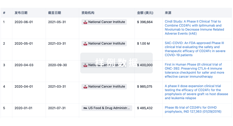
科研基金(NIH)
访问超过 200 万项资助和基金信息,以提升您的研究之旅。
登录
或
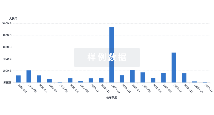
投资
深入了解从初创企业到成熟企业的最新公司投资动态。
登录
或
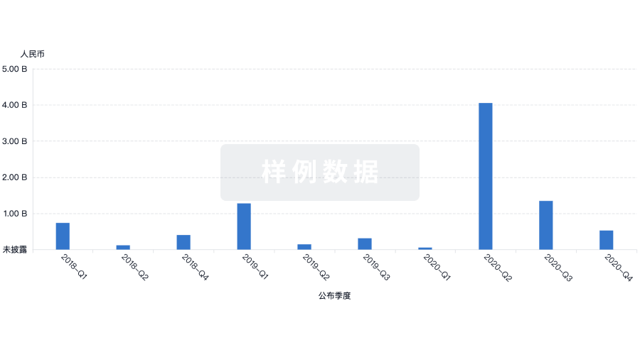
融资
发掘融资趋势以验证和推进您的投资机会。
登录
或
生物医药百科问答
全新生物医药AI Agent 覆盖科研全链路,让突破性发现快人一步
立即开始免费试用!
智慧芽新药情报库是智慧芽专为生命科学人士构建的基于AI的创新药情报平台,助您全方位提升您的研发与决策效率。
立即开始数据试用!
智慧芽新药库数据也通过智慧芽数据服务平台,以API或者数据包形式对外开放,助您更加充分利用智慧芽新药情报信息。
生物序列数据库
生物药研发创新
免费使用
化学结构数据库
小分子化药研发创新
免费使用