预约演示
更新于:2026-07-04
Cilmostim
更新于:2026-07-04
概要
基本信息
药物类型 集落刺激因子 |
别名 Cilmostim (USAN/INN)、CSF-1、M-CSF + [4] |
作用方式 激动剂 |
作用机制 AChR 激动剂(乙酰胆碱受体 激动剂) |
在研适应症- |
原研机构 |
在研机构- |
权益机构- |
最高研发阶段终止临床2期 |
首次获批日期- |
最高研发阶段(中国)- |
特殊审评- |


登录后查看时间轴
结构/序列
Sequence Code 16765080
来源: *****
关联
100 项与 Cilmostim 相关的临床结果
登录后查看更多信息
100 项与 Cilmostim 相关的转化医学
登录后查看更多信息
100 项与 Cilmostim 相关的专利(医药)
登录后查看更多信息
1,444
项与 Cilmostim 相关的文献(医药)2026-12-01·MOLECULAR BIOLOGY REPORTS
Lutein reverses M2 macrophages polarization and exhibits antitumor effects on human triple-negative breast cancer cells
Article
作者: Ramli, Redzyque Ramza ; Fauzi, Agustine Nengsih ; Nafi, Siti Norasikin Mohd ; Nurul, Asma Abdullah ; Muhammad, Siti Nur Hasyila ; Azlan, Maryam
BACKGROUND:
Triple-negative breast cancer (TNBC) remains a major therapeutic challenge due to the lack of established molecular targets and the presence of highly infiltrating tumor-associated macrophages (TAMs), particularly the immunosuppressive M2 phenotype that drives tumor progression. This study investigated the potential of lutein, a plant-derived carotenoid with anticancer properties, to repolarize human monocyte-derived M2 macrophages into the antitumor M1 phenotype and thereby modulate the TNBC tumor microenvironment.
METHODS AND RESULTS:
CD14 + monocytes isolated from peripheral blood mononuclear cells were differentiated into M1 or M2 macrophages using GM-CSF/IFN-γ/lipopolysaccharide or M-CSF/IL-4, respectively. Lutein's effects on macrophage polarization, cell proliferation, cell migration and cytokine secretion were evaluated using flow cytometry, MTT proliferation assays, migration assays and enzyme-linked immunosorbent assays (ELISAs) in MDA-MB-231 cells. Additionally, tumor-related protein expressions from MDA-MB-231 cells were analyzed using protein arrays. Lutein significantly reduced the proliferation and migration of MDA-MB-231 cells when co-cultured with M1/M2 macrophages compared to the untreated group. IL-4 stimulation increased the expression of M2 macrophage marker (CD206) and protumor cytokines (IL-10 and TGF-β1), but lutein effectively reduced these elevated molecules in IL-4-activated macrophages. Proteomic profiling revealed that lutein downregulated several protumor proteins (survivin, VEGF, BCL-X, M-CSF, MMP-9) and upregulated antitumor markers (FKHR and GM-CSF). Furthermore, lutein markedly reduced the secretion of proinflammatory and protumorigenic cytokines and chemokines (IL-6, IL-18BPa, CCL2, CCL8, CCL7, CCL3, CCL20, and CXCL8).
CONCLUSION:
These findings suggest that lutein reprograms M2 macrophages toward an M1-like phenotype and disrupts tumor-promoting signaling within the TNBC microenvironment, highlighting its potential as an adjunct therapeutic agent.
2026-05-01·Asian Pacific journal of cancer prevention : APJCP
Diagnostic Performance of Serum VEGF and M-CSF in Cervical Cancer and, Their Association with Stage
Article
作者: Alzerfi, Hussein Sattar Rebat ; Salman, Suhad Abdulhussein ; Abd Alredha, Redha Dawud ; Chyad, Abbas ; Mohammed, Ahmed Jasim
BACKGROUND:
Cervical cancer continues to be one of the most prevalent malignancies affecting women globally. Circulating biomarkers may provide added value by complementing HPV-based screening and enhancing risk stratification. The aim of this study was to investigate vascular endothelial growth factor (VEGF) and macrophage colony-stimulating factor (M-CSF) as candidate serum biomarkers for the diagnosis and disease monitoring of cervical cancer.
METHODS:
In a single-center case-control study, 45 women with cervical cancer and 45 healthy controls were enrolled. Clinical variables included FIGO 2018 stage, symptoms, smoking status, and HPV vaccination status. Serum VEGF and M-CSF levels were measured using ELISA. Group and stage differences were assessed, and diagnostic performance was evaluated using ROC analysis, Youden's index, and logistic regression.
RESULTS:
Both biomarkers were significantly elevated in cancer patients compared with controls (M-CSF: 1457 ± 582 vs. 504 ± 250 pg/mL; VEGF: 399.7 ± 136 vs. 106.7 ± 53.2 pg/mL; both p < 0.001). Concentrations increased with advancing stage (M-CSF: p = 0.0006; VEGF: p = 0.0073). Vaccinated patients exhibited lower VEGF levels (p = 0.047). Diagnostic performance was excellent (AUC: M-CSF, 0.95; VEGF, 0.97). At optimal cut-offs, VEGF achieved 95% sensitivity (95% CI: 83.1%-99.4%) and 93% specificity (95% CI: 81.3%-98.5%). The combined model (AUC: 0.974) outperformed M-CSF alone (p = 0.038). Multivariate analysis confirmed that both VEGF and M-CSF remained significant independent predictors after adjusting for age, smoking status, and HPV vaccination status.
CONCLUSIONS:
Serum VEGF and M-CSF demonstrate diagnostic utility and stage association in cervical cancer. Their combined use enhances discriminatory power. These findings support their potential as adjunct biomarkers; however, external validation and longitudinal studies are needed.
2026-04-01·Alzheimers & Dementia-Translational Research & Clinical Interventions
A potential multimodal biomarker – cognitive signature associated with the conversion from subjective cognitive decline to mild cognitive impairment
Article
作者: Mohamed Raâfet Ben Khedher ; Tamás Fülöp ; Mohamed Haddad ; Chadi Ouechtati ; Charles Ramassamy ; the Consortium for the early identification of Alzheimer's disease-Quebec (CIMA-Q)
Abstract:
INTRODUCTION:
The disruption of key mechanisms involved in amyloid beta (Aβ) clearance during the early stages of dementia may contribute to the progression of cognitive decline toward irreversible brain damage. In this study, we investigated multiple immune‐related pathways implicated in the management and clearance of Aβ within circulating extracellular vesicles (cEVs) and serum from individuals with subjective cognitive decline (SCD) who later progressed to mild cognitive impairment (MCI).
METHODS:
A cytokine panel and the levels of Aβ
1–42
were quantified in both cEVs and serum from a longitudinally followed cohort of elderly with SCD, using mesoscale and Luminex technologies. We investigated associations with Aβ burden, cognitive performance,
APOE
ε4 allele status, and the likelihood of conversion to MCI.
RESULTS:
In SCD patients, the concentrations of Aβ
1–42
and macrophage‐colony stimulating factor (M‐CSF) were higher, respectively, in cEVs and serum. No difference was observed for fraktaline, interleukin (IL)‐4, IL‐13, interferon gamma (IFN‐γ), and sCD40L in either cEVs or serum between SCD and control patients. Based on receiver operating characteristic curve analysis, regression modeling, and correlations with cognitive performance, M‐CSF levels in serum effectively distinguished individuals with SCD who converted to MCI from those who remained stable. Interestingly, combining M‐CSF and cEVs Aβ
1–42
with the Rey Auditory Verbal Learning Test (RAVLT) cognitive scores provided an excellent classification for SCD converted to MCI up to 2 years prior to clinical diagnosis.
DISCUSSION:
Our findings support the potential value of integrating serum M‐CSF levels with RAVLT performance and cEVs Aβ
1–42
concentrations into a multimodal biomarker panel for longitudinal monitoring of progressive neurocognitive impairment.
26
项与 Cilmostim 相关的新闻(医药)2026-06-30
临床痛点切入,直击骨科科研人痛点
做骨质疏松基础研究的小伙伴,几乎都绕不开同一个难题:现有抗骨吸收药物 “治标难治本,副作用拉满”。双膦酸盐长期使用易引发下颌骨坏死、非典型股骨骨折;地诺单抗价格高昂,停药后骨量快速反弹;特立帕肽存在肿瘤、心血管风险。面对庞大的绝经后骨质疏松人群,开发低毒、低成本、全新机制的小分子药物,是骨科、代谢领域科研圈持续火热的攻坚方向。
胆固醇代谢靶点 ACAT1 早已在动脉粥样硬化、肿瘤领域被反复验证,其特异性抑制剂 Avasimibe(AVA)作为经典降脂候选药,抗炎、抑癌活性已有大量报道,但它能否调控骨代谢、干预破骨细胞分化,此前完全是空白。
陆军军医大学新桥医院骨科团队另辟蹊径,依托 RNA-seq 转录组多组学测序、GO/KEGG 通路富集、GSEA 基因集富集 AI 生信分析,层层筛选关键差异基因,锁定肌酸激酶 B(CKB)作为核心中介分子,完整阐明 Avasimibe 抑制破骨生成、改善去卵巢小鼠骨丢失的分子通路,给骨质疏松新药研发提供了一条全新可落地的思路。今天就带大家拆解这篇《International Immunopharmacology》高分论文的完整逻辑、实验设计与生信挖掘亮点。
生信临床研究感兴趣,请为小骨点点关注,持续解读骨科前沿生信文献和思路~
研究核心发现 & 五大硬核亮点>>>核心总结论
ACAT1 在 RANKL 诱导破骨分化过程中显著上调;ACAT1 抑制剂 Avasimibe 通过下调 CKB,阻断 ATP 依赖的 PI3K-AKT 信号通路,在体外抑制破骨细胞成熟、体内逆转 OVX 小鼠雌激素缺失型骨流失,且无肝肾心毒性,是极具潜力的抗骨质疏松小分子药物。>>>五大独家研究亮点老药新用创新:原本用于动脉粥样硬化的降脂药 Avasimibe,首次被证实具备抗骨质疏松活性,省去新药合成、毒理初筛成本,临床转化门槛更低;多组学 AI 生信精准筛靶:RNA-seq 差异基因筛选 + GO/KEGG/GSEA 富集 AI 分析,从 500 + 差异基因中精准锁定 CKB 这一全新骨代谢调控分子,打破传统单基因盲筛模式;完整上下游调控链条:从上游靶点 ACAT1 抑制剂→中间能量代谢蛋白 CKB→下游核心成骨破骨通路 PI3K-AKT,形成完整可验证调控轴,且体外 PCr 回补实验完美反向验证机制;体外 + 体内双向完整验证:细胞层面(BMM 破骨诱导、TRAP、免疫荧光、qPCR、WB)+ 动物 OVX 绝经骨质疏松模型(Micro-CT、多色组织染色、血清生化、IHC),证据链闭环;高安全性加持:高低剂量 AVA 干预 8 周后,小鼠肝、肾、心肌生化指标无异常,无脏器病理损伤,对比现有临床药物安全性优势突出。
文章标题:ACAT1 inhibitor Avasimibe suppresses osteoclastogenesis and alleviates ovariectomy-induced bone loss via CKB/PI3K-AKT signaling
中文标题:ACAT1抑制剂Avasimibe抑制破骨细胞生成,并通过CKB/PI3K-AKT信号缓解卵巢切除术引起的骨质流失
发表期刊:Int Immunopharmacol
发表时间:2026年1月
影响因子:5.6/Q2
研究背景
骨稳态失衡催生新药研发刚需
骨质疏松核心病理:破骨细胞过度活化、骨吸收>骨形成;绝经后雌激素缺失会直接放大破骨活性,诱发脆性骨折,老龄化下疾病负担持续加重。
现有药物短板:临床主流抗骨吸收药物均存在不可逆副作用,亟需安全新型小分子抑制剂。
前期研究伏笔:ACAT1 调控胆固醇酯化,抑制剂 Avasimibe 兼具抗炎、抑癌作用,且有文献提示其可调控 PI3K-AKT 通路(骨代谢核心通路),但ACAT1、Avasimibe 与破骨分化的关联无任何报道,本研究填补空白。
科学假设:破骨分化过程 ACAT1 表达升高;抑制 ACAT1 可通过能量代谢分子 CKB 下调 PI3K-AKT,抑制破骨生成、缓解雌激素缺乏骨丢失。
研究方法
细胞、动物、多组学生信全套体系
体外细胞实验
→细胞来源:6 周龄 C57BL/6 雌鼠骨髓巨噬细胞 BMM,M-CSF+RANKL 诱导成熟破骨细胞;→干预手段:ACAT1 siRNA 敲低、梯度浓度 Avasimibe(0/3/5μM)给药、磷酸肌酸 PCr 回补拯救实验;→检测技术:CCK-8 细胞毒性、TRAP 破骨染色、F-actin / 纽蛋白免疫荧光、qRT-PCR 破骨标志基因、Western blot 蛋白定量。
RNA-seq 多组学生信 AI 分析
→分组:RANKL 诱导组、RANKL+Avasimibe 给药组;→分析流程:差异基因 DEG 筛选→聚类热图、火山图可视化→GO 生物功能富集→KEGG 通路富集→GSEA 通路打分验证,AI 工具锁定 PI3K-AKT、CKB 核心靶点。
体内 OVX 骨质疏松动物模型
→造模:8 周龄雌性 C57BL/6 小鼠双侧去卵巢模拟绝经后骨质疏松;→分组:假手术 Sham、OVX 模型组、低剂量 AVA (15mg/kg)、高剂量 AVA (30mg/kg)、AVA+PCr 回补组、阿仑膦酸钠阳性药组;→干预:腹腔注射给药 8 周,每 3 天一次;→检测:Micro-CT 骨微结构定量(BMD、BV/TV、Tb.N、Tb.Th、Tb.Sp)、股骨 H&E/Masson/TRAP 组织染色、CKB 免疫组化、血清肝肾功能心肌酶毒性检测。
统计学方法
GraphPad Prism 10,双组 t 检验、多组单因素 ANOVA+Tukey 事后检验,P<0.05 为差异显著。
结果分析
1. ACAT1 是破骨分化正向调控基因
实验流程:分离小鼠骨髓 BMM,RANKL 诱导 7 天形成成熟多核破骨;
TRAP 染色可见 RANKL 组大量多核阳性破骨;
RNA-seq 热图、qPCR、WB 同步证实:破骨分化全程 ACAT1 mRNA、蛋白持续上调,同时 TRAP、CTSK、NFATc1 等经典破骨标志物同步升高;结论:ACAT1 表达与破骨成熟高度正相关,可能是促破骨关键基因。
2. 敲低 ACAT1 直接抑制破骨细胞成熟
使用 3 条 siRNA 沉默 ACAT1:
qPCR 显示破骨标志基因 TRAP、MMP9、CTSK、c-FOS、NFATc1 全部显著下调;
WB 验证蛋白水平同步降低;
TRAP 染色:si-ACAT1 组多核破骨数量、体积大幅减少;
免疫荧光:破骨功能性 F-actin 环结构破碎、黏附斑减少;结论:内源性 ACAT1 是 RANKL 诱导破骨分化必不可少的调控因子。
3. ACAT1 抑制剂 Avasimibe 体外安全、剂量依赖性抑制破骨生成
CCK-8 毒性实验:≤5μM AVA 处理 48/72/96h,BMM 细胞活力无下降,无细胞毒;
qPCR+WB:3μM、5μM AVA 梯度给药,ACAT1 及所有破骨标志物呈剂量依赖性下降;
TRAP 染色:随 AVA 浓度升高,多核破骨数量、面积逐步减少;
免疫荧光:F-actin 环完整性被破坏,破骨骨架组装受阻;结论:小分子药物 Avasimibe 可安全阻断 RANKL 介导的破骨细胞分化。
4. 转录组 AI 生信挖掘锁定 CKB/PI3K-AKT 核心通路
RNA-seq 共筛选 536 个下调、113 个上调差异基因,热图、火山图直观区分两组表达谱;
GO 富集:差异基因集中于细胞外基质、胶原代谢(骨重塑核心功能);
KEGG+GSEA 通路 AI 打分:PI3K-AKT 通路是 AVA 干预后抑制最显著的信号轴;
差异基因重点标注 CKB:AVA 处理后 CKB 表达大幅下调,作为后续机制核心靶点。
5. CKB 是 Avasimibe 抑制破骨的关键中介,PCr 回补可逆转药效
CKB 功能:催化肌酸与 ATP 生成磷酸肌酸 PCr,维持细胞能量供给;PI3K 激活高度依赖 ATP。
WB 证实 AVA 显著下调 CKB 蛋白;
外源添加 PCr 回补实验:PCr 可逆转 AVA 对 TRAP、CTSK、NFATc1 等破骨基因的抑制;
TRAP 染色 + 免疫荧光:PCr 处理后,被 AVA 抑制的多核破骨、F-actin 环结构全部恢复;结论:Avasimibe 依靠下调 CKB、降低胞内 PCr/ATP 储备,发挥抑破骨作用。
6.Avasimibe 通过 CKB 阻断 PI3K-AKT 磷酸化激活
时间梯度 WB:RANKL 刺激后 PI3K、AKT 持续磷酸化,AVA 可随时间逐步降低 P-PI3K、P-AKT 水平;
PCr 拯救实验:PCr 补充后,被 AVA 抑制的 PI3K-AKT 磷酸化完全恢复;完整分子链条:AVA 抑制 ACAT1→下调 CKB→PCr/ATP 生成不足→ATP 依赖的 PI3K 活化受阻→AKT 磷酸化下降→下游 NFATc1 等破骨转录因子下调→破骨分化阻断。
7.体内 OVX 模型证实 Avasimibe 改善雌激素缺失型骨流失
Micro-CT 骨微结构:OVX 组骨密度 BMD、骨小梁数量 Tb.N、厚度 Tb.Th 显著下降,骨小梁间隙 Tb.Sp 升高;高低剂量 AVA 干预后剂量依赖性逆转骨微结构损伤,高剂量药效接近阳性药阿仑膦酸钠;
H&E、Masson 染色:AVA 提升骨小梁占比、胶原沉积面积;
TRAP 组织染色:AVA 显著减少骨组织内 TRAP 阳性破骨细胞数量(Oc.S/BS 比值下降);
CKB 免疫组化:OVX 模型骨组织 CKB 高表达,AVA 给药后 CKB 蛋白表达显著降低,完美复刻体外机制;
安全评价:血清 ALT/AST、CK-MB、肌酐、尿素氮无异常,心肝肾组织无病理损伤;
反向验证:AVA+PCr 联合给药会抵消 Avasimibe 的骨保护效果,再次佐证 CKB - 磷酸肌酸通路是体内药效核心。
文章小结
>基础表型层面:ACAT1 是促破骨分化新靶点;小分子抑制剂 Avasimibe 体外无毒性,强效抑制 RANKL 诱导的破骨成熟;OVX 小鼠体内可显著缓解雌激素缺乏引发的骨量丢失、改善骨微结构,无脏器毒副作用。
>生信机制层面:依托 RNA-seq 多组学 AI 通路分析,首次鉴定 CKB 作为 Avasimibe 下游关键能量代谢分子;明确CKB/PI3K-AKT是调控破骨分化的核心信号轴,PCr 回补实验从细胞、动物两层反向验证通路真实性。
>转化价值层面:将成熟降脂老药 Avasimibe 拓展至骨质疏松领域,提供低毒、低成本全新候选药物,为绝经后骨质疏松、溶骨性骨病治疗开辟新方向。
局限性本研究存在的 4 项不足
能量代谢直接证据缺失:仅通过 PCr 回补间接佐证 ATP 不足,未直接检测 AVA 干预后胞内 ATP、PCr 实时含量,缺少能量代谢直接定量数据;
仅聚焦破骨细胞,忽略成骨细胞:骨稳态是成骨 - 破骨耦合平衡,本研究未探究 Avasimibe 对成骨细胞增殖、矿化的影响,无法完整评价药物对整体骨重塑的双向作用;
缺少药物药代动力学(PK)数据:未检测 Avasimibe 体内骨组织富集度、半衰期、组织分布,不利于后续给药方案优化;
ACAT1 与 CKB 之间的直接调控中间分子未挖掘:仅证实 AVA 同时下调 ACAT1、CKB,但 ACAT1 胆固醇代谢如何直接调控 CKB 转录 / 翻译,中间衔接通路仍待深挖。
全文逻辑深度解读
整篇文章采用 “靶点发现→抑制剂表型验证→多组学生信筛下游分子→通路机制验证→回补反向证明→体内动物转化→安全性评价”标准高分代谢类文章逻辑:
正向观察:破骨分化 ACAT1 上调→敲除 ACAT1 抑破骨,锁定靶点;
药物干预:用已知 ACAT1 抑制剂 Avasimibe 重复表型,确认药物有效性;
生信筛靶:RNA-seq AI 富集缩小下游候选分子,锁定 CKB、PI3K-AKT;
机制闭环:PCr 功能回补实验,反向证明 CKB 是药效必需分子;
通路上下游:WB 验证 PI3K-AKT 磷酸化变化,完善分子链条;
体内转化:OVX 疾病模型重复体外机制,Micro-CT + 组织学表型支撑临床潜力;
安全兜底:补充脏器毒性检测,提升药物转化说服力。
整套实验设计无逻辑断点,体外细胞分子机制 + 体内动物药效双向互证,转录组多组学 AI 分析作为机制挖掘工具大幅降低盲筛工作量,非常适合骨质疏松、骨代谢、小分子药物研发方向的科研人员借鉴实验思路与生信分析流程。
如果您对生信分析和公共数据库挖掘感兴趣,但时间和精力有限或者缺乏相关经验,小骨非常乐意为您提供如下服务:免费思路评估、付费方案设计和生信分析等,有意向的老师欢迎联系小骨哦!
2026-06-24
·生物探索
引言
CAR-T细胞(chimeric antigen receptor T cells)改变了血液肿瘤治疗的想象力,但进入实体瘤后,它常常遭遇三个老问题:进不去、留不住、打不透。于是,研究人员把目光转向巨噬细胞(macrophage):这种细胞天生擅长进入组织、吞噬目标、重塑炎症环境。问题是,成熟巨噬细胞并不擅长体外扩增,也不容易做复杂基因工程,输回体内后还常被肺、肝、脾“截留”。
6月19日,《Cell》的研究报道“Expansion and CAR engineering of granulocyte-monocyte progenitors for cellular immunotherapy”,提出了一个更上游的解法:不要只改造终末分化的巨噬细胞,而是改造它的前体——粒单核祖细胞(granulocyte-monocyte progenitors, GMPs)。这项研究的核心,不只是做出一种新的CAR细胞,而是重新追问:细胞免疫治疗真正应该从哪个发育阶段开始设计?
这项研究的关键不是把巨噬细胞本身“再强化一次”,而是把工程化位置前移到粒单核祖细胞(GMPs),让细胞治疗同时获得可扩增性、可工程化性、体内髓系输出和宿主免疫接力。
问题不只是“杀伤力”,而是细胞从哪里出发
传统CAR-T强调识别和杀伤,CAR巨噬细胞(CAR macrophages, CAR-Ms)则强调抗原定向吞噬(antigen-directed phagocytosis)和肿瘤微环境改造。这个方向很有吸引力,因为巨噬细胞进入组织的能力通常优于T细胞,也能通过吞噬和抗原呈递参与后续免疫激活。
但成熟巨噬细胞有天然短板。研究中提到,系统输注后,巨噬细胞往往优先滞留在肺、肝、脾,肿瘤内积累有限;更关键的是,成熟巨噬细胞没有“上游补给站”。它们输进去多少,基本就消耗多少,不能在体内持续产生新的髓系效应细胞。制造端也不轻松:体外扩增能力弱,病毒转导和非病毒递送效率低,冻存后功能还可能受影响。
所以,真正的问题变成:能不能找到一种既能扩增、又能工程化、还能在体内持续产出髓系细胞的起点?
GMPs正好处在这个位置。它们不是造血干细胞(hematopoietic stem cells, HSCs)那样全能,也不是成熟巨噬细胞那样“终点化”。它们已经偏向髓系命运,却仍保留增殖和分化潜力。过去的常识是,这类命运受限的祖细胞寿命短、扩增有限。但这篇研究挑战了这一点。
一套培养条件,把短命GMP推向长期扩增
研究人员首先建立了小鼠GMP扩增条件,称为SCF/2i,由干细胞因子(stem cell factor, SCF)、BRAF抑制剂GDC-0879,以及小分子SKL2001组成。在这一条件下,小鼠骨髓来源细胞可以维持超过50天的对数期扩增,总量接近扩大8个数量级,达到约10¹³个细胞。
这不是简单“长得多”,更重要的是“长得像谁”。单细胞RNA测序(single-cell RNA sequencing, scRNA-seq)显示,扩增细胞的转录图谱与GMP相匹配;染色质开放性分析(ATAC-seq)也显示其接近GMP状态。流式结果显示,长期传代后超过90%的细胞保持Lin⁻Sca1⁻CD48⁺c-Kit⁺FcγR⁺表型,符合小鼠GMP特征。
功能上,这些细胞仍能形成巨噬细胞、粒细胞(granulocyte)以及粒-巨噬细胞混合克隆。在单细胞克隆形成实验中,SCF/2i扩增细胞每192个单细胞可形成110±8.66个克隆,新鲜分选的原代GMP为96±5.57个,二者没有显示出明显劣势。也就是说,扩增没有明显牺牲祖细胞功能。
再往下看,它们可以在M-CSF刺激下分化为CD11b⁺F4/80⁺巨噬细胞;在G-CSF刺激72小时后,CD115⁻Ly6G⁺粒细胞比例达到83.9±3.5%,并呈现典型多叶核形态。这说明,体外扩增并没有把它们推成一团失去方向的细胞,而是保留了可用的髓系分化程序。
人源GMP的难点,被一个新小分子打开
小鼠系统成立并不等于人源系统可用。研究人员发现,原始SCF/2i对人源GMP(human GMPs, hGMPs)的支持有限,问题集中在SKL2001在人细胞中的活性不足。于是,他们进行了结构优化,获得SKL2001类似物TN2-30,并将其与SCF、GDC-0879、IL-3和G-CSF组合,形成SCF/2i-IG条件。
在SCF/2i-IG中,人脐带血GMP能够克隆性扩增。单细胞水平上,156个单独评估的人源GMP中,有51个形成未分化克隆,比例为32.7%。成人骨髓和动员外周血来源细胞也能在这一条件下扩增。经过27天培养后,来自脐带血、动员外周血和骨髓的扩增细胞在单细胞图谱上都与GMP聚类,而不是偏离到其他血液谱系。
安全性方面,研究人员对28天SCF/2i-IG培养物进行了全基因组测序,未检测到体细胞突变负荷增加,也没有发现显著优势克隆扩张。这个结果不能等同于长期临床安全,但至少说明,在测试窗口内,扩增并未伴随明显基因组异常信号。
这些hGMP也保留了分化能力。M-CSF诱导后,CD14⁺巨噬细胞比例达到97.4±2.3%;G-CSF诱导后,CD15⁺CD66B⁺粒细胞比例达到83.6±3.2%。换句话说,研究人员不是得到了一种“长得快但身份模糊”的细胞,而是得到了一种可扩增、可工程化、仍像GMP的细胞平台。
进入体内后,GMP和成熟巨噬细胞走向完全不同
真正有意思的部分出现在体内分布实验。研究人员将表达荧光素酶的人源hGMP或人源巨噬细胞静脉输注到NSG小鼠体内,每只小鼠输入5×10⁶个细胞。结果显示,hGMP来源信号在体内增加,定位到多个造血相关生态位,约第21天达到峰值,随后重新分布到脾脏和腹部,并至少持续180天。拟合得到的衰减半衰期为150.81天。
相比之下,输注的成熟巨噬细胞在24小时内主要积累于肺部,第7天已基本不可检测,衰减半衰期只有5.31天。第14天外周血检测也显示,hGMP组可见约2.06%的GFP⁺供者来源细胞,而巨噬细胞组仅0.01%。
在免疫相容的小鼠体系中,差异同样明显。小鼠GMP输注后半衰期为142.28天,而GMP来源巨噬细胞只有3.92天。GFP标记实验进一步显示,输注后第7天,供者来源细胞占骨髓白细胞46.5%,其中20.4%的骨髓GFP⁺细胞处于增殖状态。这说明GMP并非只是“暂时停留”,而是能在体内形成一波供者来源髓系生成(donor-derived myelopoiesis)。
这个差异很关键。成熟巨噬细胞像一次性效应细胞;GMP则更像上游生产单元,能在体内持续补给单核细胞、巨噬细胞和粒细胞。
从抗感染到抗肿瘤:先证明这些细胞真的能工作
如果GMP在体内扩增和分布,但产出的细胞功能不行,平台价值就会大打折扣。研究人员用慢性肉芽肿病(chronic granulomatous disease, CGD)小鼠测试其抗感染功能。CGD模型的吞噬细胞氧化杀菌能力有缺陷,因此对细菌感染高度敏感。
在金黄色葡萄球菌(S. aureus)感染模型中,GMP输注显著减少肝脓肿数量,差异达到P<0.0001;脾脏重量也明显下降,P=0.0037。生存曲线有改善趋势,但P=0.124,不能过度解读为明确生存获益。在洋葱伯克霍尔德菌(B. cepacia)感染模型中,GMP输注明显降低血液细菌负荷,P<0.0001,并显著提高生存率,P=0.0076。
这一步证明了一个基础事实:扩增GMP在体内产生的髓系细胞不是摆设,而能恢复先天免疫(innate immunity)功能。只有跨过这一关,后续CAR工程才有意义。
给GMP装上CAR:工程化效率是成熟巨噬细胞难以比拟的
成熟巨噬细胞很难高效基因改造,是CAR-M发展的瓶颈之一。研究人员直接比较了人源GMP与CD14⁺单核细胞来源巨噬细胞的递送效率。结果很清楚:成熟巨噬细胞无论逆转录病毒、慢病毒还是质粒递送,效率都很低;hGMP则对慢病毒转导非常友好,MOI为20时转导效率约超过60%。
这意味着,祖细胞状态本身可能是工程化成功的关键变量。研究人员还展示了GMP可接受mRNA递送、CRISPR-Cas9敲除,以及连续慢病毒工程化,例如先导入αCD19 CAR,再敲除SIRPα。这为多重工程设计留下了空间。
功能测试中,αCD19 CAR-GMP分化来的巨噬细胞可以有效吞噬CD19⁺ B急性淋巴细胞白血病(B-ALL)细胞;加入抗CD47抗体后,吞噬率进一步提高,约接近90%。在特异性测试中,αCD19 CAR-M主要吞噬B-ALL细胞,αHER2 CAR-M则主要吞噬HER2⁺ SK-BR-3细胞。也就是说,这不是非特异炎症吞噬,而是抗原依赖的识别与吞噬。
体内结果同样有分量。在CD19⁺ B-ALL异种移植模型中,研究人员在建模后21天输注1×10⁷个αCD19 CAR-hGMP。到第80天,10只治疗小鼠中有6只外周血疾病负荷维持在极低或不可检测水平;对照组10只小鼠在第38天前全部死亡,治疗组生存显著改善,P<0.0001。
在HER2⁺ SKOV3实体瘤模型中,αHER2 CAR-hGMP也显示强效。治疗组肿瘤发光信号在观察期内接近完全被压制,生存显著优于对照,P<0.0001。更重要的是,hGMP来源细胞能大量进入肿瘤,也能在脾和肝中被检测到。这正是成熟巨噬细胞治疗常常难以做到的部分。
CAR-Fc:让宿主免疫系统接过抗原
常规CAR-GMP可以吞噬肿瘤,但还有一个更深的问题:如果抗原处理只停留在供者工程细胞内部,宿主抗原呈递细胞(antigen-presenting cells, APCs)网络就很难被充分调动。尤其在同种异体(allogeneic)治疗中,供者和受者主要组织相容性复合体(major histocompatibility complex, MHC)不匹配,供者细胞即使吞噬了肿瘤,也未必能有效激活宿主T细胞。
为了解决这个问题,研究人员设计了CAR-Fc结构:在CAR胞外区加入免疫球蛋白G1 Fc结构域(IgG1 Fc domain)。这个设计保留CAR细胞自身吞噬能力,同时让附近表达Fc受体(Fc receptor)的宿主吞噬细胞能够识别CAR-Fc与靶细胞形成的复合界面,从而发生“跨细胞”吞噬(trans phagocytosis)。
混合品系共培养实验把这个机制拆得很清楚。BALB/c来源的CAR-GMP巨噬细胞作为供者,C57BL/6来源的巨噬细胞和OT-II CD4 T细胞作为宿主侧。目标细胞表达HER2和OVA肽。只有当C57BL/6宿主APC摄取并呈递OVA肽时,OT-II T细胞才会被激活。结果显示,常规CAR和CAR-Fc都能进行自身吞噬(cis phagocytosis),但只有CAR-Fc能诱导宿主巨噬细胞跨细胞吞噬,并带来明显OT-II T细胞增殖和细胞因子释放。CAR-Fc组干扰素γ(interferon γ, IFN-γ)约达到600 pg/ml,IL-2约接近400 pg/ml,而常规CAR组和对照组都很低。
这个设计的意义不只是“多吞一点”。它把肿瘤抗原从供者细胞手里转交给宿主APC网络,让宿主T细胞有机会参与进来。
在同种异体A20淋巴瘤模型中,常规αCD19 CAR-GMP只能 modestly 延缓肿瘤进展,而αCD19 CAR-Fc-GMP显著压低肿瘤负荷。到第60天,CAR-Fc组9只动物中有5只仍存活;CAR-Fc相对常规CAR的生存优势P=0.007。
在HER2⁺ 4T1乳腺癌原位模型中,CAR-Fc组肿瘤内CD3⁺ T细胞和颗粒酶B⁺(granzyme B, GZB⁺)细胞密度更高,肿瘤体积更小,生存也更长。研究人员报告,CAR-Fc组中位生存期大约是对照组的两倍。这里传递出的信息很明确:CAR-Fc并没有让供者细胞永久存在,却让短暂存在的供者细胞更有效地启动宿主免疫。
一个意外靶点:MPO限制了GMP的增殖寿命
这篇研究还有一个容易被忽略但很有启发性的部分:为什么SCF/2i能让GMP长期扩增?
GDC-0879是BRAF抑制剂,这一点相对清楚。其他BRAF抑制剂如SB590885和PLX8394也能部分替代其作用。但SKL2001原本被认为与Wnt/β-catenin通路相关,结果却不符合预期:CHIR99021、Wnt agonist 1和Wnt3a都不能复制SKL2001效果,Wnt通路抑制剂IWR1、FH535也不能阻断扩增。β-catenin缺失的mGMP仍能在SCF/2i中正常扩增。
研究人员进一步用化学蛋白质组学寻找SKL2001相关靶点,发现髓过氧化物酶(myeloperoxidase, MPO)是主要富集蛋白,分子量约81 kDa。MPO通常被认为是成熟粒细胞杀菌相关酶,但这里它出现在GMP增殖控制中。
竞争培养实验更直接:Mpo敲除GMP与野生型GMP按1:1混合培养。没有SKL2001时,Mpo敲除细胞逐渐占优势,野生型MPO阳性细胞比例降到约5.75%;加入SKL2001后,这种优势消失,MPO阳性细胞维持在约54.8%。这说明药物抑制MPO在功能上类似于遗传敲除MPO。
两个结构不同的MPO抑制剂verdiperstat和mitiperstat也能复制GMP持续扩增效果。表面等离子共振(surface plasmon resonance, SPR)显示SKL2001可逆结合小鼠MPO,TN2-30可逆结合人MPO;酶活实验显示SKL2001和TN2-30分别抑制小鼠和人MPO的氯化活性。这把“可扩增GMP平台”从经验配方推进到了机制层面:MPO可能是限制GMP增殖寿命和状态稳定性的关键内在刹车之一。
这项研究真正打开的问题
小鼠和人源GMP可以在定义条件下长期扩增,同时保留GMP身份和髓系分化能力;GMP比成熟巨噬细胞更容易基因工程化;CAR-GMP能在小鼠模型中产生抗原特异性抗肿瘤效果;CAR-Fc能把抗原处理进一步接入宿主APC-T细胞轴。
未来髓系细胞治疗的关键,可能不是把成熟巨噬细胞改得越来越强,而是把工程化位置前移到可扩增、可分化、可持续供给的祖细胞阶段。这个逻辑一旦成立,细胞治疗设计的核心参数就会从“单个效应细胞有多强”转向“体内髓系输出如何被编程”。
但也必须保持谨慎,人源hGMP的主要体内疗效是在NSG免疫缺陷小鼠中完成的,不能完全代表真实患者免疫环境;肿瘤模型集中在CD19和HER2两个靶点,尚不能说明对更多抗原和复杂肿瘤微环境都有效;同种异体GMP在免疫完整宿主体内仍会被逐步清除,CAR-Fc解决的是抗原转交和T细胞激活问题,并不是彻底解决免疫排斥。祖细胞产品还涉及长期安全性、罕见克隆事件、异位炎症、组织重塑等风险,这些都需要更大队列、更长观察和更严格释放标准。
这项研究没有只给CAR细胞治疗增加一个新构件,而是提出了一个更底层的问题:我们究竟应该工程化一个已经走到终点的效应细胞,还是工程化一个能够在体内继续产生效应细胞的发育节点?
如果CAR-T代表“给T细胞装上雷达”,那么GMP平台代表的是另一种设计思路:给髓系生成过程本身写入方向。真正值得期待的,也许不是某一种细胞瞬间变强,而是免疫系统被重新组织起来,持续、定向、可控地把肿瘤抗原交给该接手的细胞。
参考文献
Yue S, Guo Z, Pan C, Jing XA, Tao L, Nguyen T, Tang J, Chan Y, Contreras-Trujillo H, Jiang D, Yan X, Xiang H, Liu X, Bloom C, Ediriwickrema A, Koschade S, Wang X, Wang Z, Shu N, Shi Y, McKim DB, Lu R, Majeti R, Zhang C, Ying QL. Expansion and CAR engineering of granulocyte-monocyte progenitors for cellular immunotherapy. Cell. 2026 Jun 19:S0092-8674(26)00643-4. doi: 10.1016/j.cell.2026.05.043. Epub ahead of print. PMID: 42320470.
声明:本文仅用于分享,不代表平台立场,如涉及版权等问题,请尽快联系我们,我们第一时间更正,谢谢!
往期热文:
Nature Genetics | 父母的基因,也会“绕路”影响孩子吗?一项家系研究给多基因评分装上了“防偏差雷达”
Cell | 肺癌发生前5年,血液可能已经听见了风声
Nature Methods | 把“读脑”和“控脑”装进同一根探针:Neuropixels Opto为什么值得关注?
Cell | 一个细胞里,转录因子究竟坐在基因组哪里?
Nature | 肿瘤细胞为何怕“铁”?一个小分子,改写了铁死亡的故事
Nature | 两百万个细胞告诉我们:IBD风险不只藏在基因里,还藏在“哪一种细胞”里
Nature Biotechnology | 抗体进了身体,为什么到不了肿瘤深处?单细胞空间图谱揭开实体瘤递送屏障
Nature Biotechnology | 细胞不是“拼图”:它们如何自己长出形状?
Nature Genetics | 阿尔茨海默病的遗传拼图,又多了16块
Nature Biotechnology | 基因编辑的下一道门槛:不是剪得准,而是把“修好的细胞”挑出来
2026-05-26
集落刺激因子(Colony-Stimulating Factor,CSF)是一类功能十分关键的糖蛋白细胞因子,能够有效激活骨髓造血祖细胞增殖、分化,诱导形成成熟的功能性血细胞集落。在机体内,它主要负责调控造血发育、维持免疫稳态,同时参与炎症应答与组织修复等一系列生理过程,是髓系免疫细胞生长、成熟、活化的核心调控蛋白。凭借稳定且独特的生物活性,CSF广泛应用于造血基础研究、免疫机制探索、干细胞实验及生物医药研发等多个领域。
01
CSF的几个主要类型
在目前的科研与生物医药应用中,集落刺激因子主要分为三大主流亚型。三类因子各司其职、相互配合,完整覆盖髓系免疫细胞的整个发育周期,能够轻松适配基础科研、临床研究、药物研发等多种场景,各亚型的具体特点如下:
• 粒细胞集落刺激因子(G-CSF):是一款靶向性极强的造血调控因子,主要作用于骨髓粒系祖细胞,专一推动中性粒细胞增殖、成熟并释放至外周血,基本不会干扰巨噬细胞、淋巴细胞等其他免疫细胞。它作用精准、功能稳定,也是目前研究最透彻、应用最普遍的CSF亚型。
• 粒细胞-巨噬细胞集落刺激因子(GM-CSF):拥有更加广谱的生物活性,可同时促进粒细胞、巨噬细胞增殖分化,兼具造血调控与免疫激活双重功能。能够有效提升免疫细胞的抗原呈递、吞噬与杀菌能力,是连接机体造血稳态与免疫应答的重要因子,在免疫激活、抗感染研究中应用广泛。
• 巨噬细胞集落刺激因子(M-CSF):专门靶向单核-巨噬细胞体系,主要调控单核细胞分化、巨噬细胞活化与稳态维持。在炎症机制探究、组织重塑、纤维化疾病研究、药物效果验证等常规科研实验中,都是非常常用的核心工具因子。
• 白细胞介素 - 3(IL-3):亦称作多集落刺激因子(multi-CSF),是作用范围最广的集落刺激因子。它几乎可诱导所有血细胞生成,包括巨噬细胞、中性粒细胞、嗜酸性粒细胞、嗜碱性粒细胞、肥大细胞、血小板以及红细胞。
02
CSF应用领域
1、基础科研应用
对于广大科研人员而言,集落刺激因子是非常实用的基础研究工具,通用性高、适配场景丰富,是高校实验室、科研院所、生物医药研发团队的常用核心试剂。目前主流的基础科研应用方向主要集中在免疫调控、造血发育、炎症病理及前沿模型构建等领域,具体可分为以下几类:
• 造血干细胞发育与分化研究:G-CSF、GM-CSF及IL-3可以很好地模拟体内造血微环境,支持造血干细胞体外扩增、髓系谱系分化与造血体系重建。非常适合用于造血发育机制、干细胞干性调控、造血损伤修复等基础课题,实验体系稳定、重复性佳,是造血方向研究的标配试剂。
• 免疫细胞体外构建与功能研究:GM-CSF是体外诱导树突状细胞分化、成熟的关键因子,常被用于抗原呈递、肿瘤免疫激活、疫苗机制等研究;M-CSF可稳定诱导单核细胞分化为巨噬细胞,为巨噬细胞极化、固有免疫调控、肿瘤免疫逃逸等实验提供标准化细胞模型,是免疫科研的常用核心工具。
• 炎症与病理机制研究:GM-CSF、M-CSF积极参与体内炎症微环境调节、组织重塑与纤维化进程,可轻松搭建体外炎症损伤、组织纤维化、自身免疫损伤等实验模型,方便科研人员开展疾病机制分析、靶点验证、药理探究等各类基础实验。
• 前沿科研模型构建:在类器官培养、干细胞工程、免疫共培养体系等前沿实验中,CSF系列因子能够稳定维持细胞分化状态与模型生理活性,有效保障基因编辑、细胞改造、类器官构建等复杂实验的稳定性,助力各类交叉创新研究顺利推进。
2、生物制药与工业研发应用
在生物制药与产业研发领域,集落刺激因子主要衔接基础科研与药物转化,是创新药前期研发的重要配套原料。广泛用于细胞治疗、免疫药物、抗炎靶向药的机制验证、药效筛选与模型构建,可辅助完成种子细胞驯化、体外扩增工艺摸索与药物细胞水平评价,为新药研发和工艺优化提供可靠的实验支撑。
3、临床医疗应用
集落刺激因子也具备成熟的临床应用价值,作用机制清晰、应用效果稳定。其中rhG-CSF常被用于肿瘤放化疗、骨髓移植后的造血恢复工作,能够快速提升中性粒细胞数量,降低患者感染风险;GM-CSF与M-CSF则多用于重症感染辅助干预、创面修复、慢性炎症相关研究。整体来看,临床应用场景相对集中,远不如基础科研场景丰富多元。
推
荐
产
品
随着干细胞技术、肿瘤免疫研究、类器官模型等前沿领域快速发展,集落刺激因子已经成为基础医学研究中不可或缺的工具试剂,科研端对高品质、高稳定性细胞因子原料的需求也在持续增长。
针对各类基础实验对纯度、活性、稳定性和批次一致性的严苛要求,乔默生物重组集落刺激因子系列产品,采用无动物源成分生成工艺制备,具备活性验证、高纯度、低内毒素、批次稳定的突出特点,能够有效避免普通原料常见的活性不足、数据波动、批次差异大等实验问题。未来,乔默生物将持续深耕细胞因子赛道,持续优化产品质控与性能,以适配各类高端实验体系的优质科研级细胞因子,助力全球科研团队的基础研究与创新转化工作。
100 项与 Cilmostim 相关的药物交易
登录后查看更多信息
外链
| KEGG | Wiki | ATC | Drug Bank |
|---|---|---|---|
| D03498 | - | - | - |
研发状态
10 条进展最快的记录, 后查看更多信息
登录
| 适应症 | 最高研发状态 | 国家/地区 | 公司 | 日期 |
|---|---|---|---|---|
| 高胆固醇血症 | 临床2期 | 美国 | - | |
| 高胆固醇血症 | 临床2期 | - | - | |
| 肿瘤 | 临床2期 | 美国 | - |
登录后查看更多信息
临床结果
临床结果
适应症
分期
评价
查看全部结果
| 研究 | 分期 | 人群特征 | 评价人数 | 分组 | 结果 | 评价 | 发布日期 |
|---|
No Data | |||||||
登录后查看更多信息
转化医学
使用我们的转化医学数据加速您的研究。
登录
或

药物交易
使用我们的药物交易数据加速您的研究。
登录
或

核心专利
使用我们的核心专利数据促进您的研究。
登录
或

临床分析
紧跟全球注册中心的最新临床试验。
登录
或

批准
利用最新的监管批准信息加速您的研究。
登录
或

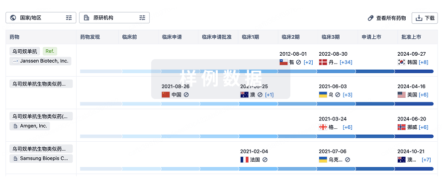
生物类似药
生物类似药在不同国家/地区的竞争态势。请注意临床1/2期并入临床2期,临床2/3期并入临床3期
登录
或
特殊审评
只需点击几下即可了解关键药物信息。
登录
或

芽仔
全新生物医药AI Agent 覆盖科研全链路,让突破性发现快人一步
立即开始免费试用!
智慧芽新药情报库是智慧芽专为生命科学人士构建的基于AI的创新药情报平台,助您全方位提升您的研发与决策效率。
立即开始数据试用!
智慧芽新药库数据也通过智慧芽数据服务平台,以API或者数据包形式对外开放,助您更加充分利用智慧芽新药情报信息。
生物序列数据库
生物药研发创新
免费使用
化学结构数据库
小分子化药研发创新
免费使用