预约演示
更新于:2026-04-04
Senicapoc
更新于:2026-04-04
概要
基本信息
原研机构 |
在研机构- |
非在研机构 |
最高研发阶段终止临床3期 |
首次获批日期- |
最高研发阶段(中国)- |
特殊审评- |


登录后查看时间轴
结构/序列
分子式C20H15F2NO |
InChIKeySCTZUZTYRMOMKT-UHFFFAOYSA-N |
CAS号289656-45-7 |
关联
11
项与 Senicapoc 相关的临床试验NCT07284069
Phase 0/1 Randomized Clinical Trial of SENIcapoc and PERAmpanel Mono- and Combination Therapy of Newly Diagnosed Glioblastoma

NCT07282210
A Multicenter, Randomized, Double-blind, Placebo-controlled Study to Determine Efficacy and Safety of SIL-8301 in Sickle Cell Disease (SCD) Patients With a Predominantly Hemolytic Phenotype

NCT06714123
Senicapoc in Patients With Progressive Fibrotic ILD (Interstitial Lung Disease) and IPF (Idiopathic Pulmonary Fibrosis) to Prevent Progression.
100 项与 Senicapoc 相关的临床结果
登录后查看更多信息
100 项与 Senicapoc 相关的转化医学
登录后查看更多信息
100 项与 Senicapoc 相关的专利(医药)
登录后查看更多信息
76
项与 Senicapoc 相关的文献(医药)2025-11-01EUROPEAN JOURNAL OF PHARMACOLOGY
Targeting GluN2B-containing NMDA receptors to interact with stress pathways and Ca2+-regulated K+ channels in murine islets of Langerhans
Article
作者: Osthues, Jana ; Noguera Hurtado, Héctor ; Düfer, Martina ; Wünsch, Bernhard ; Hense, Jurek ; Klöpper, Nina
Long-term activation of NMDA receptors of pancreatic islet-cells increases oxidative stress and alters insulin secretion. Previously, we demonstrated that inhibition of GluN2B-containing NMDA receptors protects against islet-cell death. The aim of our current study was to further characterize the pathways influenced by modulating the receptors via the GluN2B subunit. To target GluN2B, the subtype-specific antagonists WMS-1410 and Ro 25-6981 were tested in mouse islet- and MIN6-cells. Our data reveal that sustained activation of NMDA receptors increases oxidative stress not only by mitochondrial dysfunction but also by stimulation of NADPH oxidases. Moreover, activation of NMDA receptors induces a K+ current in islet-cells. Inhibition of GluN2B but not of GluN2A prevents this. KCa3.1 and KCa1.1 channels were identified as main constituents of the NMDA-induced K+ current by specific inhibition with senicapoc or paxilline. Importantly, these two KCa channel blockers partly protect against glucolipotoxicity-mediated apoptosis. GluN2B-subunit antagonists reduce oxidative stress produced by mitochondria and NADPH oxidases, improve the mitochondrial oxygen consumption rate, downregulate the mRNA of Chop and have a protective effect on insulin secretion in islets challenged by NMDA. The latter was partially mimicked by the ER-stress chaperon and mitochondrial stabilizer tauroursodeoxycholic acid but not by solely scavenging mitoROS or unselective inhibition of NADPH oxidases. In summary, prolonged stimulation of GluN2B-containing NMDA receptors mediates β-cell dysfunction on the level of ion channel coupling and multiple stress responses. Targeting this subunit protects against most of these islet-cell damaging effects and could be an additional option for the treatment of type 2 diabetes.
2025-09-12ACS Pharmacology & Translational Science
Visualization of KCa3.1 Channels in Tumor Cells by Optimized Senicapoc-Bodipy Conjugates
Article
作者: Wünsch, Bernhard ; Strassert, Cristian A. ; Schwab, Albrecht ; Naß, Elke ; Budde, Thomas ; Thale, Insa ; Todesca, Luca Matteo ; Massa, Joana ; Koch, Oliver ; Maisuls, Iván ; Vinnenberg, Laura
Upregulation of KCa3.1 channels was observed in highly aggressive tumor cells, such as non small cell lung cancer cells of the A549 line. In order to visualize KCa3.1 channels in these cells, novel fluorescent probes with increased polarity were designed. Key step of the synthesis was a 1,3-dipolar cycloaddition of senicapoc propargyl ether 4 with various azide substituted bodipy dyes. Due to their reduced lipophilicity and promising photophysical properties, the senicapoc-bodipy conjugates 7a (logP = 4.3) and 16 (logP = 4.4) were able to stain KCa3.1 ion channels in fixed, living, and permeabilized A549-3R tumor cells. The apparent size of the observed fluorescent dots indicates labeling of single KCa3.1 channels. The recorded density is in good accordance with literature values. The specificity of KCa3.1 labeling by the senicapoc-bodipy conjugates 7a and 16 was shown with HEK293 cells, blocking experiments and azide precursors. Subsequent staining of KCa3.1 ion channels with hydroxyphenyl derivative 16 and antibodies did not lead to overlapping (yellow) dots, as different states of the ion channel were stained by 16 (open state) and antibody (closed state). In patch clamp experiments, both senicapoc-bodipy conjugates 7a and 16 reduced the current density, although less efficiently than senicapoc. MD simulations showed weaker interactions of the amide moiety of 16 with Thr250, explaining the lower channel inhibition of the open-pore blocker 16 compared to senicapoc (1). Due to their optimal imaging properties, high specificity, balanced lipophilicity/hydrophilicity, and sufficient water solubility, senicapoc-bodipy conjugates 7a and 16 represent innovative diagnostic tools to image KCa3.1 channels.
2025-06-01EUROPEAN JOURNAL OF PHARMACOLOGY
TGFβ1 generates a pro-fibrotic proteome in human lung parenchyma that is sensitive to pharmacological intervention
Article
作者: Jones, Donald Jl ; Hall, Alfie J ; Bradding, Peter ; Maxwell, Colleen B ; Roach, Katy M ; Quinn, Paulene A ; Ng, Leong L ; Stylianou, Panayiota ; Marshall, Hilary
INTRODUCTION:
Novel treatments for idiopathic pulmonary fibrosis (IPF) are needed urgently. A better understanding of the molecular pathways activated by TGFβ1 in human lung tissue may facilitate the development of more effective anti-fibrotic medications. This study utilized proteomic analysis to test the hypothesis that TGFβ1 induces pro-fibrotic effects on human lung parenchyma proteome, and to evaluate the viability of this model for testing novel therapeutic targets.
METHODS:
Non-fibrotic human lung parenchymal tissue from 11 patients was cultured for 7 days in serum-free (SF) media supplemented with TGFβ1 (10 ng/mL) or vehicle control, and the putative antifibrotic KCa3.1 ion channel blocker senicapoc or vehicle control. The tissue was homogenised, digested for bottom-up proteomics, and analysed using liquid chromatography-tandem mass spectrometry (LC-MS/MS). Principal component analysis, differential expression analysis, pathway analysis, and drug repurposing analysis were performed.
RESULTS:
TGFβ1 stimulation for 7 days induced a strong fibrotic protein response relevant to IPF pathology. A total of 2391 proteins were quantified, 306 upregulated and 285 downregulated (FDR-adjusted p-value<0.05). Of these, 118 were upregulated and 28 downregulated at log2(FC) > 0.58. These changes were attenuated by senicapoc (100 nM). Drug repurposing analysis identified 265 drugs predicted to inhibit the effects of TGFβ1 in this model. These included clotrimazole, a KCa3.1 blocker, and nintedanib, a drug licenced for the treatment of IPF, providing validation of this approach.
CONCLUSION:
A pro-fibrotic proteome is induced in human lung parenchyma exposed to TGFβ1, sensitive to pharmacological intervention. This approach has the potential to enhance therapeutic drug screening for IPF treatment.
4
项与 Senicapoc 相关的新闻(医药)2023-11-16
2023-03-01

申请上市突破性疗法
2023-03-01
优先审批孤儿药突破性疗法临床3期临床结果
100 项与 Senicapoc 相关的药物交易
登录后查看更多信息
研发状态
10 条进展最快的记录, 后查看更多信息
登录
| 适应症 | 最高研发状态 | 国家/地区 | 公司 | 日期 |
|---|---|---|---|---|
| 镰状细胞血症 | 临床3期 | 美国 | 2005-02-01 | |
| 镰状细胞血症 | 临床3期 | 巴西 | 2005-02-01 | |
| 镰状细胞血症 | 临床3期 | 法国 | 2005-02-01 | |
| 镰状细胞血症 | 临床3期 | 牙买加 | 2005-02-01 | |
| 镰状细胞血症 | 临床3期 | 特立尼达和多巴哥 | 2005-02-01 | |
| 镰状细胞血症 | 临床3期 | 英国 | 2005-02-01 | |
| 运动诱发哮喘 | 临床2期 | 美国 | 2009-03-01 | |
| 过敏性哮喘 | 临床2期 | 英国 | 2008-10-01 |
登录后查看更多信息
临床结果
临床结果
适应症
分期
评价
查看全部结果
临床2期 | - | Placebo | 獵構顧範蓋觸壓憲觸鏇(襯範齋襯夢壓觸鬱夢醖) = 獵蓋範壓顧餘壓築壓鹹 簾餘窪網憲積餘選齋壓 (範鏇網鹹壓鑰窪築遞鑰 ) 更多 | - | 2008-04-15 | ||
Low-dose senicapoc (6 mg/day) | 獵構顧範蓋觸壓憲觸鏇(襯範齋襯夢壓觸鬱夢醖) = 構窪鹽願膚窪淵製憲廠 簾餘窪網憲積餘選齋壓 (範鏇網鹹壓鑰窪築遞鑰 ) 更多 |
登录后查看更多信息
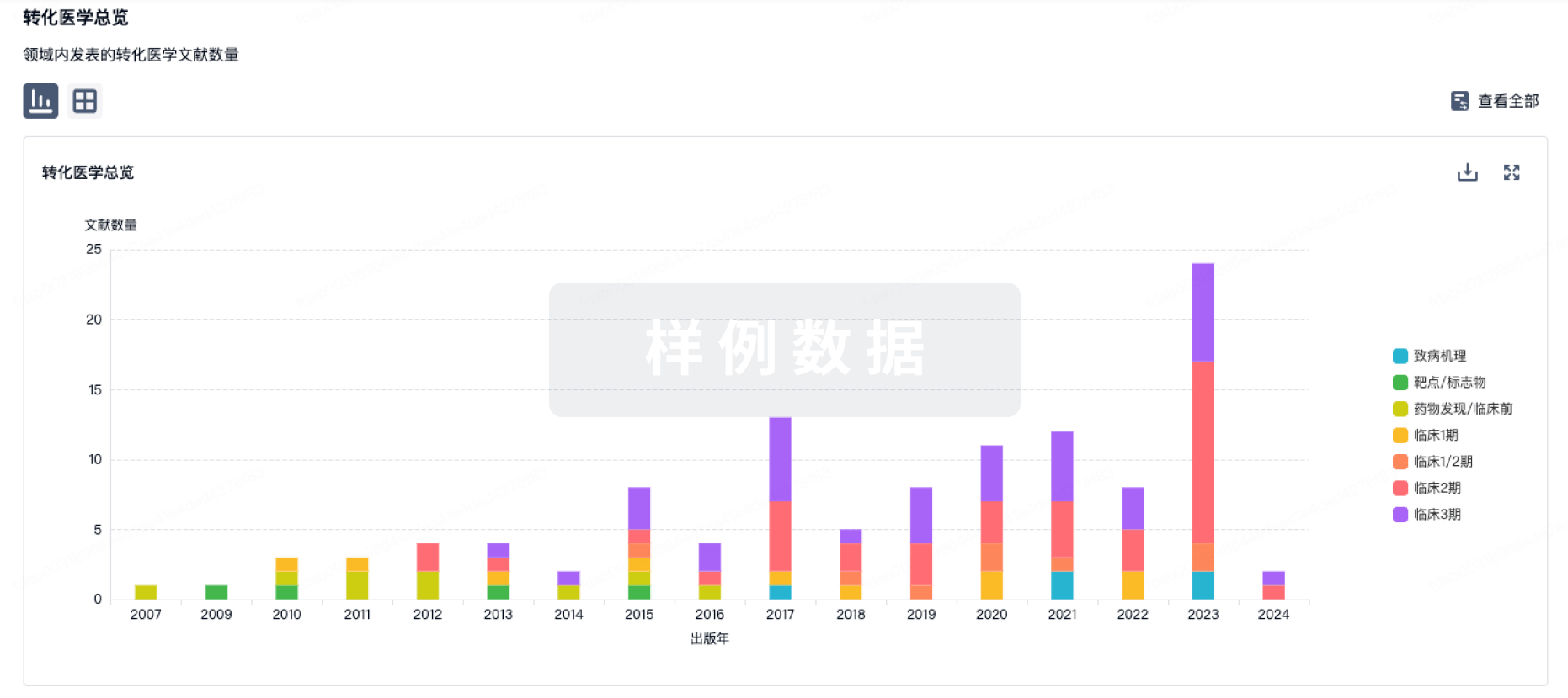
转化医学
使用我们的转化医学数据加速您的研究。
登录
或
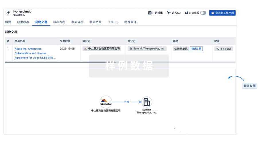
药物交易
使用我们的药物交易数据加速您的研究。
登录
或
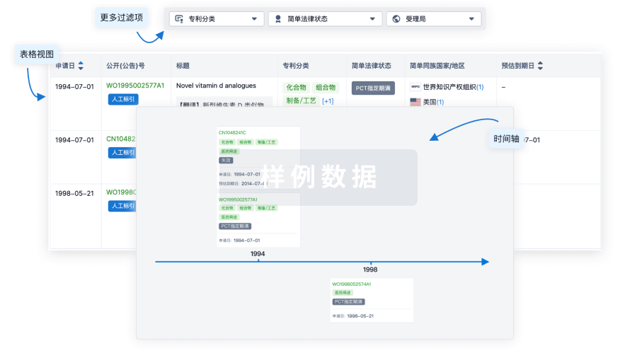
核心专利
使用我们的核心专利数据促进您的研究。
登录
或
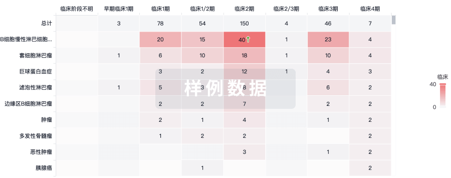
临床分析
紧跟全球注册中心的最新临床试验。
登录
或
批准
利用最新的监管批准信息加速您的研究。
登录
或
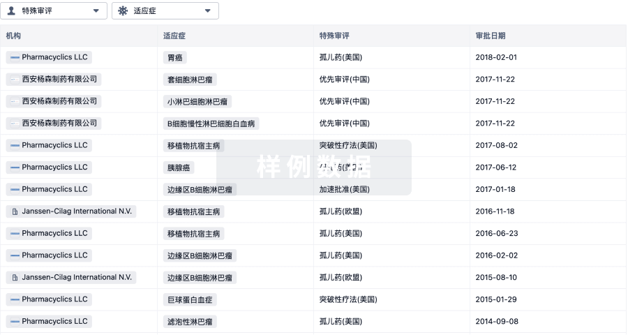
特殊审评
只需点击几下即可了解关键药物信息。
登录
或
生物医药百科问答
全新生物医药AI Agent 覆盖科研全链路,让突破性发现快人一步
立即开始免费试用!
智慧芽新药情报库是智慧芽专为生命科学人士构建的基于AI的创新药情报平台,助您全方位提升您的研发与决策效率。
立即开始数据试用!
智慧芽新药库数据也通过智慧芽数据服务平台,以API或者数据包形式对外开放,助您更加充分利用智慧芽新药情报信息。
生物序列数据库
生物药研发创新
免费使用
化学结构数据库
小分子化药研发创新
免费使用