预约演示
更新于:2025-06-06
LP-003
更新于:2025-06-06
概要
基本信息
关联
100 项与 LP-003 相关的临床结果
登录后查看更多信息
100 项与 LP-003 相关的转化医学
登录后查看更多信息
100 项与 LP-003 相关的专利(医药)
登录后查看更多信息
3
项与 LP-003 相关的文献(医药)2017-04-01·Materials science & engineering. C, Materials for biological applications
Stavudine loaded gelatin liposomes for HIV therapy: Preparation, characterization and in vitro cytotoxic evaluation
Article
作者: Ashe, Sarbani ; Boxi, Ankita ; Nayak, Bismita ; Thathapudi, Neethi Chandra ; Nayak, Debasis
Despite continuous research and availability of 25 different active compounds for treating chronic HIV-1 infection, there is no absolute cure for this deadly disease. Primarily, the residual viremia remains hidden in latently infected reservoir sites and persistently release the viral RNA into the blood stream. The study proposes the dual utilization of the prepared stavudine-containing nanoformulations to control the residual viremia as well as target the reservoir sites. Gelatin nanoformulations containing very low dosage of stavudine were prepared through classical desolvation process and were later loaded in soya lecithin-liposomes. The nanoformulations were characterized through dynamic light scattering (DLS), Transmission electron microscopy (TEM), X-ray diffraction (XRD) and ATR-FTIR. All the formulations were in nano regime with high hemocompatibility and exhibited dose-dependent cytotoxicity towards Raw 264.7 macrophages. Among the various formulations, SG-3 (Stavudine-Gelatin Nanoformulation sample 3) and SG-LP-3 (Stavudine-Gelatin Nano-Liposome formulation sample 3) showed the best results in terms of yield, size, charge, encapsulation efficiency, hemocompatibility and % cell viability. For the first time, liposomal delivery of antiretroviral drugs using nanocarriers has been demonstrated using very low dosage (lower than the recommended WHO dosage) showing the prominent linear release of stavudine for up to 12h which would reduce the circulatory viremia as well as reach the sanctuary reservoir sites due to their nanosize. This method of liposomal delivery of antiretroviral drugs in very low concentrations using nanocarriers could provide a novel therapeutic alternative to target HIV reservoir sites.
1994-05-01·Cellular immunology3区 · 医学
A Unique Murine CD43 Epitope Lp-3: Distinct Distribution from Another CD43 Epitope S7
3区 · 医学
Article
作者: Sachiko Hirose ; Takashi Okada ; Hiroshi Okamoto ; Masaaki Abe ; Susumu Hattori ; Hiroyuki Nishimura ; Toshikazu Shirai ; Hiromichi Tsurui ; Jun Shiota ; Bo Yu ; Shingo Nozawa
In foregoing studies, we found a unique B cell differentiation antigen Lp-3 which is expressed on pre-B and premature B cells in the bone marrow, but is negative on bone marrow mature B cells and peripheral resting B cells. Nonetheless, Lp-3 was clearly positive on the majority of CD5 B(B1) cells. When we examined the biochemical nature and partial amino acid sequences of purified 132-kDa Lp-3 molecules and the nucleotide sequence of the cDNA clones, we found that Lp-3 is an epitope of CD43. Thus, the monoclonal antibody (mAb) Lp-3 may be the first mAb to murine CD43 defined by primary target structure analysis. Comparison of tissue distribution of Lp-3 and S7, an epitope previously suggested to associate with murine CD43, showed that they were similarly distributed on thymocytes, peripheral B and T cells, granulocytes, and platelets. In the bone marrow, while both Lp-3 and S7 were negative on mature B cells, the former was positive on all B lineage cells at an early ontogeny and the latter was positive only on the minor population of pre-B cells and pro-B cells. Lp-3 and S7 epitopes also showed different distributions on basement membranes of renal glomerulus, bronchus, and endometrium, lining cells of choroid plexus and muscular cells of arterioles in a variety of tissues. As CD43 has various isoforms generated by different degrees of glycosylation of the common core peptide, it is likely that Lp-3 and S7 are associated with different CD43 isoforms.
1989-01-01·International immunology3区 · 医学
The novel murine B cell differentiation antigen LP-3
3区 · 医学
Article
作者: Jun Shiota ; Kiyoshi Matsumoto ; Hidetoshi Sato ; Yasuo Ishida ; Sachiko Hirose ; Masaaki Abe ; Hiroyuki Nishimura ; Takashi Okada ; Toshikazu Shirai
The unique murine lymphocyte differentiation antigen, Lp-3, with a mol. wt of approximately 125 kd, was found using a rat monoclonal antibody. The Lp-3 antigen was distributed on a wide variety of myeloid, T cell, and B cell lineages in mice. However, the expression was only found in B cells at certain stages of differentiation. The pre-B and virgin B cells in the bone marrow from 2-month-old BALB/c mice were weakly positive for Lp-3, while the resting B cells in the spleen and lymph node were Lp-3 negative. In contrast, the majority of B cells in the peritoneal cavity, mostly Ly-1 (CD5) B cells, had a brighter fluorescence for Lp-3 than did bone marrow B cells. The Lp-3 antigen could be induced in a high density in approximately one-half of lipopolysaccharide-stimulated, large, blastic spleen B cells. Cell cycle analysis showed that Lp-3 is an early B cell activation antigen which is first expressed at the G1A phase of the cell cycle. Therefore this novel B cell differentiation antigen will be useful for differentiating pre-B and virgin B cells in the bone marrow, resting B cells, and a population of activated B cells in the periphery. In contrast to findings in BALB/c mice, there was an elevated population of B cells with a bright Lp-3 expression in the spleen of autoimmune-prone NZB x NZW F1 mice.
5
项与 LP-003 相关的新闻(医药)2025-03-17
Recently, Novartis has made significant strides in the chronic spontaneous urticaria (CSU) market, announcing a priority review application for its BTK inhibitor Remibrutinib and a $830 million deal to acquire Kyorin's clinical-stage MRGPRX2 antagonist.
The announcement highlights Novartis' commitment to maintaining its leadership position in the CSU treatment landscape, which is marked by a prevalence rate of approximately 0.05% to 3%, with CSU accounting for about 68% of chronic urticaria cases. The condition primarily manifests as hives and itching, and can lead to deeper systemic issues, increasing the risk of autoimmune diseases and cancers. First-line treatment typically involves H1 antihistamines, but nearly half of patients struggle to achieve adequate control, necessitating additional therapies.
The global CSU market is projected to grow from $779.28 million in 2024 to $1.54141 billion by 2032, with a compound annual growth rate (CAGR) of 8.90% from 2025 to 2032. As awareness and treatment demands increase, this market is poised for continued growth.
As a market leader, Novartis has long relied on its collaboration with Roche to dominate the CSU field with Xolair (omalizumab), an anti-IgE drug that has generated over $4.3 billion in sales in 2024, despite emerging competition. However, with the expiration of Xolair's compound patent and an impending expiry of its formulation patent in November 2025, Novartis faces pressure from competitors like Sanofi/Regenron's Dupixent (dupilumab), which has recently gained approval for CSU in Japan and is challenging Xolair's dominance.
In light of these developments, Novartis has shifted its focus to exploring next-generation therapies, particularly BTK inhibitors. Bruton’s tyrosine kinase (BTK) plays a crucial role in CSU's pathogenesis, and inhibiting BTK can prevent mast cell activation and the production of autoantibodies, addressing two primary pathological mechanisms of CSU.
Last year, Novartis reported positive results from two pivotal Phase III trials of Remibrutinib, demonstrating significant symptom improvement over 52 weeks. The recent submission of its New Drug Application (NDA) positions Remibrutinib as a potential successor to Xolair, aiming to solidify Novartis' standing in the CSU market.
In addition to Remibrutinib, Novartis is strategically acquiring promising candidates like KRP-M223, an MRGPRX2 antagonist, which is anticipated to enhance its portfolio in this therapeutic area. MRGPRX2, a G protein-coupled receptor expressed predominantly on mast cells, is activated during allergic responses, leading to the release of mediators such as histamine. Although KRP-M223 is still in preclinical stages, its unique mechanism holds the potential for significant advancements in treating CSU and related allergic conditions.
In the domestic market, several promising therapies targeting CSU are advancing through clinical trials. Notably, Tianchen Biotech's LP-003, a next-generation anti-IgE antibody, recently reported mid-stage Phase II clinical data that surpassed Xolair's efficacy. With a dosing regimen of every 8 weeks, LP-003 could significantly improve patient compliance compared to Xolair's more frequent administration.
Another contender is Jimin Xinke's JYB1904, a long-acting anti-IgE antibody that boasts a half-life more than twice that of Xolair, reducing injection frequency and potentially enhancing patient adherence to treatment.
As Novartis strengthens its position in the CSU market with innovative therapies and strategic acquisitions, the competitive landscape continues to evolve. The battle for dominance in this lucrative market is intensifying, with Novartis poised to defend its title against emerging threats while exploring new growth avenues.
临床结果临床3期临床2期优先审批申请上市
2025-03-02
SAN DIEGO, March 2, 2025 /PRNewswire/ -- Longbio Pharma (Suzhou) Co., Ltd. (referred to as "Longbio"), a leading biotech company dedicated to developing innovative biologic treatments for allergy, respiratory, dermatology, hematology, ophthalmology, and other autoimmune and rare diseases, proudly announced the release of interim analysis of CSU (Chronic Spontaneous Urticaria) Phase II data for LP-003, the novel long-acting anti-IgE antibody, at the 2025 American Academy of Allergy, Asthma & Immunology annual meeting (AAAAI 2025). LP-003 demonstrated superior improvement compared to Omalizumab in mean change of UAS7 (urticaria activity score 7) from baseline and percentage% of patients with UAS7=0 at week 4 and week 12.
The presentation of the poster titled "An interim analysis of Phase II study of LP-003, a novel high-affinity, long-acting anti-IgE antibody for CSU" by LongBio marked a significant moment during the conference.
Title: An interim analysis of Phase II study of LP-003, a novel high-affinity, long-acting anti-IgE antibody for CSU
ClinicalTrials.gov ID: NCT06228560
Date and Time: March 2nd, 2025, 9:45 am (UTC -5)
Location: Convention Center, Ground Level, Hall A.
Poster Number: 685
Methods: This is a multi-center, double-blind, placebo-controlled Phase II clinical study, randomized to receive 100 mg Q8W, 200 mg Q8W or 200 mg Q4W of LP-003 or 300mg Omalizumab or placebo (n=40/group). The primary endpoint is the proportion of participants achieving complete absence of wheals and itching (UAS7 = 0) at week 12. Secondary endpoints include mean change of UAS7 from baseline at week 4 and 12, and other efficacy and safety endpoints.
Comparing to the first-generation anti-IgE antibody, omalizumab, LP-003 shows:
superior improvement in mean change of UAS7 from baseline at week 12 as well as the percentage% of patients with UAS7=0 at week 4 and week 12.
rapid and sustained relief in CSU symptoms in a dose-dependent manner.
favorable safety profile, no statistically significant difference in adverse events were found between LP-003 and placebo groups.
These results suggested that LP-003 might become a
best-in-class anti-IgE therapy.
In addition, LP-003's efficacy result was also comparable to barzolvolimab without raising any new safety concerns. Barzolvolimab is an anti-C-KIT mAB developed by Celldex, and intended to explore its application in CSU field. Together with its long-acting profile, LP-003 stands as a strong challenger in CSU market, positioning it at the forefront of the CSU treatment.
CSU stands as a prevalent skin disorder affecting approximately 1–2% of the global population. Not only that, ~35% patients are not well controlled or refractory to the first-line treatment (anti-histamines). The US market size will reach $2 billion USD in 2026 and increase by 8-10% CAGR in next five years. According to the market research from RAPT, even with efficacy 20% below omalizumab, prescribers still prefer less frequent dosing. LP-003 not only with achieving the longer dosing interval than omalizumab, but also showed superior efficacy. It marks a major step forward, positioning the LP-003 at the forefront of the anti-IgE field.
This achievement also marks a significant milestone for LP-003 following its initial success in allergic rhinitis, and LongBio is planning to continue exploring its promising potential in food allergy and asthma. The Phase II trial of asthma has started and the agency (NMPA, China FDA) approved a dosing interval of every 3 months (Q12W). LP-003 also got the IND approval of food allergy last year and planning to initial the clinical trial in the near future.
Food allergy is huge market with great potential (>$10 billion USD) but less competitors. Based on the epidemiology research, there are more than 13.5 million adults and 3.5 million children are suffering food allergy in US and anti-IgE therapy is the only approved MOA by FDA.
With ongoing studies in allergic rhinitis, CSU, asthma and food allergy, Longbio's innovative approach to biologic therapies continues to show remarkable potential for addressing unmet medical needs across allergic diseases. LP-003 would stand poised to make a significant impact on the global allergy and immunology markets.
About LongBio
LongBio is a biotech company located in Shanghai/Changshu, China. The company, which was founded in 2018, focuses on autoimmune and rare diseases, serving patients and society.
LongBio aims to bring our drugs to both domestic and international markets with our partners, especially to bring patients with more affordable and high-quality bio-medicines. Our leading pipeline is a next generation of anti-IgE antibody (LP-003), which is already in Phase III stage, and prepare for pre-BLA in 2025H2. LP-003 has started clinical studies of several indications, like allergic rhinitis, CSU and asthma. Another pipeline is a bifunctional complement inhibitor, LP-005, with fully inhibition of all three pathways in complement system. The Phase II of PNH of LP-005 is on-going at present, intending to explore renal diseases and neuroinflammations in the near future.
For more information, please visit: or please contact: [email protected].
SOURCE LongBio
WANT YOUR COMPANY'S NEWS FEATURED ON PRNEWSWIRE.COM?
440k+
Newsrooms &
Influencers
9k+
Digital Media
Outlets
270k+
Journalists
Opted In
GET STARTED
临床2期临床结果上市批准临床3期
2024-09-24
SHANGHAI, Sept. 24, 2024 /PRNewswire/ --
LongBio Pharma (Suzhou) Co., Ltd. ("LongBio" or the "Company"), a Phase III-stage biotech company focused on the R&D of innovative antibody and fusion protein drugs for allergies and complement-mediated diseases, announced the completion of its Series B2 financing round, raising tens of millions of RMB. The round was led by Qiming Venture Partners, with continued support from existing investors. The funds will primarily be used to advance late-stage clinical studies in the company's pipeline, strengthen team capabilities, and bolster working capital.
Leveraging its proprietary technology platform, LongBio has developed a range of innovative biologics with independent intellectual property rights.
LP-003, a best-in-class, high-affinity anti-IgE antibody, has demonstrated the potential for superior efficacy, lower dosing, longer dosing intervals, and greater cost-effectiveness. Existing data indicates that:
LP-003 can extend the dosing frequency of current drugs targeting the same target from once every 2-4 weeks to once every 3-6 months.
100 mg of LP-003 showed comparable efficacy to 300 mg of omalizumab.
Subgroup analysis in Phase II study further revealed that LP-003 may offer superior efficacy compared to omalizumab under similar pollen density conditions.
LongBio has also initiated clinical studies in China for LP-003 in various other indications, including chronic spontaneous urticaria (CSU), asthma, and food allergy. The company aims to present the topline results of the LP-003 Phase II study for CSU, using omalizumab as a positive control, at the 2025 American Academy of Allergy, Asthma & Immunology (AAAAI) meeting in San Diego, US, in February 2025. The US FDA IND filing for CSU and food allergy is currently in preparation, and will be submitted later this year.
LP-005, a potential first-in-class bi-functional anti-C3&C5 antibody, has entered Phase II clinical trials for paroxysmal nocturnal hemoglobinuria (PNH) in China. By fully inhibiting all three pathways of the complement system, LP-005 may offer superior efficacy in diseases involving multiple pathways compared to other complement drugs that target only one. LongBio is also exploring LP-005 as a potential treatment for complement-mediated nephritis, eye diseases, and neurological disorders.
For more information, please visit or contact [email protected]
About Qiming Venture Partners
Qiming Venture Partners was founded in 2006. Currently, Qiming Venture Partners manages eleven US Dollar funds and seven RMB funds with $9.5 billion in capital raised. Since our establishment, we have invested in outstanding companies in the Technology and Consumer (T&C) and Healthcare industries at the early and growth stages.
Since our debut, we have backed over 530 fast-growing and innovative companies. Over 200 of our portfolio companies have achieved exits through IPOs at the NYSE, NASDAQ, HKEX, Shanghai Stock Exchange, or Shenzhen Stock Exchange, or through M&A or other means. There are also over 70 portfolio companies that have achieved unicorn or super unicorn status.
SOURCE LongBio
WANT YOUR COMPANY'S NEWS FEATURED ON PRNEWSWIRE.COM?
440k+
Newsrooms &
Influencers
9k+
Digital Media
Outlets
270k+
Journalists
Opted In
GET STARTED

临床2期临床结果
100 项与 LP-003 相关的药物交易
登录后查看更多信息
研发状态
10 条进展最快的记录, 后查看更多信息
登录
| 适应症 | 最高研发状态 | 国家/地区 | 公司 | 日期 |
|---|---|---|---|---|
| 尤文肉瘤 | 临床申请 | - | 2023-11-22 | |
| 神经母细胞瘤 | 临床申请 | - | 2023-11-22 | |
| 小细胞肺癌 | 临床申请 | - | 2023-11-22 | |
| 卵巢癌 | 临床前 | 中国 | 2023-10-19 | |
| 白血病 | 临床前 | 中国 | - | |
| 黑色素瘤 | 临床前 | 中国 | - |
登录后查看更多信息
临床结果
临床结果
适应症
分期
评价
查看全部结果
| 研究 | 分期 | 人群特征 | 评价人数 | 分组 | 结果 | 评价 | 发布日期 |
|---|
No Data | |||||||
登录后查看更多信息
转化医学
使用我们的转化医学数据加速您的研究。
登录
或

药物交易
使用我们的药物交易数据加速您的研究。
登录
或

核心专利
使用我们的核心专利数据促进您的研究。
登录
或

临床分析
紧跟全球注册中心的最新临床试验。
登录
或

批准
利用最新的监管批准信息加速您的研究。
登录
或

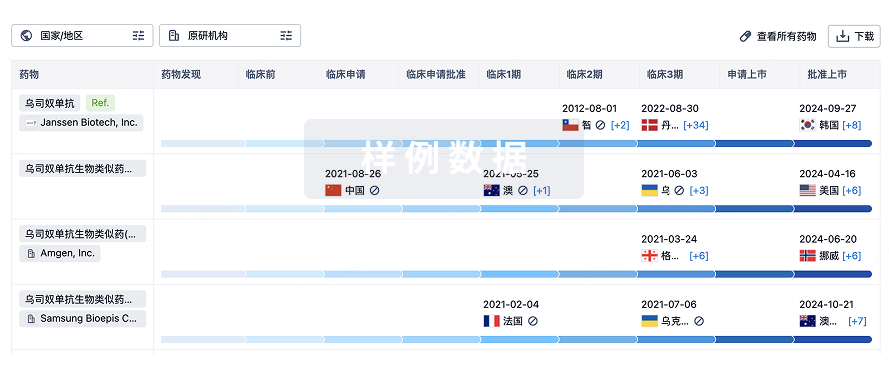
生物类似药
生物类似药在不同国家/地区的竞争态势。请注意临床1/2期并入临床2期,临床2/3期并入临床3期
登录
或

特殊审评
只需点击几下即可了解关键药物信息。
登录
或

生物医药百科问答
全新生物医药AI Agent 覆盖科研全链路,让突破性发现快人一步
立即开始免费试用!
智慧芽新药情报库是智慧芽专为生命科学人士构建的基于AI的创新药情报平台,助您全方位提升您的研发与决策效率。
立即开始数据试用!
智慧芽新药库数据也通过智慧芽数据服务平台,以API或者数据包形式对外开放,助您更加充分利用智慧芽新药情报信息。
生物序列数据库
生物药研发创新
免费使用
化学结构数据库
小分子化药研发创新
免费使用
