预约演示
更新于:2025-10-25
Gonadotropin, Chorionic(Bristol Myers)
绒毛膜促性腺激素(美国百时美施贵宝)
更新于:2025-10-25
概要
基本信息
药物类型 激素 |
别名 Follutein |
作用方式 激动剂 |
作用机制 FSHR激动剂(卵泡刺激素受体激动剂)、LHCGR激动剂(促黄体激素/绒毛膜促性腺激素受体激动剂) |
治疗领域- |
在研适应症- |
非在研适应症- |
在研机构- |
权益机构- |
最高研发阶段撤市 |
首次获批日期 美国 (1974-12-26) |
最高研发阶段(中国)- |
特殊审评- |
登录后查看时间轴
关联
100 项与 绒毛膜促性腺激素(美国百时美施贵宝) 相关的临床结果
登录后查看更多信息
100 项与 绒毛膜促性腺激素(美国百时美施贵宝) 相关的转化医学
登录后查看更多信息
100 项与 绒毛膜促性腺激素(美国百时美施贵宝) 相关的专利(医药)
登录后查看更多信息
2
项与 绒毛膜促性腺激素(美国百时美施贵宝) 相关的文献(医药)1970-03-01Journal of reproduction and fertility
A STUDY OF EGG TRANSPORT IN THE RABBIT USING A FREEZING--CLEARING TECHNIQUE
Article
作者: HOWE, G. R.
Summary. The utilization of a freezing\p=m-\clearingtechnique with rabbit oviducts produced data which differed from those obtained by tubal flushings or autoradiography. The current study indicated that the primary site for sphincteric activity was the tubo-uterine junction rather than the ampullary\p=m-\isthmicjunction. Previous studies have suggested that ova are retained in the ampullary portion of the oviduct before continuing through the isthmus (Burdick & Pincus, 1935; Aiden, 1942; Black & Asdell, 1958). Attempts by Greenwald (1961) to reveal the existence of a sphincter between the ampulla and isthmus have failed. In the earlier studies, ovum transport data were obtained by such techniques as tubai flushing (Greenwald, 1959) and autoradiography (Harper, Bennett, Boursnell & Rowson, 1960), both of which required manipulation of the oviduct. More recently, Hafez (1963) has suggested that tubai secretions may play a role in egg transport. If this is correct, it is possible that manipulation of the oviducts during flushing or autoradiography could result in the displace¬ ment of ova from their normal position. The purpose of this study was to reveal the position of eggs within the oviduct through the utilization of a technique that minimizes tubai manipulation. Sexually mature, New Zealand does were killed 15 to 65 hr after ovulation (induced by an intravenous injection of 75 i.u. hcg, Squibb, Follutein) by an overdose of Nembutal. Within 1 min, the animals were opened and liquid nitrogen was gently poured over both uterine horns and oviducts. Following gradual thawing, the entire oviducts with a 5-mm segment of the proximal uterine horn were dissected free of fat and subjected to a benzyl benzoate clearing technique described by Orsini (1962). The exact positions of the ova with respect to the ampullary-isthmic junction (AIJ) and the tubo-uterine junction (TUJ) were determined by microscopic examination. The length of the different tubai segments of seventy cleared oviducts was as follows: ampullary portion, 30 to 80 mm (average 58 mm) ; isthmic segment, 40 to 85 mm (average 54 mm) ; total oviduct length including the TUJ, 70 to 160 mm (average 112 mm). The position ofall AIJ was 40 to 65% (average52%) of the distance from the fimbria to the TUJ. Such variation was noted not only among animals but between oviducts in the same doe.
1937-02-01The Journal of experimental medicine1区 · 医学
THE EFFECTS OF GONADOTROPIC HORMONES IN THE TREATMENT OF EXPERIMENTAL TUBERCULOSIS
1区 · 医学
Article
作者: Steinbach, M. Maxim ; Klein, Sidney J.
Experimental tuberculosis in rabbits and guinea pigs was favorably influenced by the administration of antuitrin-S, pregnant mare serum, and, to a lesser extent, follutein. No retardation of disease was obtained by the use of either anterior pituitary extract or emmenin.The results suggest that the gonadotropic hormone may be a factor in the temporary amelioration of symptoms observed in tuberculous women during pregnancy.
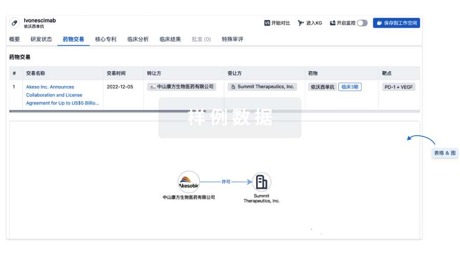
100 项与 绒毛膜促性腺激素(美国百时美施贵宝) 相关的药物交易
登录后查看更多信息
研发状态
登录后查看更多信息
临床结果
临床结果
适应症
分期
评价
查看全部结果
登录后查看更多信息
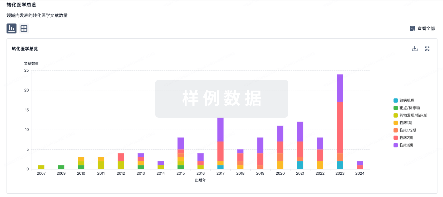
转化医学
使用我们的转化医学数据加速您的研究。
登录
或
药物交易
使用我们的药物交易数据加速您的研究。
登录
或
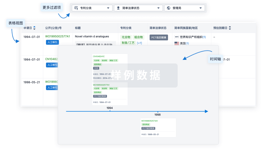
核心专利
使用我们的核心专利数据促进您的研究。
登录
或
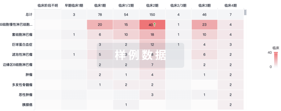
临床分析
紧跟全球注册中心的最新临床试验。
登录
或
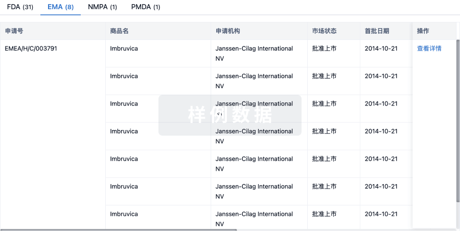
批准
利用最新的监管批准信息加速您的研究。
登录
或
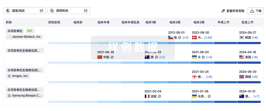
生物类似药
生物类似药在不同国家/地区的竞争态势。请注意临床1/2期并入临床2期,临床2/3期并入临床3期
登录
或
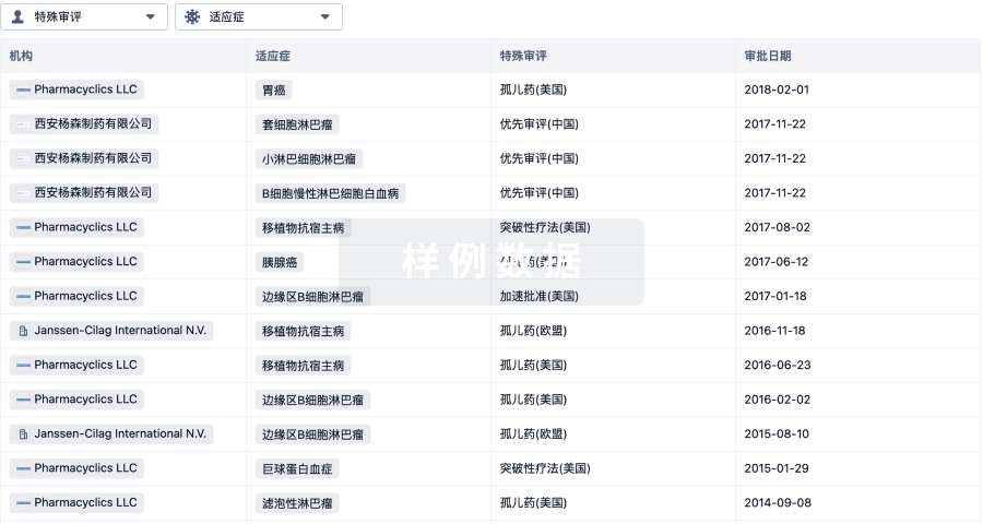
特殊审评
只需点击几下即可了解关键药物信息。
登录
或
生物医药百科问答
全新生物医药AI Agent 覆盖科研全链路,让突破性发现快人一步
立即开始免费试用!
智慧芽新药情报库是智慧芽专为生命科学人士构建的基于AI的创新药情报平台,助您全方位提升您的研发与决策效率。
立即开始数据试用!
智慧芽新药库数据也通过智慧芽数据服务平台,以API或者数据包形式对外开放,助您更加充分利用智慧芽新药情报信息。
生物序列数据库
生物药研发创新
免费使用
化学结构数据库
小分子化药研发创新
免费使用