预约演示
更新于:2026-06-26
GA-001
更新于:2026-06-26
概要
基本信息
原研机构 |
在研机构 |
非在研机构- |
权益机构- |
最高研发阶段临床1期 |
首次获批日期- |
最高研发阶段(中国)临床1期 |
特殊审评快速通道 (美国)、孤儿药 (美国) |
登录后查看时间轴
关联
2
项与 GA-001 相关的临床试验ChiCTR2300079040
A single-center, open-label, dose-escalation study of the safety, tolerability, immunogenicity, and efficacy of a single intravitreal injection of GA001 in the population of primary and total blindness caused by retinal degeneration.
CTR20262482
一项评价GA001注射液在视网膜色素变性患者中的安全性、耐受性和有效性的I期临床研究
100 项与 GA-001 相关的临床结果
登录后查看更多信息
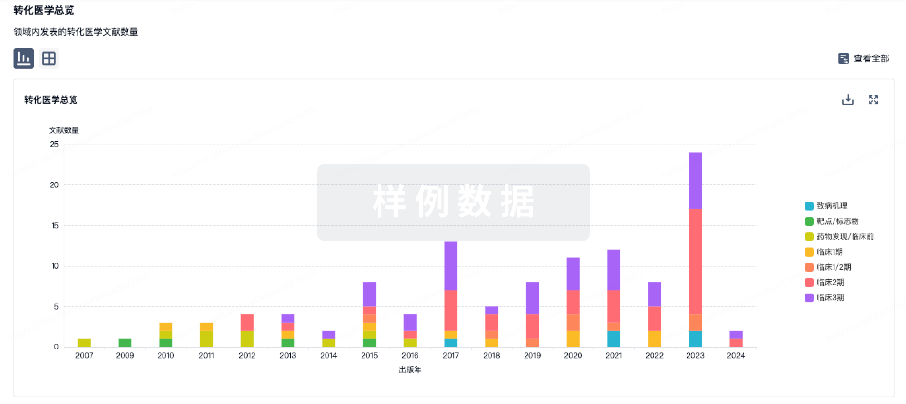
100 项与 GA-001 相关的转化医学
登录后查看更多信息
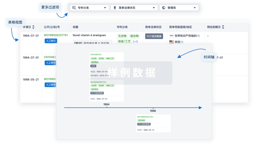
100 项与 GA-001 相关的专利(医药)
登录后查看更多信息
97
项与 GA-001 相关的文献(医药)2026-02-01Quantitative Imaging in Medicine and Surgery
Clinical and neuroradiologic spectrum of glutaric acidemia type 1 in children: insights from a retrospective cohort in Guangdong Province, China
Article
作者: Wang, Rui ; Fang, Liguang ; Yin, Xuntao ; Zhou, Yan ; Liu, Hongsheng ; Wu, Yu ; Cao, Yaxian ; Liu, Shuyi ; Zheng, Haige
Background:
Glutaric acidemia type 1 (GA-1) is a rare autosomal recessive metabolic disorder resulting from a deficiency in glutaryl-CoA dehydrogenase (GCDH). Current evidence indicates that GA-1 remains under-recognized by clinicians, a factor that may contribute to delayed diagnosis. The aim of this study was to retrospectively analyze the clinical manifestations and imaging characteristics of GA-1.
Methods:
This study enrolled patients diagnosed with GA-1 at the Guangzhou Women and Children's Medical Center between April 2014 and April 2024. Clinical data related to GA-1 were retrieved through the electronic medical record system, and magnetic resonance imaging (MRI) scans were collected for all patients. Cranial MRI images were independently evaluated by two radiologists (with 10 and 6 years of experience in pediatric neuroimaging diagnosis, respectively) using a blinded approach. Blood acylcarnitine levels were analyzed using tandem mass spectrometry, urinary organic acid concentrations were quantified via gas chromatography-mass spectrometry, and GCDH gene analysis was performed in a subset of patients.
Results:
This study enrolled 24 GA-1 children (8 males, 16 females) from Guangdong Province, China. Diagnosis was confirmed by elevated glutaric acid (GA), 3-hydroxyglutaric acid (3-HGA), and glutarylcarnitine (C5DC) levels, with increased C5DC/octanoylcarnitine (C8) and C5DC/propionylcarnitine (C3) ratios. Genetic analysis identified 12 GCDH mutations in 11 patients, including 5 novel variants (c.395G>A, c.271+1G>A, c.1156C>G, c.146_149delACTG, and c.1011A>G). Neuroimaging revealed abnormal brain MRI findings in all patients (100%), predominantly featuring frontotemporal extracerebral space widening (75.0%, 18/24) and symmetric basal ganglia hyperintensity (83.3%, 20/24). These findings align with the established GA-1 phenotypes.
Conclusions:
This study underscores the need for heightened awareness of GA-1 among clinicians and radiologists, characterizes its MRI signature, and expands the GCDH mutation spectrum with five novel variants, thereby offering valuable guidance for imaging-based diagnosis and genetic counselling.
2025-09-01PLANT SCIENCE
S-domain receptor-like protein kinase OsGRSK1 participates in regulating plant height and grain size via gibberellin pathway in rice
Article
作者: Du, Haitao ; Sun, Hongzheng ; Li, Junzhou ; Dong, Quanshi ; Hu, Xin ; Chen, Cong ; Li, Ying ; Fu, Yihan ; Duanmu, Fanqing ; Du, Changqing
Gibberellin (GA) has been extensively confirmed to mainly be involved in the regulation of plant height. In the present study, we characterized the function of an S-domain receptor-like protein kinase, OsGRSK1 (GA-related S-locus-like receptor protein kinase 1), in GA-mediated plant height regulation. Expression profile analysis revealed that OsGRSK1 which encodes a plasma membrane-localized protein is mainly expressed in both young and mature rice roots. At the seedling stage, the Osgrsk1 mutant exhibited a semidwarf phenotype with shorter roots and shoots than those of corresponding wild type HY (Hwayoung). At maturity, the Osgrsk1 mutant also displayed the semidwarf phenotype with an increased grain size. Cas9-OsGRSK1 lines mimicked the phenotypes of plant height and grain size in the Osgrsk1 mutant. Transcriptomic analysis indicated that most of GA pathway-related genes were differentially expressed between the Osgrsk1 mutant and its corresponding wild type HY. Further expression analyses also confirmed these differences, implying that the phenotypic changes in the Osgrsk1 mutant may be related to the GA pathway. Furthermore, the expression of OsGRSK1 was significantly inhibited by exogenous GA. The contents of endogenous GAs (GA1 and GA4) were significantly lower in the Osgrsk1 mutant than that of corresponding wild type HY, and the semidwarf phenotype of the Osgrsk1 mutant was restored with exogenous spraying of GA. Collectively, our findings reveal the biological function of S-domain receptor-like protein kinase OsGRSK1 and provide novel insights into the GA-mediated plant height regulation mechanisms in plants.
2025-03-01MOLECULAR GENETICS AND METABOLISM
Brain morphometric analysis in patients with glutaric aciduria type 1
Article
作者: Liu, ChengXiang ; Liu, Peng ; Tian, Pu ; Zhu, XiaoNa ; Li, Dan ; Zhang, Lei ; Liu, ZhuoHang ; Bian, BingYang
PURPOSE:
Cerebral structural changes in both cortical and subcortical regions were detected in the pathology of glutaric aciduria type 1 (GA-1) patients. Conventional magnetic resonance imaging was limited by radiologist-inter variability in evaluating its severity. This cross-sectional study aimed to identify the affected brain structures and their functional correlations within the cortical and subcortical regions of patients with GA-1.
METHODS:
Seventeen patients with GA-1 and 17 healthy controls (HCs) were included (mean age, 38 ± 17 months; both groups contained 6 males). Three-dimensional T1-weighted imaging data were acquired, and voxel and surface-based morphometry were used to quantitatively investigate differences in gray matter volume (GMV) and cortical thickness (CT). Two-sample t-tests were performed.
RESULTS:
Patients with GA-1 had lower GMV in the bilateral basal ganglia, thalamus, limbic system, default mode network, and right cerebellum, as well as lower CT in the bilateral insula, lateral occipital cortex, right inferior parietal cortex, inferior temporal gyrus, and posterior cingulate cortex than HCs. Patients with GA-1 had higher CT in the bilateral lingual gyrus, parahippocampal gyrus, superior frontal gyrus, left postcentral gyrus, right precuneus, precentral gyrus, middle temporal gyrus, and inferior temporal gyrus than HCs. In patients with GA-1, urinary glutaryl-carnitine levels were significantly negatively correlated with the GMV in the left inferior temporal gyrus.
CONCLUSION:
Our brain morphological analyses revealed quantitative differences in the GMV and CT of GA-1 patients compared to HCs and provided useful information about normal and abnormal neuroanatomy.
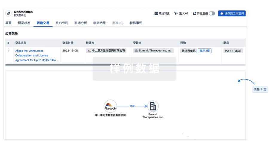
100 项与 GA-001 相关的药物交易
登录后查看更多信息
研发状态
10 条进展最快的记录, 后查看更多信息
登录
| 适应症 | 最高研发状态 | 国家/地区 | 公司 | 日期 |
|---|---|---|---|---|
| 视网膜色素变性 | 临床1期 | 中国 | 2026-06-26 | |
| 失明 | 临床1期 | 中国 | 2023-09-01 |
登录后查看更多信息
临床结果
临床结果
适应症
分期
评价
查看全部结果
登录后查看更多信息
转化医学
使用我们的转化医学数据加速您的研究。
登录
或
药物交易
使用我们的药物交易数据加速您的研究。
登录
或
核心专利
使用我们的核心专利数据促进您的研究。
登录
或
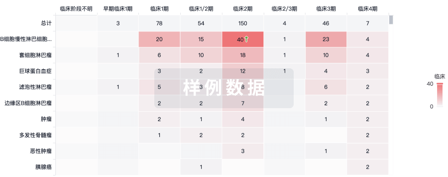
临床分析
紧跟全球注册中心的最新临床试验。
登录
或
批准
利用最新的监管批准信息加速您的研究。
登录
或
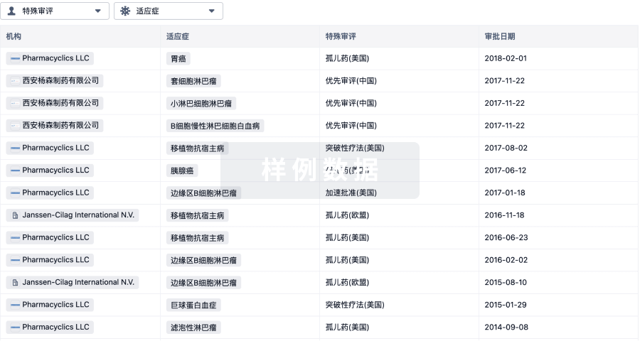
特殊审评
只需点击几下即可了解关键药物信息。
登录
或
生物医药百科问答
全新生物医药AI Agent 覆盖科研全链路,让突破性发现快人一步
立即开始免费试用!
智慧芽新药情报库是智慧芽专为生命科学人士构建的基于AI的创新药情报平台,助您全方位提升您的研发与决策效率。
立即开始数据试用!
智慧芽新药库数据也通过智慧芽数据服务平台,以API或者数据包形式对外开放,助您更加充分利用智慧芽新药情报信息。
生物序列数据库
生物药研发创新
免费使用
化学结构数据库
小分子化药研发创新
免费使用