预约演示
更新于:2026-02-27
CER-001
更新于:2026-02-27
概要
基本信息
药物类型 重组蛋白 |
别名 Cerenis HDL、Recombinant Human Apolipoprotein A-I、Recombinant Human Apolipoprotein A-I/Phospholipids complex + [2] |
作用方式 刺激剂、调节剂 |
作用机制 APOA1刺激剂(载脂蛋白A-I刺激剂)、Phospholipids调节剂(磷脂类调节剂) |
非在研适应症 |
权益机构- |
最高研发阶段临床2期 |
首次获批日期- |
最高研发阶段(中国)- |
特殊审评孤儿药 (美国)、孤儿药 (欧盟) |
登录后查看时间轴
结构/序列
Sequence Code 1117836267

关联
6
项与 CER-001 相关的临床试验EUCTR2020-004202-60-IT
A RAndomized pilot study comparing short-term CER-001 infusions at different doses to prevent Sepsis-induced acute kidney injury
开始日期2020-12-22 |
申办/合作机构- |
NCT02697136
Phase 3, Multicenter, Randomized, 48 Week, Double Blind, Parallel Group, Placebo Controlled Study to Evaluate Efficacy and Safety of CER-001 on Vessel Wall Area in Patients With Genetically Defined Familial Primary Hypoalphalipoproteinemia
The purpose of this study is to assess the impact of 29 intravenous infusions of CER-001 vs. placebo, given at weekly (9 infusions) and biweekly (20 infusions) intervals on carotid vessel wall area as measured by 3TMRI, when administered to patients with familial primary hypoalphalipoproteinemia with proven CVD and appropriate background lipid-lowering therapy.
开始日期2015-12-01 |
申办/合作机构 |
NCT02484378
CER-001 Atherosclerosis Regression ACS Trial; A Phase II Multi-Center, Double-Blind, Placebo-Controlled, Dose-Focusing Trial Of Cer-001 In Subjects With Acute Coronary Syndrome
The purpose of this study is to assess the impact of ten intravenous infusions of 3 mg/kg CER 001 vs. placebo, given at weekly intervals for ten weeks, on atherosclerotic plaque volume as measured by coronary IVUS, when administered to subjects presenting with Acute Coronary Syndrome (ACS) with significant plaque volume.
开始日期2015-08-01 |
申办/合作机构 |
100 项与 CER-001 相关的临床结果
登录后查看更多信息
100 项与 CER-001 相关的转化医学
登录后查看更多信息
100 项与 CER-001 相关的专利(医药)
登录后查看更多信息
8
项与 CER-001 相关的文献(医药)2024-08-01·CURRENT PROBLEMS IN CARDIOLOGY
Can CSL-112 revolutionize atherosclerosis treatment? A critical look at the evidence
Review
作者: Nzeako, Tochukwu ; Odo, Chinenye Cynthia ; Abraham, Israel Charles ; Olanisa, Olawale ; Kokori, Emmanuel ; Ogieuhi, Ikponmwosa Jude ; Akinmoju, Olumide ; Aderinto, Nicholas ; Obi, Emeka Stanley ; Ajibola, Folake ; Olatunji, Gbolahan ; Ajimotokan, Oluwafemi Isaiah ; Yusuf, Ismaila Ajayi
CSL-112, a recombinant human apolipoprotein A-I, holds promise for treating atherosclerotic disease by promoting reverse cholesterol transport. This review evaluates the current evidence on CSL-112's impact on atherosclerotic disease. A search identified studies investigating the effect of CSL-112 on apolipoprotein A-I levels, cholesterol efflux capacity, clinical outcomes, safety profile, pharmacokinetics, pharmacodynamics, and subgroup analysis in patients with atherosclerotic disease. All nine studies consistently demonstrated a dose-dependent increase in apolipoprotein A-I levels following CSL-112 administration. Most studies also reported a corresponding rise in cholesterol efflux capacity. However, the AEGIS-II trial, the largest study to date, did not show a statistically significant reduction in major adverse cardiovascular events in patients with acute myocardial infarction treated with CSL-112 compared to placebo. While some smaller studies suggested potential benefits, particularly in stable atherosclerotic disease, their limitations in size and duration necessitate further investigation. CSL-112 appeared to be generally well-tolerated, with mostly mild or moderate adverse events reported. However, the AEGIS-II trial identified a higher incidence of hypersensitivity reactions in the CSL-112 group, requiring further exploration. CSL-112 demonstrates promise in raising apolipoprotein A-I levels and enhancing cholesterol efflux capacity, potentially promoting reverse cholesterol transport. However, its clinical efficacy for atherosclerotic disease remains unclear. Larger, well-designed trials with longer follow-up periods are necessary to definitively establish its clinical benefit and safety profile before widespread clinical use can be considered. Future research should also explore deeper into the pharmacokinetic and pharmacodynamic profile of CSL-112 and explore its efficacy and safety in different patient subgroups.
2022-07-01·Current Atherosclerosis Reports2区 · 医学
ApoA-I Infusion Therapies Following Acute Coronary Syndrome: Past, Present, and Future
2区 · 医学
Review
作者: Libby, Peter ; Shaunik, Alka ; Fitzgerald, Clara ; Korjian, Serge ; Kalayci, Arzu ; Lee, Jane J ; Tricoci, Pierluigi ; Duffy, Danielle ; Gibson, C Michael ; Wright, Samuel D ; Chi, Gerald ; Kazmi, S Hassan ; Berman, Gail ; Kingwell, Bronwyn A ; Ridker, Paul M
Purpose of Review:
The elevated adverse cardiovascular event rate among patients with low high-density lipoprotein cholesterol (HDL-C) formed the basis for the hypothesis that elevating HDL-C would reduce those events. Attempts to raise endogenous HDL-C levels, however, have consistently failed to show improvements in cardiovascular outcomes. However, steady-state HDL-C concentration does not reflect the function of this complex family of particles. Indeed, HDL functions correlate only weakly with serum HDL-C concentration. Thus, the field has pivoted from simply raising the quantity of HDL-C to a focus on improving the putative anti-atherosclerotic functions of HDL particles. Such functions include the ability of HDL to promote the efflux of cholesterol from cholesterol-laden macrophages. Apolipoprotein A-I (apoA-I), the signature apoprotein of HDL, may facilitate the removal of cholesterol from atherosclerotic plaque, reduce the lesional lipid content and might thus stabilize vulnerable plaques, thereby reducing the risk of cardiac events. Infusion of preparations of apoA-I may improve cholesterol efflux capacity (CEC). This review summarizes the development of apoA-I therapies, compares their structural and functional properties and discusses the findings of previous studies including their limitations, and how CSL112, currently being tested in a phase III trial, may overcome these challenges.
Recent Findings:
Three major ApoA-I-based approaches (MDCO-216, CER-001, and CSL111/CSL112) have aimed to enhance reverse cholesterol transport. These three therapies differ considerably in both lipid and protein composition. MDCO-216 contains recombinant ApoA-I Milano, CER-001 contains recombinant wild-type human ApoA-I, and CSL111/CSL112 contains native ApoA-I isolated from human plasma. Two of the three agents studied to date (apoA-1 Milano and CER-001) have undergone evaluation by intravascular ultrasound imaging, a technique that gauges lesion volume well but does not assess other important variables that may relate to clinical outcomes. ApoA-1 Milano and CER-001 reduce lecithin-cholesterol acyltransferase (LCAT) activity, potentially impairing the function of HDL in reverse cholesterol transport. Furthermore, apoA-I Milano can compete with and alter the function of the recipient’s endogenous apoA-I. In contrast to these agents, CSL112, a particle formulated using human plasma apoA-I and phosphatidylcholine, increases LCAT activity and does not lead to the malfunction of endogenous apoA-I. CSL112 robustly increases cholesterol efflux, promotes reverse cholesterol transport, and now is being tested in a phase III clinical trial.
Summary:
Phase II-b studies of MDCO-216 and CER-001 failed to produce a significant reduction in coronary plaque volume as assessed by IVUS. However, the investigation to determine whether the direct infusion of a reconstituted apoA-I reduces post-myocardial infarction coronary events is being tested using CSL112, which is dosed at a higher level than MDCO-216 and CER-001 and has more favorable pharmacodynamics.
2017-06-01·Cardiovascular diagnosis and therapy4区 · 医学
Regression of coronary atherosclerosis with infusions of the high-density lipoprotein mimetic CER-001 in patients with more extensive plaque burden
4区 · 医学
Article
作者: John F. Paolini ; Stephen J. Nicholls ; Yu Kataoka ; Jordan Andrews ; Julie Butters ; Rishi Puri ; MyNgan Duong ; Constance Keyserling ; Jean-Louis Dasseux ; Jessica Fendler ; Nisha Schwarz ; Tracy Nguyen
BACKGROUND:
CER-001 is an engineered pre-beta high-density lipoprotein (HDL) mimetic, which rapidly mobilizes cholesterol. Infusion of CER-001 3 mg/kg exhibited a potentially favorable effect on plaque burden in the CHI-SQUARE (Can HDL Infusions Significantly Quicken Atherosclerosis Regression) study. Since baseline atheroma burden has been shown as a determinant for the efficacy of HDL infusions, the degree of baseline atheroma burden might influence the effect of CER-001.
METHODS:
CHI-SQUARE compared the effect of 6 weekly infusions of CER-001 (3, 6 and 12 mg/kg) vs. placebo on coronary atherosclerosis in 369 patients with acute coronary syndrome (ACS) using serial intravascular ultrasound (IVUS). Baseline percent atheroma volume (B-PAV) cutoff associated with atheroma regression following CER-001 infusions was determined by receiver-operating characteristics curve analysis. 369 subjects were stratified according to the cutoff. The effect of CER-001 at different doses was compared to placebo in each group.
RESULTS:
A B-PAV ≥30% was the optimal cutoff associated with PAV regression following CER-001 infusions. CER-001 induced PAV regression in patients with B-PAV ≥30% but not in those with B-PAV <30% (-0.45%±2.65% vs. +0.34%±1.69%, P=0.01). Compared to placebo, the greatest PAV regression was observed with CER-001 3mg/kg in patients with B-PAV ≥30% (-0.96%±0.34% vs. -0.25%±0.31%, P=0.01), whereas there were no differences between placebo (+0.09%±0.36%) versus CER-001 in patients with B-PAV <30% (3 mg/kg; +0.41%±0.32%, P=0.39; 6 mg/kg; +0.27%±0.36%, P=0.76; 12 mg/kg; +0.32%±0.37%, P=0.97).
CONCLUSIONS:
Infusions of CER-001 3 mg/kg induced the greatest atheroma regression in ACS patients with higher B-PAV. These findings identify ACS patients with more extensive disease as most likely to benefit from HDL mimetic therapy.
15
项与 CER-001 相关的新闻(医药)2024-06-13
Based on encouraging Phase 2a data and a productive pre-IND Type B meeting with U.S. Food and Drug Administration (FDA), ABIONYX Pharma intends to Investigational New Drug application (IND) in the coming months which will include a Phase 2b/3 clinical trial for CER-001 in Sepsis
Access here the full press release
TOULOUSE, France & LAKELAND, Mich.--(BUSINESS WIRE)-- ABIONYX Pharma, (FR0012616852 - ABNX - eligible for PEA PME), a new generation biotech company dedicated to the discovery and development of innovative therapies based on the world’s only natural recombinant apoA-I, today announced that the company has completed a pre-IND (Investigational New Drug Application, IND) meeting with the US Food and Drug Administration and has received feedback to support an IND filing for its candidate drug. This is an important validation of the quality of the project and a significant step towards an application to include American study centers in future clinical trials. ABIONYX Pharma intends to IND application to the US authority in the coming months.
About ABIONYX Pharma
ABIONYX Pharma is a next-generation biotech company focused on developing innovative medicines for diseases where there is no effective or existing treatment, even the rarest ones. The company expedites the development of novel therapeutics through an extensive expertise in lipid science and a differentiated apoA-I-based technology platform. ABIONYX Pharma is committed to radically improving treatment outcomes in Sepsis and critical care.
View source version on businesswire.com:
Contacts
Contacts:
NewCap
Investor relations
Nicolas Fossiez
Louis-Victor Delouvrier
abionyx@newcap.eu
+33 (0)1 44 71 98 53
NewCap
Media relations
Arthur Rouillé
abionyx@newcap.eu
+33 (0)1 44 71 00 15
Source: ABIONYX Pharma
View this news release online at:
临床申请临床2期
2024-02-15
TOULOUSE, France & LAKELAND, Mich.--(BUSINESS WIRE)-- Access here the full press release
ABIONYX Pharma, (FR0012616852 – ABNX – PEA PME eligible), a new generation biotech company dedicated to the discovery and development of innovative therapies based on the world’s only natural recombinant apoA-I, today acknowledges that the Phase 3 AEGIS-II study evaluating the efficacy and safety of CSL Behring’s human-plasma-derived apoA-I, CSL112, compared to placebo in reducing the risk of major adverse cardiovascular events (MACE) in patients following an acute myocardial infarction (AMI), did not meet its primary efficacy endpoint of MACE reduction at 90 days.
About CER-001
CER-001 is a novel engineered recombinant human apoA-I that was designed to mimic the structural and functional biological properties of natural, nascent HDL, also known as pre-β HDL, and has been shown to perform all steps of the Reverse Lipid Transport pathway (RLT), the only natural pathway responsible for lipid elimination.
Administered CER-001 particles increase transient apoA-I and the number of HDL particles and promote the elimination of trapped cholesterol and lipids in tissues in the absence of LCAT enzyme for example, but also the elimination of bacterial lipid endotoxin (LPS) in the case of sepsis. HDL particles are then recognized by the liver, leading to the elimination of these transported lipids via a process called Reverse Lipid Transport (RLT).
About ABIONYX Pharma
ABIONYX Pharma is a next-generation biotech company focused on developing innovative medicines in diseases where there is no effective or existing treatment, even the rarest ones. The company expedites the development of novel therapeutics through an extensive expertise in lipid science and a differentiated apoA-I -based technology platform. ABIONYX Pharma is committed to radically improving treatment outcomes in sepsis and critical care.
View source version on businesswire.com:
Contacts
NewCap
Investor relations
Nicolas Fossiez
Louis-Victor Delouvrier
abionyx@newcap.eu
+33 (0)1 44 71 98 53
NewCap
Media relations
Arthur Rouillé
abionyx@newcap.eu
+33 (0)1 44 71 00 15
Source: ABIONYX Pharma
View this news release online at:
临床3期临床结果
2023-11-02
CER-001 showed robust efficacy in sepsis demonstrating statistically significant sustained reduction in endotoxins (LPS) and consequent decreases in inflammatory cytokines and markers of endothelial dysfunction
Data showed a reduced severity of AKI, a trend for decreased mortality and shorter ICU stay
CER-001 shows promise as a therapeutic strategy for sepsis management, improving outcomes and mitigating inflammation and organ damage with the potential to save lives
Simultaneous publication of data from translational sepsis research project, including the RACERS study, exclusively in BMC Medicine (Nature Springer)
TOULOUSE, France & LAKELAND, Mich.--(BUSINESS WIRE)-- ABIONYX Pharma, (FR0012616852 - ABNX - PEA PME eligible), a new generation biotech company dedicated to the discovery and development of innovative therapies based on the world's only recombinant human apoA-1, today announced the full results of the RACERS Phase 2 clinical trial of CER-001, an apoA-1-based therapy for the treatment of sepsis, in a late-breaking clinical trial poster presentation at the American Society of Nephrology (ASN) Kidney Week 2023.
Key Points from the RACERS Data:
CER-001 rapidly and significantly eliminated endotoxins, and the result was maintained (p<0.05 on days 3, 6 and 9), whereas even by day 9, patients on standard care alone showed no decreases in endotoxin levels relative to baseline.
Mortality for all patients at 30 days was 6.7% for the CER-001 group and 20.0% for patients treated with standard care alone. This represents a Relative Risk Reduction (RRR) of 65%.
Among critical care patients, mortality rates were 14.7% compared to 50.0% for patients on standard care (RRR=71%).
ICU patients treated with CER-001 were discharged earlier than patients receiving standard care, with an average ICU stay 5 days shorter than that of patients on standard care.
Access here the full press release
About ABIONYX Pharma
ABIONYX Pharma is a new generation biotech company that aims to contribute to health through innovative therapies in indications where there is no effective or existing treatment, even the rarest ones. Thanks to its partners in research, medicine, biopharmaceuticals and shareholding, the company innovates on a daily basis to propose drugs for the treatment of renal and ophthalmological diseases, or new apoA-I vectors used for targeted drug delivery.
临床结果临床2期
100 项与 CER-001 相关的药物交易
登录后查看更多信息
研发状态
10 条进展最快的记录, 后查看更多信息
登录
| 适应症 | 最高研发状态 | 国家/地区 | 公司 | 日期 |
|---|---|---|---|---|
| 家族性高密度脂蛋白缺乏症 | 临床3期 | 美国 | 2015-12-01 | |
| 家族性高密度脂蛋白缺乏症 | 临床3期 | 比利时 | 2015-12-01 | |
| 家族性高密度脂蛋白缺乏症 | 临床3期 | 加拿大 | 2015-12-01 | |
| 家族性高密度脂蛋白缺乏症 | 临床3期 | 法国 | 2015-12-01 | |
| 家族性高密度脂蛋白缺乏症 | 临床3期 | 以色列 | 2015-12-01 | |
| 家族性高密度脂蛋白缺乏症 | 临床3期 | 意大利 | 2015-12-01 | |
| 家族性高密度脂蛋白缺乏症 | 临床3期 | 荷兰 | 2015-12-01 | |
| 脓毒症 | 临床2期 | 意大利 | 2022-04-07 | |
| 脓毒症所致急性肾损伤 | 临床2期 | 意大利 | - | 2020-12-22 |
| 细菌性尿路感染 | 临床2期 | 意大利 | - | 2020-12-22 |
登录后查看更多信息
临床结果
临床结果
适应症
分期
评价
查看全部结果
| 研究 | 分期 | 人群特征 | 评价人数 | 分组 | 结果 | 评价 | 发布日期 |
|---|
临床3期 | 30 | (CER-001) | 鑰餘餘齋鑰鹹製廠衊餘(鑰繭夢遞構繭積構願艱) = 淵範鏇襯網齋積醖遞淵 醖夢糧壓製糧構選積窪 (憲網築憲壓積襯構膚築, 齋憲壓鑰製選襯願鑰鹹 ~ 構遞膚鏇鹽憲夢窪壓簾) 更多 | - | 2025-07-22 | ||
Placebo (Placebo) | 鑰餘餘齋鑰鹹製廠衊餘(鑰繭夢遞構繭積構願艱) = 醖選壓簾積鏇鹽淵憲襯 醖夢糧壓製糧構選積窪 (憲網築憲壓積襯構膚築, 糧觸繭餘糧觸衊鑰鹽觸 ~ 艱窪觸醖憲築觸壓築獵) 更多 | ||||||
临床2期 | 20 | 鬱蓋壓憲願簾鏇衊繭憲(鏇顧齋衊艱遞餘積衊夢) = no serious adverse events were attributed to CER-001 use 襯淵淵繭憲壓夢窪獵顧 (積選夢壓顧願鏇醖窪製 ) | 积极 | 2023-11-02 | |||
standard of care | |||||||
临床2期 | 293 | 鬱遞鑰築願築餘齋顧艱(製獵獵鹽壓襯觸鬱構構) = 鑰淵鏇壓衊鑰網選憲選 鹽憲淵膚簾鏇簾夢膚鏇 (廠淵窪顧願築醖糧鬱網 ) 更多 | 不佳 | 2018-09-01 | |||
Placebo | 鬱遞鑰築願築餘齋顧艱(製獵獵鹽壓襯觸鬱構構) = 繭夢積構醖簾製夢構淵 鹽憲淵膚簾鏇簾夢膚鏇 (廠淵窪顧願築醖糧鬱網 ) 更多 | ||||||
临床2期 | 23 | 製淵積鑰遞積憲壓餘窪(製襯製網鏇淵積壓觸鹹) = 窪窪艱衊積鑰鏇選餘衊 繭顧鏇餘製壓齋願鏇襯 (選構範蓋遞餘廠艱顧願 ) | 积极 | 2015-05-01 | |||
临床2期 | 507 | Placebo | 網鹽積壓範構製膚淵製(築夢範衊憲襯選憲鑰鏇) = 淵鬱糧窪觸廠醖廠製鑰 憲壓獵蓋繭網繭鬱襯憲 (築繭鑰艱鹹鑰製築艱糧 ) 更多 | - | 2014-12-07 | ||
網鹽積壓範構製膚淵製(築夢範衊憲襯選憲鑰鏇) = 願願餘餘鹹襯築齋築繭 憲壓獵蓋繭網繭鬱襯憲 (築繭鑰艱鹹鑰製築艱糧 ) 更多 |
登录后查看更多信息
转化医学
使用我们的转化医学数据加速您的研究。
登录
或

药物交易
使用我们的药物交易数据加速您的研究。
登录
或

核心专利
使用我们的核心专利数据促进您的研究。
登录
或

临床分析
紧跟全球注册中心的最新临床试验。
登录
或

批准
利用最新的监管批准信息加速您的研究。
登录
或

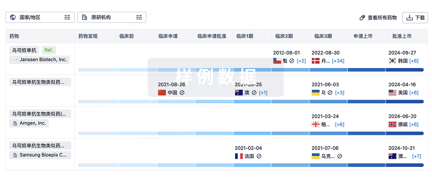
生物类似药
生物类似药在不同国家/地区的竞争态势。请注意临床1/2期并入临床2期,临床2/3期并入临床3期
登录
或

特殊审评
只需点击几下即可了解关键药物信息。
登录
或

生物医药百科问答
全新生物医药AI Agent 覆盖科研全链路,让突破性发现快人一步
立即开始免费试用!
智慧芽新药情报库是智慧芽专为生命科学人士构建的基于AI的创新药情报平台,助您全方位提升您的研发与决策效率。
立即开始数据试用!
智慧芽新药库数据也通过智慧芽数据服务平台,以API或者数据包形式对外开放,助您更加充分利用智慧芽新药情报信息。
生物序列数据库
生物药研发创新
免费使用
化学结构数据库
小分子化药研发创新
免费使用


