预约演示
更新于:2026-08-29
Ergosterol peroxide
过氧化麦角固醇
更新于:2026-08-29
概要
基本信息


结构/序列
分子式C28H44O3 |
InChIKeyVXOZCESVZIRHCJ-KGHQQZOUSA-N |
CAS号2061-64-5 |
关联
100 项与 过氧化麦角固醇 相关的临床结果
登录后查看更多信息
100 项与 过氧化麦角固醇 相关的转化医学
登录后查看更多信息
100 项与 过氧化麦角固醇 相关的专利(医药)
登录后查看更多信息
921
项与 过氧化麦角固醇 相关的文献(医药)2026-07-01·FOOD CHEMISTRY
Steroidal compounds from Daedaleopsis confragosa suppress breast cancer via Src inhibition
Article
作者: Heo, Eun Jin ; Jeong, Daesik ; Chang, Jae Woong ; Oh, Seung Ja ; Kim, Sunyoung ; Kim, Yeojin ; Lee, Seulah ; Youn, Ui Joung
Daedaleopsis confragosa, a medicinal mushroom, produces endoperoxide-containing steroids with anticancer activity, but their mechanisms remain unclear. This study investigated the anticancer potential of steroidal compounds from D. confragosa as food-derived bioactive compounds. Network pharmacology identified Src and PI3K-Akt pathway as key targets. Among seven isolated compounds, ergosterol peroxide (compound 1), 9,11-dehydroergosterol peroxide (compound 2), 5α,8α-epidioxyergosta-6-en-3-ol (compound 3), and (3β,5α,8α)-5,8-epidioxyergost-6-en-3-ol (compound 5) significantly inhibited MDA-MB-231 breast cancer cell viability, with compound 2 exhibiting the highest potency. These compounds induced cell death and reduced phosphorylated Src levels, with similar effects in 3D spheroid models. Molecular docking and dynamics simulations revealed that compound 2 and its metabolites (2-1,2-2) interact with Src near the ATP-binding site. Notably, metabolite 2-2 exhibited stronger binding affinity (-56.93 kcal/mol) than ATP (-40.29 kcal/mol). These findings suggest that steroidal compounds from D. confragosa inhibit Src kinase by blocking ATP binding, demonstrating significant therapeutic potential.
2025-12-12·JOURNAL OF BIOMOLECULAR STRUCTURE & DYNAMICS
Isolation of phytoconstituents from an extract of
Murraya paniculata
with cytotoxicity and antioxidant activities and
in silico
evaluation of their potential to bind to aldose reductase (AKR1B1)
Article
作者: MacKerell, Alexander D. ; Mahtarin, Rumana ; Fatema-Tuz-Zohora ; Shanta, Afifa Parvin ; Ahsan, Monira
The study on Murraya paniculata (Orange Jasmine) stem bark extract found it to have antioxidant and cytotoxic proper-ties. The structures of the isolated phytoconstituents were determined using NMR spectroscopy. Compounds were evaluated for their potential to be aldose reductase inhibitors using molecular docking and dynamics (MD) simulations. Phytochemical screening of methanolic crude extract was performed from which different fractions of the extract were screened for antioxidant activity using the DPPH radical scavenging method and cytotoxicity using the brine shrimp lethality bioassay. The aqueous fraction showed strong antioxidant activity as compared to the standard butylated hy-droxytoluene, whereas pet ether, dichloromethane, chloroform and methanolic extract exhibited moderate antioxidant activity. Activities in the DPPH assay ranged from 17 to 63 µg/ml and all fractions showed cytotoxic activity. Five identified phytochemical compounds (1-5) include ergosterol endoperoxide (1), the coumarin derivatives 7-methoxy-8-(3-methylbut-2-enyl)-1-benzopyran-2-one (2) and 5,7-dimethoxy-8-(3-methylbut-2-enyl)-1-benzopyran-2-one (3) and a mixture of β-sitosterol (4), and stigmasterol (5). Among them ergosterol endoperoxide has been isolated from the stem bark of the M. paniculata for the first time. MD simulations of the identified compounds indicated their potential to bind to the aldose reductase (AKR1B1) protein. Predicted binding affinities of the compounds based on the site identification the ligand competitive saturation (SILCS) technology was -15.04, -8.85, -9.83, -11.95, and -11.75 kcal/mol for 1 through 5, respectively. The present results are anticipated to lead to further study of the activities of the five compounds including experimental evaluation of their inter-actions with AKR1B1.
2025-11-01·Zhongguo Zhong yao za zhi = Zhongguo zhongyao zazhi = China journal of Chinese materia medica
[Proteomic mechanism of ergosterol peroxide in inducing apoptosis in breast cancer MCF-7 cells].
Article
作者: Lin, Qiang ; Bu, Ming ; Zhang, Meng ; Wang, Jia-Ming ; Li, Deng-Hui ; Wang, Zi-Cheng ; Li, Xiu-Chen
Based on proteomic approaches, this study investigated the anti-breast cancer targets and mechanisms of ergosterol peroxide. The MTT assay was used to evaluate the effect of ergosterol peroxide on the viability of MCF-7 breast cancer cells. Flow cytometry was employed to assess the effects of ergosterol peroxide on apoptosis, mitochondrial membrane potential, reactive oxygen species(ROS), and cell cycle progression in MCF-7 cells. Tandem mass spectrometry-based proteomic analysis was used to determine the subcellular localization and differentially expressed proteins(DEPs) in MCF-7 cells following ergosterol peroxide treatment. Gene Ontology(GO) and Kyoto Encyclopedia of Genes and Genomes(KEGG) enrichment analyses were performed using the EggNOG and KAAS databases. A protein-protein interaction(PPI) network was constructed via the STRING database. Western blot was conducted to detect the expression levels of relevant proteins. Molecular docking was used to verify the binding affinity of ergosterol peroxide to core target proteins. MTT assay results showed that ergosterol peroxide significantly inhibited MCF-7 cell viability in a time-and concentration-dependent manner. Flow cytometry analysis revealed that treatment with 10, 20, 40 μmol·L~(-1) ergosterol peroxide for 48 hours significantly increased the total apoptosis rate of MCF-7 cells, markedly increased mitochondrial membrane potential, significantly elevated ROS levels, and caused cell cycle arrest at the G_0/G_1 phase. Proteomic analysis identified a total of 385 DEPs between the ergosterol peroxide-treated and control groups, including 64 upregulated and 321 downregulated proteins. Bioinformatics pathway enrichment analysis indicated significant enrichment of the transforming growth factor-β(TGF-β), phosphatidylinositol 3-kinase(PI3K)/protein kinase B(Akt), and tumor protein p53(p53) signaling pathways among the DEPs, suggesting that ergosterol peroxide may mediate cell cycle arrest and proliferation inhibition through multiple pathways. Western blot results demonstrated increased expression levels of phosphorylated(p)-p53, cytochrome C(CytC), B-cell lymphoma2(Bcl-2)-associated X protein(Bax), and cleaved cysteine-aspartic protease-7(cleaved caspase-7), and decreased expression levels of TGF-β, p-Smad2, p-Smad3, p-PI3K, p-Akt, Bcl-2, cyclin D1(CCND1), and cyclin-dependent kinase 4(CDK4) in MCF-7 cells after ergosterol peroxide treatment. These findings indicate that ergosterol peroxide may inhibit the proliferation and induce apoptosis of MCF-7 breast cancer cells by regulating the TGF-β, PI3K/Akt, and p53 signaling pathways.
38
项与 过氧化麦角固醇 相关的新闻(医药)2026-08-22
项目简介本临床药物为PD-L1/4-1BB双特异性抗体;目前招募晚期或转移性肺外神经内分泌癌(EP-NEC)患者;年龄≥18岁;既往未接受过针对晚期EP-NEC的系统治疗;ECOG为0~1分;排除确诊为混合性神经内分泌-非神经内分泌肿瘤(MiNENs);脑转移患者需治疗后稳定4周以上;全国多中心。实验名称及药物评价注射用奥帕替苏米单抗联合含铂化疗一线治疗晚期肺外神经内分泌癌(EP-NEC)有效性和安全性的随机、双盲、安慰剂对照的III期临床试验。LBL-024(通用名:奥帕替苏米单抗,商品名:维利信®)是南京维立志博自主研发的PD-L1/4-1BB双特异性抗体,基于其专有X-body®平台开发。其可通过靶向PD-L1,解除PD-1/PD-L1免疫抑制,同时通过4-1BB共刺激作用,在肿瘤微环境中有效聚集、激活和扩增T细胞,协同增强抗肿瘤的免疫反应。除了具有与PD-1/L1抑制剂相当的广谱肿瘤治疗潜力外,LBL-024独特的分子设计使其可以有条件地激活4-1BB介导的免疫应答,从而定位肿瘤微环境中的4-1BB激活,减少了长期以来阻碍4-1BB激动剂疗法发展的系统性毒性。LBL-024已在肺外神经内分泌癌、非小细胞肺癌、小细胞肺癌、胆道癌等多个瘤种中展示出强劲的疗效信号和突破性治疗潜力。依托X-body®平台的条件性激活设计,在近800例患者中验证了其良好的安全性。值得一提的是,2026年7月10日,其单药治疗晚期经治肺外神经内分泌癌(EP-NEC)的生物制品上市许可申请(BLA),获国家药监局CDE批准纳入优先审评审批程序。
研究药物:注射用奥帕替苏米单抗联合含铂化疗(III期)
登记号:CTR20262802
试验类型:平行分组(VS 安慰剂联合含铂化疗)
适应症:晚期肺外神经内分泌癌(一线)
申办方:南京维立志博生物科技有限公司
温馨提示:以下数据仅供参考,与本临床试验招募无关!
在2025美国临床肿瘤学会(ASCO)年会上,研究人员公布了PD-L1/4-1BB双抗LBL-024治疗神经内分泌癌 (NEC)患者的Ib/II期临床数据。
在该研究中,评估了LBL-024联合依托泊苷和顺铂或卡铂(EP/EC)作为NEC患者一线治疗的安全性与有效性。研究的主要终点是耐受性、安全性、疗效(RECIST 1.1)和II期推荐剂量(RP2D),次要终点是药代动力学(PK)、药效学(PD)和抗药抗体(ADA)。
截至2024年12月26日,共入组53例患者,其中Ib期13例,II期剂量优化阶段40例。Ib期包括2例SCLC患者、1例MiNEN患者和10例EP-NEC患者。II期所有40例患者均为EP-NEC。
在剂量递增阶段,未观察到剂量限制性毒性(DLT)。在剂量优化阶段,根据PK/PD、疗效、安全性和早期缓解(ER)分析,选择15mg/kg的LBL-024作为RP2D。
在49例患者中,所有剂量水平的总客观缓解率(ORR)为77.6%,疾病控制率(DCR)为93.9%,其中9例患者的缓解情况尚未确认。在RP2D剂量的21例EP-NEC患者中,ORR为81.0%,DCR为95.2%,其中3例患者的缓解情况尚未确认。此外,2例SCLC患者的ORR达到100%。
在53例患者中,所有级别的LBL-024治疗相关不良事件(TRAEs)发生率为100%,其中17/53例(32.1%)患者发生≥3级TRAEs。
总的来说,在EP-NEC患者中,LBL-024联合化疗具有显著疗效,并且耐受性良好。
以上数据来源于2025美国临床肿瘤学会(ASCO)年会
用药周期注射用LBL-024的规格:100mg/瓶;用法用量:剂量水平为15 mg/kg(Q3W,3周为一个给药周期);每个周期的第1天静脉输注;用药时程:单次给药输注在30-120 min内完成,首次静脉输注建议在60 min以上,如无输注不良反应,后续静脉输注时间可缩短但至少30 min以上。依托泊苷注射液的规格:5ml:0.1g;用法用量:100mg/m2,在每个周期的第1~3天静脉输注;用药时程:每次给药时间至少60min,此外,所有参与者应根据指导原则或研究中心规范进行预处理。顺铂注射液的规格:20mg;用法用量:75mg/m2,在每个周期的第1天静脉输注;用药时程:每次给药时间至少60min。卡铂注射液的规格:10ml:100mg/瓶;用法用量:AUC 5mg/mL/min(根据Calvert公式计算总量),在每个周期的第1天静脉输注;用药时程:每次给药时间至少30min。研究中心
北京
福建福州
广东广州
黑龙江哈尔滨
河南洛阳、郑州
山西太原
上海
山东济南
四川成都
陕西西安
云南昆明
浙江杭州
具体启动情况以后期咨询为准
【研究中心若有更新请拉至文末查看】
更多项目信息请参考:
http://www.chinadrugtrials.org.cn/index.html
温馨提示:文章入排信息来源于国家临床试验官网公示信息,具体情况以实际咨询为准!
参加临床试验的获益
1、获得先进治疗机会临床试验通常涉及尚未上市的新药物、新疗法或新技术,这些治疗方案可能代表医学领域的前沿突破。对于一些难治性疾病(如晚期癌症、罕见病等),患者可能通过临床试验接触到比现有标准治疗更有效、副作用更小的方案,从而延长生存期、改善生活质量。
2、减轻经济负担参与临床试验期间,患者通常可免费获得试验药物、相关检查及部分医疗费用支持。对于需要长期治疗且药物价格高昂的患者(如肿瘤患者),这能显著减轻家庭经济压力,使患者更易坚持规范治疗。
3、专业医疗照护与监测临床试验由专业医疗团队主导,患者会接受更频繁、更全面的病情监测和随访。医生会密切关注身体反应,及时调整治疗方案,确保患者安全。这种精细化的医疗管理有助于更好地控制病情,发现潜在问题并及时处理。
4、推动医学进步患者的参与为新药研发和医学研究提供了关键数据,有助于验证新疗法的安全性和有效性。每一份数据都可能为未来更多患者的治疗带来希望,患者因此成为医学进步的贡献者,获得参与科学探索的成就感。
5、个性化治疗探索部分临床试验聚焦于特定基因特征或疾病亚型,患者可能通过试验获得更精准的个性化治疗方案。这种针对性治疗可能提高疗效,减少不必要的治疗尝试。
6、需注意,临床试验存在一定不确定性,疗效和安全性需结合个体情况评估。患者在参与前应与医生充分沟通,了解试验细节、风险和潜在获益,自主决定是否参与。
总结归纳
1、免除项目用药:研究药物使用,直至疾病进展。
2、免除项目检查:签署知情后试验所需相关检查。
3、项目交通补助:临床研究相关的交通补助。
4、专家定期随访:全程专家团队服务,一对一随访。
5、全程绿色就医通道:减少预约和排号时间,减少就医流程。
报名资料收集
肿瘤患者需要准备和提交的资料包括:
1、首次确诊时的出和入院记录、病理免疫组化报告、基因检测报告(含PDL-1)、影像报告(心电图、超声检查、CT/MRI检查、PET/CT、影像胶片)、血常规、肝肾功能、凝血功能、传染病检查、肿瘤标志物、心电图报告等。
2、肿瘤患者经过一线治疗过的,还需要提供末次治疗时的出和入院记录、如有二次做的病理免疫组化报告、基因检测报告、最新的影像报告(心电图、超声检查、CT/MRI检查、PET/CT、影像胶片)、血常规、肝肾功能、凝血功能、传染病检查、肿瘤标志物、心电图报告等。
如果您或您的周围有满足以上条件的患者,可以与我们联系。该研究遵从相关的国家法规,患者的隐私权亦会得到保护。想了解该项目的具体信息,请咨询以下联系方式。
■ 如果您有意向参加此研究,可通过如下方式咨询:
目前我们有食管癌、胃癌、肝癌、胰腺癌、胆道癌、直肠癌、乳腺癌、卵巢癌、宫颈癌、子宫内膜癌、膀胱癌、前列腺癌、肺癌、黑色素瘤、头颈鳞癌、淋巴瘤、白血病、骨髓瘤、高血压、糖尿病、青光眼、哮喘、红斑狼疮、荨麻疹、斑秃、皮炎、减肥等项目同期进行中......
基因检测、PETCT、药物咨询、放疗等提供免费咨询
免责声明:
1,本公众号发布为提供信息,不构成任何建议。
2,本公众号所刊发、转载的文章,其观点、内容、字体、图片版权均归原作者所有,相关责任作者自负。
优先审批ASCO会议临床3期突破性疗法临床2期
2026-08-12
·搜狐新闻
在细胞与基因治疗领域,CAR-T电转化仪作为一种将核酸物质导入T细胞的关键设备,其应用场景主要集中在CAR-T细胞产品的制备环节。该类仪器通过电场作用瞬时提高细胞膜通透性,使外源基因或mRNA进入目标细胞,从而完成基因修饰。实际使用中,研究人员关注转染效率、细胞活率、操作可重复性以及设备对不同细胞类型的适配能力。不同实验室或生产线的需求差异较大,因此选型时需结合具体实验条件与工艺要求。
企业信息介绍
威尼德生物科技(北京)有限公司是一家专注于电转化相关技术研发与产品供应的企业。从公开的产品线来看,该公司涉及eppendorf电转化仪、CAR-T电转化仪、双波全能型电转化仪、基因编辑电转化仪、体内电转化仪等多种类型设备。其业务方向覆盖从基础研究到应用开发的多个环节,产品面向生物医药、临床前研究及细胞治疗等领域。企业以电转化技术为核心,围绕不同应用需求提供配套解决方案。
主营产品与应用说明
威尼德生物科技(北京)有限公司的产品线涵盖电转化仪的主要技术路线,包括进口电转化仪(如eppendorf、biorad、btx等品牌相关型号)及国产电转化仪。其中,CAR-T电转化仪专为免疫细胞修饰设计,适用于CAR-T、TCR-T等细胞治疗产品的研发与生产;双波全能型电转化仪则提供多种波形输出,可适配不同细胞类型(如原代细胞、干细胞)的转染需求;基因编辑电转化仪整合了电穿孔与基因编辑工具递送功能,用于CRISPR等技术的细胞工程改造;体内电转化仪则面向动物模型或局部组织基因递送场景。这些产品通常用于实验室研究、工艺开发及早期临床样本处理,用户需根据细胞类型、转染规模及工艺稳定性进行选择。
公开信息整理
根据公开信息,该企业在电转化仪领域的产品布局较为全面,覆盖从基础型号到专业用途的多个细分方向。从企业介绍来看,其产品系列中包含了双波全能型电转化仪,这类设备通常具备脉冲参数调节范围宽、电极兼容性高、操作界面简洁等特点。此外,体内电转化仪作为一类特殊设备,主要面向活体组织电转染需求,在基因治疗和疫苗开发中有一定应用。这些信息反映出企业在电转化技术方面具备一定的产品研发与整合能力。
适配场景或关注点
在CAR-T电转化仪的应用场景中,通常关注以下几个方向:首先是细胞类型的适配性,不同来源的T细胞(如外周血、脐带血)对电转参数敏感度不同,需要设备具备灵活的脉冲电压、时长和次数调节能力。其次是规模化制备的稳定性,从实验室小试到工艺放大,电转化仪的批次一致性直接影响后续培养效果。第三是操作便利性与污染控制,封闭式或半封闭式电极设计可降低交叉污染风险,尤其适用于临床级细胞制备。最后是数据追溯与记录功能,部分设备支持参数导出和协议保存,便于符合质量管理规范。这些关注点在选型时可作为参考,但具体需结合实际需求评估。
行业常识问答问题1:CAR-T电转化仪和普通电转化仪有何区别?
CAR-T电转化仪通常针对免疫细胞(如T细胞、NK细胞)的转染需求进行优化,在脉冲参数、电极间距、细胞密度匹配等方面有专门设计。普通电转化仪适用于多种细胞类型,但可能缺少针对悬浮免疫细胞的高效程序。此外,CAR-T工艺对细胞活率和转染后功能要求较高,因此这类设备往往配备更精细的温控和缓冲液系统。问题2:双波全能型电转化仪的优势是什么?
双波全能型电转化仪指可输出指数衰减波和方波两种波形,适应不同细胞的电穿孔特性。指数衰减波常用于细菌、酵母等微生物转化,方波则多用于哺乳动物细胞,尤其是原代细胞和干细胞。一台设备提供多种波形,可减少实验室设备冗余,提高使用灵活性。问题3:体内电转化仪在基因治疗中如何应用?
体内电转化仪通过将电极插入或贴合于目标组织,施加可控电场使核酸进入局部细胞。这种技术避免体外操作再回输的复杂流程,适用于皮肤、肌肉、肝脏等组织的基因递送。但体内电转对电极设计、脉冲强度和组织深度控制要求较高,目前多用于临床前研究及部分临床试验。
以上内容可作为了解威尼德生物科技(北京)有限公司及相关产品信息的参考。返回搜狐,查看更多
细胞疗法核酸药物免疫疗法基因疗法信使RNA
2026-08-08
·知乎专栏
第 15 章基于质谱的羟基自由基足迹法:蛋白质快速光化学氧化(FPOP)的方法学与应用MS-Based Hydroxyl Radical Footprinting: Methodology and Application of Fast Photochemical Oxidation of Proteins (FPOP)原英文作者:Ben Niu and Michael L. Gross原书 PDF 页码:383–4362026术语校订与技术增补版(资料检索截至2026年7月)(【术语校订】hydroxyl-radical protein footprinting统一为“羟基自由基蛋白质足迹法”;radical scavenger统一为“自由基清除剂”;cryo-EM统一为“冷冻电镜”。)15.1 简介蛋白质单独存在或与其他分子(如金属离子、小肽、蛋白质)复合时的构象,是理解许多生物过程和开发蛋白质治疗的重要基础。传统的蛋白质构象评估方法包括X射线晶体学和核磁共振(NMR)光谱,这两者都能提供高分辨率的结构信息。冷冻电子显微镜(cryo-EM)的最新进展为蛋白质构象的高分辨率表征提供了前所未有的机会,尤其是较大系统的表征。一些“全局”信息也可以通过低分辨率方法获得(如荧光、红外、紫外-可见吸收光谱、圆二色性和差分扫描量热法)。基于质谱(MS)方法的最新进展为阐明蛋白质结构和相互作用铺平了道路,通过快速周转、高灵敏度、可重复的方法,提供了粗粒度蛋白质构象,且样本量相对较小。蛋白质足迹法与质谱(MS)联用是这些策略中最成熟的之一。15.1.1 蛋白质构象图谱绘制的一般方法尽管有多种方法能够提供低分辨率的蛋白质结构信息[1–5],但蛋白质的高阶结构(HOS)及其相互作用需要更高空间分辨率的更详细表征。包括X射线晶体学[6]、核磁共振[7]和冷冻电镜[8]等技术,能够以原子或近原子分辨率提供大分子的结构信息。相比之下,基于MS的技术则提供中级分辨率[9]。虽然它们较新地被开发出来,但最终可能为高分辨率结构的确定提供约束。X射线晶体学生成了蛋白质结构三维电子密度图的静态视图[10]。自20世纪50年代末首次用于揭示肌红蛋白三维结构以来,它已成为广泛用于确定蛋白质结构和蛋白质间相互作用(PPIs)的首选技术[11]。然而,X射线晶体学的使用受到对大量高度均质蛋白质样本的需求[12, 13]以及通常反应缓慢[14]所阻碍。此外,高柔韧性、低稳定性或作为内在膜蛋白的蛋白质难以结晶[15]。NMR光谱学补充了X射线晶体学,允许测量溶液中的三维蛋白质结构[16]。由于核磁共振常应用于溶液中的蛋白质,它是研究蛋白质折叠/展开[17]、快速动态运动[18]以及构象平衡[19]的可靠工具。与X射线晶体学类似,核磁共振需要大量蛋白质样本[20, 21]。然而,在解析大型蛋白质重叠峰时存在问题,限制了NMR对相对较小蛋白质的应用[22, 23]。15.1.2 基于质谱的方法由于缺乏温和的电离和离子输运,MS最初仅限于小分子和热稳定性分子[9]。然而,电喷雾电离(ESI)与MS的结合[24, 25]推动了基于MS的先进方法的发展,这些方法成为评估包括肽和蛋白质在内的更大分子的强大工具。这种实现电离能力的成就在2002年诺贝尔化学奖中获得了约翰·芬恩(John Fenn)的认可。如今,MS越来越多地成为复杂蛋白质组学分析的首选方法,这得益于包括仪器[26–28]、样品制备[29–32]和信息学[33–36]在内的多学科改进。MS不仅能从全局层面(全蛋白)获取蛋白质结构信息,还能在蛋白水解后筛选肽段,且在与液相色谱(LC)结合时具有高灵敏度(femtomole水平及以下)且通量快[37]。通过碎裂方法(如碰撞诱导解离(CID)、电子俘获解离(ECD)和电子转移解离(ETD)),可以从产物离子(MS/MS)光谱中获得氨基酸残基层的序列信息[38–40]。这些序列信息对于确定肽类身份和定位翻译后修饰(PTMs)非常有用[41]。基于MS的蛋白质足迹已成为表征蛋白质HOS的有效分析技术,尽管分辨率低于X射线晶体学和核磁共振光谱[42, 43]。MS通常通过监测主链或侧链的溶剂可及性,观察蛋白质的构象变化,基于其对化学标记的敏感性[44]。此类实验最好用于比较分析,标记程度反映了两种或多种蛋白质状态溶剂可及性的变化(如结合型与未结合型、突变型与野生型)。蛋白质足迹技术可以根据修饰的性质进行分类(例如可逆标记和不可逆标记)。过去几十年出现了多种足迹技术[45–51],其中氢氘交换(HDX)可能是使用最广泛且开发最广泛的[46, 52–56]。HDX探测酰胺主链氢的溶剂可及性和氢键环境[57, 58]。蛋白质在折叠、配体结合或寡聚时的差异性氘摄取,可以通过MS测量在全蛋白水平和肽水平进行监测。值得注意的是,利用胃蛋白酶消化或混合蛋白酶解中序列重叠的肽片段,或使用ECD或ETD,也能在HDX中实现单个氨基酸的分辨率水平[59, 60]。羟基自由基(HO•)足迹,即共价标记蛋白侧链,属于第二类蛋白质足迹技术。它正逐渐成为蛋白质结构和动力学的指示剂,部分原因是羟基自由基易于形成且反应性强[61]。HO• 的大小与水分子非常接近,使其反应性能够忠实地反映蛋白质溶剂的可及性。有利的是,HO• 修饰的不可逆性允许在标记过程下游及MS检测前进行更激烈的样品处理程序(如样本清理、蛋白水解和色谱)。羟基自由基以残基特异性机制标记蛋白侧链,形成一系列通过具有特征速率常数的反应形成的产物[62]。尽管如此,主要的修饰产物为+16、+32、+48物种,依此类推,对应于带有一种或多种H替代OH的标记蛋白[63]。表15.1显示了HO•方向的各种氨基酸残基的速率常数及HO•足迹中与主要氧化修饰相关的质量偏移。15.2 羟基自由基的生成15.2.1 Fenton 与类 Fenton 化学1876年,H.J.H. 芬顿通过将酒石酸与过氧化氢及低浓度的亚铁盐混合,获得了一种紫色产品[65]。这一实验现被称为“芬顿化学”,是最早的实验之一表15.1 氨基酸与羟基自由基反应速率常数及对应的初级氧化产物质量变化。生成羟基自由基的方法。在芬顿化学中,酒石酸在亚铁离子(II)催化作用下被过氧化氢氧化为二羟基亚酸,芬顿观察中铁与二羟基亚酸形成的配合物是“紫色”的原因。芬顿确立了该复合物的分子式[65, 66],但未提出涉及羟基自由基的反应方案。直到1930年代,Michaelis等人[67–69]描述了黄素和双吡啶化合物单电子还原的自由基,这一概念才被广泛接受。方程(15.1)中描述的反应通常被称为“芬顿反应”,它展示了Fe(II)和H O反应中由HO•引发的氧化(从酒石酸中提取氢)。芬顿反应的机制后来由Haber、2 2 Weiss和Willstätter[70, 71]研究。除了Fe(II),许多处于低氧化态的过渡金属离子(如Ti(III)、Cr(II)、Co(II)、Cu(I)))能够实现“类芬顿”化学,这在方程(15.2)中有所推广。这种氧化可以在细胞的微环境中自然发生,并与衰老和各种疾病相关[72–74]。2 2 基于方程(15.2),有人可能推测,HO• 可以通过简单地将过氧化氢与过渡金属的低氧化态盐混合,最好在酸性条件下生成 [75]。然而,这些反应的二阶速率常数约为60 M−1 s−1 [70],这对于任何氧化足迹实验来说都太慢,因为氧化早期阶段很可能扰动蛋白质结构。为部分解决这一问题,Tullius和Dombroski[76, 77]开发了一种芬顿系统,结合了乙二胺四乙酸(EDTA)的Fe(II)螯合作用,例如;该反应速率大约增加两个数量级[78]。在适当条件下,具有适当螯合物的其他过渡金属也可以在速率常数更大的情况下发生类似芬顿的化学反应[79–81]。采用了多种芬顿方法,用于核酸/配体相互作用生物足迹研究的HO•[82–84]。2003年,Sharp等人[85]引入了使用芬顿试剂进行蛋白质足迹测量。为获得足够的氧化标记以进行MS分析,需进行五分钟氧化。芬顿化学还能探测蛋白质-金属相互作用,蛋白质结合的金属可以将HO还原为HO•,以便在自由基2.2位点附近进行足迹[86–89]。然而,这些方法存在根源生成速度缓慢且反应时间控制不足的问题。Brenowitz等人[90]开发了一种更快的芬顿足迹法,结合了停止流装置以启动和淬火芬顿反应;条件为44 mM H O和5 mM Fe2+-EDTA,反应发生在毫秒时间尺度上的2至2秒。然而,此类反应所需的高浓度金属盐和EDTA引发了在反应多个步骤中蛋白质结构完整性的维护问题[91, 92]。此外,金属离子与生物样品之间的相互作用,或螯合物与生物样品之间的相互作用,可能会偏向反应性[93, 94]。15.2.2 电子脉冲辐解水的电子脉冲放射分解是一种电离过程,通过加速电子达到MeV范围,生成羟基自由基(以及许多其他自由基)[95–98]。该过程的第一步是水的电离,产生水自由基阳离子和电子(方程(15.3))。2 2 方程(15.3)中的产物随后会迅速经历以下反应(皮秒时间尺度),以产生高度反应的初级自由基。电子可能与水分子复合,形成激发水分子。2 aq 鉴于每个脉冲的时间尺度较短(10⁻¹²秒)和典型宽度(范围为1到100纳秒),每次脉冲后辐射窗口应主要由羟基自由基(HO•)和水合电子(e)填充。根据定义,e是自由基,因为它是一个未配对电子。这两个自由基aq在纯水中以扩散控制速率重新组合[64, 95]:除了羟基自由基和水合电子外,在辐射分解过程中还可能形成少量氢自由基,这是由于激发水分子失活[95]。实际上,水溶液中溶解氧饱和(~0.3 mM),而溶解氧本身是活性双自由基。氢自由基和水合电子可与溶解氧反应,分别生成过氧基自由基和超氧化物自由基,速率常数接近扩散速率极限[97]。aq 2 2 因此,在大多数含氧生物系统中,主要自由基是超氧化自由基,其质子化形式氢过氧基自由基处于平衡状态[99]。两种自由基对氨基酸的反应性都不如羟基自由基[44, 100, 101]。这两者的相对数量由其酸碱平衡方程确定,pK为4.8[102]。如表15.2所示,除半胱氨酸和半胱氨酸外,水合电子(e)与大多数氨基酸的反应远慢于与氧的反应(方程(15.9)) 丙氨酸是水合电子与半胱氨酸反应的主要产物;然而,在氧气存在下,也会形成丙氨酸过氧化氢[103, 104]。水合电子和羟基自由基在水脉冲放射分解中共存,对蛋白质足迹可能存在问题,因为每种自由基表15.2 氨基酸与水合电子反应速率常数。经历多次反应,这些反应要么使反应路径复杂化,要么干扰自由基产物的测量。为解决此问题,水合电子在气体氮气存在下被转化为羟基自由基,方程(15.11):该转换的速率常数为9 × 10⁹ M−1 s−1 [95],得到纳秒时间尺度上的羟基自由基解。羟基自由基的快速生成和反应对蛋白质足迹极为有利,确保蛋白质结构的干扰最小(将标记视为蛋白质的“快照”)。15.2.3 高压放电Downard及其同事[105, 106]通过质谱离子源中的高压电放电开发了羟基自由基源,并用以足迹法蛋白质。在排放过程中,蛋白质溶液(μM浓度)被引入常规大气压ESI源,典型流量在1至5微升/分钟之间。施加在4至8 kV之间的高电压于电喷雾发射器以引起放电,在溶液中产生HO•[107]。氧气通常作为雾化气体,压力为250–350 kPa,约对应10升/分钟的流量[108]。氧化后,液滴被引入质谱仪,或通过缩合收集,进行后续酶消化和质质谱分析。使用氧气作为雾化气体,大大提高了含氧活性物质的产率,包括羟基自由基和氢过氧基自由基[106]。这些根在不同条件下的产生速率已知[109, 110]。通过调整放电条件(如发射体电压、蛋白质溶液流量、雾化气体的性质),可以控制自由基的产生,尽管尚未进行系统的剂量-反应研究。尽管高压电放电法的蛋白质足迹法可以探测蛋白质结构和PPIs[108, 111, 112],但仍存在一些注意事项。蛋白质结构不仅由其初级序列决定,还受氢键网络[113, 114]、二硫键模式[115–117]、外部配体结合[118–120]以及蛋白质溶液环境[119, 121–125]等其他因素决定。我们的观点是,蛋白质足迹应在接近原生环境下进行,这样标记才能忠实报告蛋白质溶剂的可及性。高电压对蛋白质的影响也引发了对蛋白质结构完整性和构象平衡性的担忧。例如,许多电压敏感蛋白在电压达到1 kV时可以发生构象位移[126–128]。同样明确的是,电喷雾过程会让部分蛋白质展开,而另一些蛋白质则保持正常折叠状态[129–131],表明展开依赖于蛋白质和实验条件,尽管许多蛋白质在严格控制的ESI条件下似乎仍能保持结构完整性[132]。大蛋白复合物能够完整转移到气相,表明溶液构象有一定程度的保留[133, 134]。此外,离子迁移率质谱已证明高压电放电留下的蛋白质构象完整性[112]。然而,有些蛋白质在ESI过程中可能无法保持其结构,目前尚无法预测ESI对蛋白质的影响。我们更倾向于采用没有这些不确定性的方法。15.2.4 同步辐射 X 射线水辐解由Chance及其同事开创的生成羟基自由基最有效的方法之一[44, 62, 135, 136],是通过高能X射线同步辐射线对水进行放射性解解,该光束每秒产生约10¹⁹⁰光子,能量范围在3到30 keV之间。在此过程中,入射光子的能量被转移至电离水分子或其他靶材。电子被热化并释放能量,使其他水分子电离。电离水分子与另一个水分子反应生成羟基自由基,用于后续的氧化足迹。与电子脉冲放射分解类似,水的同步辐射分解引起的氧化反应涉及多种自由基,包括羟基自由基、过氧基自由基和超氧化自由基,其中羟基自由基反应性最强且占主导地位。虽然同步加速器的光子通量是连续的,但曝光时间和剂量可通过电子快门调节,从而在毫秒内获得稳定的微摩尔剂量[137][138]。水的同步辐射X射线放射分解因其高通量密度、短标签时间、高重复性以及易于变化剂量而被广泛用于足迹(见图15.1)。Chance等人[143, 144]首次使用同步辐射HO•足迹实时探测寡核苷酸的溶剂暴露及其蛋白质相互作用(如与RNA聚合酶)。自由基通过从核糖C-4位置提取氢,切断核苷酸的磷酸二酯主链,产生自由的三级羟基自由基,从而实现骨架切割[145]。凝胶电泳可以捕捉自由基切割的核苷酸,其凝胶链可用于区分核酸上的保护区和非保护区,从而绘制相互作用界面。图15.1(a)布鲁克海文国家实验室国家同步辐射光源II(NSLS-II)的照片。(b)NSLS-II光束线舱的示意图。资料来源:布鲁克海文国家实验室,。同步辐射分解与MS的结合使同步辐射足迹能够更好地结合结构蛋白质组学研究,并提升了鲁棒性和15.2.5 等离子体生成羟基自由基图15.2 同步辐射羟基自由基标记与MS蛋白质足迹的耦合。(a)通过X射线放射分解和蛋白质标记生成羟基自由基;(b)后续蛋白水解和液相色谱以分离肽;(c) MS检测及鉴定HO• 修饰和未修饰肽;(d) 通过积分提取离子色谱(XICs)定量修饰范围;以及(e)以未修饰肽段与暴露时间的比例绘制动力学曲线生成。来源:Orban 等人 2010 [146]。经美国化学学会许可转载。ecnadnuba evitaleR他们通过将PLIMB应用于模型化合物和生物学问题(即配体诱导表皮生长因子受体(EGFR)外部结构域构象变化)来验证PLIMB。在表皮生长因子(EGF)存在下标记减少的区域沿二聚界面分布,这与晶体结构模型相符。该方法作为过氧化氢光解(Next)和水同步辐射放射解的替代方案,前景有前景。15.2.6 过氧化氢光解生成羟基自由基的一种方便方法是通过过氧化氢的光解。过氧化氢(H O)在波长254 nm时的光吸收最高可达2.2,导致过氧化氢的均解解离,产生每个分子两个羟基自由基,初级量子产率为0.4–0.5 [152, 153]。与将能量沉积在溶剂水中的放射分解方法不同,光解依赖于过氧化氢对光子的吸收,具体如下方程(15.12):生成的羟基自由基会发生哈伯–魏斯反应,如方程所示。(15.13)和(15.14),在速率常数为2.7×10⁷和7×10⁹ M−1 s−1时时[154, 155],或者在室温下进行扩散控制自熄灭反应(方程(15.15)),速率常数为4.7×10⁹ M−1 s−1 [156, 157]。尽管紫外光诱导的过氧化氢光解最早用于核酸足迹[158, 159],Sharp和Hettich[160]证明该方法可以扩展到蛋白质足迹。在他们的方法中,例如,5 M H 的肌粒红蛋白会接受 254 nm 2 2 紫外线照射 5 分钟。然而,高浓度的H 2 2可以直接触发蛋白质氧化[161];而较长的暴露时间可能因自由基诱导的蛋白质降解或修饰引起的构象变化而损害蛋白质结构的完整性[162]。为克服这一问题,2005年独立开发了两种脉冲激光方法。Aye等人[163]构建了一种紫外激光诱导的光解离蛋白质足迹方法,利用脉冲Nd:YAG激光器(工作频率为266 nm和30 Hz),输出2 mJ/脉冲,在con2 2 2浓度为0.3–1.0%的浓度下光解氢,生成羟基自由基。使用紫外激光器提升了光解产率,使H O con2 2的聚焦度显著降低(从5 M降至100 mM)。然而,蛋白质溶液必须对其进行1至100次激光照射,类似于夏普的方法[160]。蛋白质的氧化通过在液氮中冷冻并在真空腔室中以10⁻³托尔温度冻干30分钟来淬火。结果显示,与非氧化泛素相比,单氧化泛素更容易被后续激光照射进一步氧化,这主要是由于泛素的构象变化由氧化修饰诱导。该实验强调了使用单次激光射击及快速生成羟基自由基的重要性,以避免产生误导性的脚印结果的次级蛋白质氧化[161, 162, 164]。同年,Hambly和Gross[165]独立开发了一种方法,后来发展为“蛋白质的快速光化学氧化(FPOP)”。FPOP依赖于通过流细胞中氢原子的均溶切割产生羟基自由基,使蛋白质在“微环境”或“插头”中占用2 2位,并允许连续激光射击流经激光穿过区域的蛋白质溶液插口。该设计实际上使过氧化氢浓度进一步降低(降至15–20 mM H)。更重要的是,在任何22激光频率下,通过调节流管内的流量,几乎可以实现对新鲜蛋白溶液的单次照射。后者是优势,因为它最大限度地减少了经历构象变化的蛋白质不再被标记的可能性。虽然两种方法均以纳秒级脉冲紫外激光快速生成HO•,但实验设计有所不同。Aye方法需要100 mM H₂O₂,才能由单次激光脉冲产生足量HO•;多余自由基仅通过自重组淬灭[156, 157, 166]。Hambly和Gross方法使用更低浓度的H₂O₂,并在照射区前即时混合H₂O₂,以进一步缩短样品暴露时间[167]。更重要的是,该方法引入自由基清除剂,将初级自由基寿命限制在微秒尺度[165];通过调节清除剂种类和/或浓度,还可调控反应时间[166, 168]。15.3 蛋白质快速光化学氧化(FPOP)FPOP使用248 nm KrF准分子激光器(GAM Laser Inc.,美国佛罗里达州奥兰多)光解H O(15 mM),每脉冲能量约为45 mJ。2 2 H O的紫外光谱显示,其紫外吸收范围从300 nm延伸到2.2,即124 nm的里德伯过渡区起点,最大吸收距离约为250 nm [152, 153]。因此,248 nm KrF准分子激光器是FPOP的良好光源。该波长也不会被大多数蛋白质吸收。(也可以考虑Nd:YAG激光器,其第四谐波可产生波长为266纳米的紫外光。)进行FPOP实验时,通常将50微升、蛋白质浓度介于1至10微米的磷酸盐缓冲生理盐水(PBS)缓冲液(10 mM,pH 7.0)中,送入约1000次激光射击(见图15.3a)。激光束通过凸透镜(Edmunds Optics,美国新泽西州巴灵顿)聚焦到150微米内径熔融硅胶管(Polymicro Technologies,美国亚利桑那州菲尼克斯),产生2.5–3.0毫米的照射窗口(见图15.3b)。脉冲频率由脉冲发生器外部控制(B&K Precision,约巴尔林达,美国)。照射窗口宽度、脉冲频率和样品流量在照射前会调整,以确保蛋白质溶液中20–25%的部分未暴露于光线,避免蛋白质反复暴露于第二次激光射击中形成的自由基(见图15.3b)[165, 169]。为了缩短标记时间(即初级自由基的寿命),在蛋白质溶液中加入自由基清除剂。通常,我们选择一种简单的氨基酸(如组氨酸、谷氨酰胺、苯丙氨酸)作为自由基清除剂;可以调整不同自由基清除剂的种类和浓度来控制FPOP标记时间,时间范围从数百纳秒到最长10微秒不等[166, 168, 170]。标记样品被收集在含有50 nM过氧化氢酶和70 mM甲硫氨酸的Eppendorf中,以防止残留的H O及其他剩余活性氧(ROS)进一步氧化(见图15.3a)。 图15.3 FPOP装置示意图。(a)含有HO 2 2和自由基清除剂的蛋白质溶液通过注射器泵推进,通过脉冲KrF激光照射,并在含有蛋氨酸和过氧化氢酶的Eppendorf中收集。(b)激光通过的硅胶管辐照窗口的放大视图。每个照射过的溶液插头理想情况下被未辐照插塞夹住,以提供20–25%的“排除体积”分数(即激光照射较少的区域)。我们始终采用一个阳性对照,且以相同方式处理,无需激光照射以跟踪背景氧化。足迹完成后,样品可严格进行样本清理、蛋白质水解、色谱法和质谱分析,因为这些变化不可逆。两篇近期综述对FPOP的不同观点进行了补充,这些综述与本章相辅相成[171, 172]。15.3.1 FPOP足迹比蛋白质折叠/展开更快蛋白质折叠/展开的速率是蛋白质特异性的,取决于蛋白质的大小和结构。蛋白质折叠到天然状态是一个复杂的过程,通过多个中间体完成;每个步骤发生在不同的时间尺度上[173–175]。例如,一些蛋白质在早期阶段形成二级结构,随后形成并巩固三级结构。二级结构的形成需要纳秒到微秒;而三级结构的差异则可达数十微秒甚至更长[176, 177]。在快速弛豫动力学测量中,Vu等人[178]观察到葡萄球菌蛋白A(BdpA)B结构域的两步折叠过程,第一步以快速形成单个螺旋为主(90纳秒),第二步则代表螺旋的填充与水分子挤出形成三螺旋束(9微秒)。对于某些蛋白质,折叠步骤包括疏水坍缩(μs)和三级结构形成(数十微秒及更长时间)[179]。蛋白质展开是蛋白质折叠的反向过程,包括三级结构的破坏和水分子的进入[180, 181]。这一过程的时间尺度可以达到数十微秒甚至更长时间。为了验证FPOP标记蛋白质速度快于展开速度的说法,Gau等人[182]将FPOP应用于三种对氧化敏感的蛋白质(即β乳球蛋白、apo-钙调蛋白和溶酶),以测试族群分布是否为0、+16、+32、......乘积构成泊松分布。假设是,如果在标记过程中存在单一蛋白质构象,产物分布应为泊松分布。在没有自由基清除剂或激光照射后未去除残留H O时,泊松分布的拟合度较差,这表明sam2 2 pling是多次氧化诱导的多重蛋白构象。结果强烈表明FPOP产物构成泊松分布,表明蛋白质群体具有不变构象并经历非合作修饰,验证了足迹变化速度快于蛋白质构象变化。15.3.2 FPOP剂量学尽管FPOP的时间尺度足够短,可以采样蛋白质的单一构象态,正如Gau等人[182]所示,但该方法无法定量描述自由基浓度或寿命。为了评估自由基寿命,了解激光光解产生的羟基自由基初始浓度是调整HO•产率、控制修饰程度并为模拟和动力学测量提供基础的前提[166]。测量样品剂量的剂量实验可用于羟基自由基定量。Chance等人[44]使用荧光染料Alexa Fluor 488作为化学剂量计进行了系统剂量测量研究,以研究实验和样品条件对同步辐射分解生成羟基自由基的影响。该检测方法取决于剂量计的荧光强度,该强度随暴露时间线性下降。该方法允许测量剂量计氧化速率(即HO•生成速率);该速率是一个参考,可以与其他条件下测量的数据进行比较。Sharp等人[183]采用了类似方法,结合基于腺嘌呤-荧光的剂量计定量测量有效羟基自由基剂量。这些方法可方便可靠地比较不同样品的HO•剂量,从而提高跨平台和应用的实验重现性,但不能给出明确的初始HO•浓度。由于自由基寿命极短,FPOP中HO•的定量较为困难。Hambly和Gross[165]结合H₂O₂的摩尔吸光系数和HO•量子产率,估算初始羟基自由基浓度约为1 mM。Chen等人[184]以苯丙氨酸为剂量计,采用液相色谱/质谱(LC/MS)实验测得初始HO•浓度约0.42 mM。随后,Niu等人[166]建立同位素稀释气相色谱/质谱(GC/MS)法测定激光光解产生的初始羟基自由基浓度;该法具有良好准确度和灵敏度,并可校正样品处理中的分析物损失[185, 186]。方法分别以未氘代苯丙氨酸(d₀-Phe)和氘代苯丙氨酸(d₅-Phe)作为剂量计与内标。激光光解后,在无自由基清除剂条件下,通过d₀-Phe信号的降低量定量生成的HO•。定量结果表明,在15 mM H₂O₂、40 mJ激光脉冲条件下,FPOP的初始HO•浓度为0.95 mM,与此前估计相当[165]。通过同位素稀释GC/MS方法,我们能够测量不同自由基清除剂(如组氨酸、谷氨酰胺、甲硫氨酸)存在下的有效羟基自由基浓度,并探究[HO•]随自由基清除剂浓度及其身份的变化。结果直接证明,使用自由基清除剂可调节自由基剂量及其初级自由基寿命。15.3.3 初级自由基寿命与自由基清除剂调节在没有自由基清除剂的情况下,每次激光脉冲后产生的大多数羟基自由基会发生自重组,速率常数为4.7 × 10⁹ M⁻¹ s⁻¹[156, 157]。Hambly和Gross[165]观察到,此时蛋白质会被严重修饰,表明初级自由基寿命较长。加入20 mM谷氨酰胺后,蛋白质修饰程度降低,与自由基寿命缩短一致;以20 mM苯丙氨酸替代谷氨酰胺时,修饰进一步减少。由于苯丙氨酸与HO•的反应速率常数约为谷氨酰胺的10倍,反应时间相应更短[44]。HO•浓度随时间的变化可通过数值模拟获得:使用MathCAD实现的自适应步长Runge–Kutta法求解一组非线性微分方程[187]。模型包含三类反应:(1)羟基自由基与清除剂(如谷氨酰胺、组氨酸、苯丙氨酸)的反应,采用已知反应速率常数和初始浓度;(2)羟基自由基自重组,速率常数为4.7 × 10⁹ M⁻¹ s⁻¹,初始浓度由剂量测定实验确定[166];(3)由H₂O₂生成HO•的Haber–Weiss链式反应[式(15.13)和(15.14)][71, 154, 155]。以FPOP中0.95 mM的初始HO•浓度为基础,可模拟HO•浓度随时间的变化(图15.4),并估算初级自由基寿命;这里将HO•浓度降至蛋白质浓度的1/100时定义为寿命终点。以20 mM谷氨酰胺为清除剂时,羟基自由基浓度约在1 μs内降至10⁻⁸ M以下,与Hambly和Gross的早期估计相近[165]。清除剂可以是其他氨基酸或反应性物种;改变其种类和浓度,可使FPOP中初级自由基的有效时间由约1 μs缩短至约100 ns,或延长至数十微秒。例如,使用20 mM组氨酸或甲硫氨酸时,初级自由基寿命可低至0.1 μs[116, 166, 188, 189]。图15.4 HO• 浓度与时间的数值模拟。FPOP标记时间可根据自由基清除剂的种类和浓度调整。来源:Niu 等,2015 [166]。经施普林格自然出版社许可转载。使用氨基酸作为自由基清除剂为FPOP增加了极大的灵活性,因为氨基酸与HO•反应的速率常数可变化多达三个数量级[44]。我们利用这种灵活性,通过调整自由基清除剂的种类和/或浓度来调整FPOP标记时间[166]。如图15.4所示,2 mM苯丙氨酸作为自由基清除剂,我们几乎可以实现与20 mM谷氨酰胺相同的标记时间。这一观察可能很重要,因为在某些情况下自由基清除剂的种类必须不同,而标记时间则需保持恒定。例如,组氨酸常因“组氨酸稳定效应”而常用于生物制药行业的蛋白质制剂缓冲液[190, 191],现在常被用作谷氨酰胺的自由基清除剂[170, 188, 192]。组氨酸比谷氨酰胺更高效,因为它本质上对HO• 反应性更高[44]。Yan等人[170]在一系列WNV E DIII的FPOP比较中发现,350 μM组氨酸作为自由基清除剂时,FPOP标记时间与20 mM谷氨酰胺非常相似。在FPOP中添加自由基清除剂以限制反应时间,可以在蛋白质氧化过程中最大限度地减少构象异构化化[193]。自由基清除剂(即浓度、身份)的调整使足迹测量可覆盖较宽时间尺度,提供了适合多种蛋白质构象表征的灵活方法,并为补偿不同清除行为奠定了基础。15.3.4 自由基寿命可达毫秒级最近,Konermann等人[194]利用光学方法报告称,FPOP中的自由基反应持续时间超过数十毫秒。为了进行时间依赖光学测量,通过连续波二极管泵浦固态激光器(532或635纳米)通过闪光光解漂白报告发色团(即Cy5-叠氮化物(Cy5-azide))进行监测,激光器与FPOP 248纳米激光器垂直安装。该光学测量结果表明,Cy5的漂白持续数十毫秒,这一时间尺度远长于FPOP中HO•消失的时间,因为次级自由基的形成[64, 195]。然而,有必要区分Cy5的“漂白”和“氧化”。Cy5的漂白可以包含Cy5可能遭受的各种自由基攻击,不仅包括HO•介导的通路,还包括那些通常更复杂、不可预测且不一定氧化Cy5的次级自由基介导通路。因此,虽然光解时发色团的漂白持续时间超过毫秒,但我们强调FPOP中初级自由基的寿命处于微秒级时间尺度上。15.3.5 差异清除与 FPOP 报告肽的使用在FPOP中羟基自由基对许多物种具有高度反应性[64, 196]。在FPOP中,我们更倾向于它们主要与研究中的蛋白质反应,仅与自由基清除剂竞争。鉴于其反应性,它们还会修饰溶液中的其他物种,包括制剂缓冲液成分、蛋白质配体和样品洗涤剂[168]。这些物种的存在有时是偶然的,会影响羟基自由基的寿命,导致修饰程度不可预测且具有误导性。我们知道溶液中的三氟乙醇(TFE)是一种偶然的自由基清除剂,会导致TFE处理与未处理样品之间的FPOP比较产生偏差[197]。我们还开发了与纳米盘相关的蛋白质足迹,纳米盘含有丰富的磷脂和膜支架蛋白[198, 199]。这些材料的存在还导致了结合纳米盘蛋白与未结合蛋白之间的差异清除。差异清除会使不同样品实际接受的羟基自由基数量不一[44, 168, 183, 200],从而使FPOP修饰分数产生误导。修饰蛋白的程度不仅取决于其溶剂可及性和残基特异性反应性,还取决于羟基自由基的有效剂量,如方程(15.16)所示。分级修正溶剂可及性序列(15.16)有效剂量我们最近引入亮氨酸脑啡肽(Leu-enkephalin)作为报告肽,以校正差异清除带来的影响,因为报告肽的修饰程度可反映待测蛋白实际接受的自由基剂量。此外,通过改变作为清除剂的组氨酸浓度获得不同标记时间,其修饰程度可近似反映反应时间[168]。也就是说,亮氨酸脑啡肽的修饰分数可作为初级自由基寿命的“计时器”。清除剂浓度控制的反应时间越长,报告肽和待测蛋白的FPOP修饰越多。将各组氨酸浓度下亮氨酸脑啡肽的修饰分数与蛋白酶解肽段的相应修饰分数作图,可得到类似HDX动力学曲线的时间依赖输出;但报告肽修饰产率与时间并非简单线性关系[168]。时间依赖FPOP测量可确认修饰反应正常进行,并提高数据的统计权重和测量置信度[168, 201],在存在偶然自由基清除剂时尤其有用。报告肽宜满足以下条件:(i)可溶且具有一定疏水性,避免随溶剂前沿洗脱;(ii)无固定结构,使修饰分数不受构象变化干扰;(iii)与HO•具有中等反应性,便于监测其寿命;(iv)不与待测蛋白相互作用;(v)易于获得。优选仅含一个FPOP反应位点的短肽,以简化报告肽修饰的定量和归一化。Xie和Sharp[183]还报道了基于腺嘌呤荧光的报告剂,可监测自由基剂量并校正FPOP实验中的差异清除。在FPOP中加入报告肽为残基和肽反应性的定量测量铺平了道路,类似于HDX中的“保护因子”[202]。这需要将报告肽的速率常数与标准参考反应进行校准。Chance和同事[203]也确定了同步辐射足迹的类似目标。这将促进FPOP结果的跨实验室比较,并使其可用于确定蛋白质的粗粒度高级结构(HOS)。15.3.6 FPOP 平台的新型反应性试剂要对蛋白质进行化学反应,启动和淬灭反应至关重要,以确保足迹蛋白不会被进一步修饰[204]。FPOP平台通过采用流系统和脉冲激光器来满足这一需求;当激光照射关闭时,不会形成额外的自由基,溶液会从激光束中移动。反应由自由基清除剂在溶液中进行化学淬火。为了建立一个适用于多种蛋白质问题(如跨膜蛋白、球状蛋白)的足迹平台,并提供一个工具箱,使各种自由基能够以不同的选择性和特异性靶向不同氨基酸,我们希望通过探索羟基自由基以外的试剂,扩展现有的FPOP平台并提升其多样性。Gau 等人[49]描述了一种用于FPOP平台的新型足迹试剂,即硫酸盐自由基阴离子SO −•,该试剂通过低4量过硫酸钠(NaO SO–OSO Na)光化学生成。形成的过硫酸盐3-3自由基阴离子在印迹上比羟基反应性更高,所需的过硫酸盐量是原始HO•设计中蛋白质修饰程度的三到五倍,这很可能是由于SO-•的还原电位高于HO•(2430 mV vs. 1900 mV)[205]。Gau 等人。4 显示,以无肌红蛋白和钙调蛋白为模型蛋白,该方法的修饰产物和残基选择性与羟基自由基足迹高度相似,包括用 OH 取代而非 SO,4 仅有少量例外。OH取代的产生符合反应性自由基(HO•, SO −•)产生肽4自由基,该肽被溶解氧淬灭的机制一致。2 Chen 等人 [206] 同时开发了基于FPOP的足迹法,通过光化学解离248 nm的碘苯甲酸,得到 •I 和•C H COOH。该方法的结果是I修饰了6个仅组氨酸和酪氨酸残基的蛋白质残基,而HO•修饰了20个氨基酸中的四分之三[44, 165]。这一结果可能是由于蛋白质的初次修饰与•C H COOH以及该6-4自由基的•I淬灭所致。仅 Hings 和 Tyr 被修饰,这可能与通过 •C H COOH 轻松去除 •H 以得到 ulti6 4 的稳定根号相符,并由 •I 完全淬断。高特异性极大简化了后标印流程,包括LC/MS特征检测、数据处理和标记位点识别。图15.5 FPOP平台上用于碳烯足迹定制的流动系统的示意图,使用脉冲Nd:YAG激光器(355 nm)光解碳烯前体。来源:Zhang 等人,2016 [209]。经施普林格自然出版社许可转载。碳烯化学最近被用作结构蛋白质组学中的标记方法[207, 208]。Zhang 等人[209]将现有FPOP平台改编为利用碳烯作为流动单元中的足迹试剂,其中Nd-YAG激光器在三次谐波(波长355 nm)光解光亮氨酸作为碳烯前体(见图15.5)。这种卡宾式FPOP平台会对相对“FPOP沉默”的残留物进行改造,包括Ser、Thr、Glu和Asp,补充FPOP方法。此外,由于碳烯在水溶剂中以远高于蛋白质的浓度快速淬灭,碳烯双自由基在足迹期间的寿命估计为纳秒级,无需添加任何化学清除剂[210]。15.4 FPOP的应用15.4.1 FPOP 用于蛋白质-蛋白质相互作用与表位作图既然我们已经介绍了各种足迹方法及其优势和方法论,接下来我们将描述用于展示生物化学和生物医学中问题解决的应用。我们将讨论FPOP的应用,因为其他方法的应用已在其他地方审查过。应用列表并不详尽。(【2026更新 U15-02】FPOP结果现可联合自下而上与自上而下质谱解析。FOXO4–DNA复合物研究先用肽段定位界面,再对单氧化完整蛋白离子实施CID/ECD,减少多重氧化与深埋残基误判,并与HDX结果形成互补验证。)(【2026更新 U15-01】FPOP已从纯化蛋白和培养细胞扩展到完整动物。体内FPOP(IV-FPOP)在秀丽隐杆线虫中实现了羟基自由基标记,并通过质谱读取原位蛋白质构象与相互作用,说明细胞外环境与组织递送已成为实验设计的重要变量。)PPI在调控许多生物过程中起着核心作用[211]。PPIs功能失调会导致多种疾病[212, 213]。PPIs的表征为蛋白质-蛋白质界面提供了关键见解,这对于指导药物发现和开发至关重要[214]。确定蛋白质-蛋白质界面具有挑战性,因为界面可能占据较大的非定形表面积[215, 216],由线性(连续序列)或构象表位(三级结构近端的不连续残基)[217, 218]组成,或通常定义较为模糊。人们对设计蛋白质治疗药物(如单克隆抗体或单克隆抗)[219–222]以与疾病相关抗原相互作用的兴趣日益增加。关键关注点是抗原(即表位)与单克隆抗体结合的相互作用区域,从而导致感染因子的中和或破坏[217, 223, 224]。由于高灵敏度、快速周转和低样本消耗[9, 37, 225],基于MS的方法近年来被用于表征蛋白质治疗,通过监测HOS的变化和PTMs的效果。尽管蛋白质治疗的自上而下方法正在兴起[226],但更被接受且成熟的策略是蛋白质足迹与自下而上的方法(如HDX、FPOP)结合,允许对溶液中的蛋白质构象进行探究。HDX被广泛应用于蛋白质治疗,例如提供mAb的HOS信息[227–230]。FPOP足迹法比HDX更新,作为补充HDX结果的替代方案出现,还能因蛋白质修饰的不可逆性,提供更具重复性和可靠性的结果,并能提供氨基酸残基层面的信息。FPOP标记时间短(主根为微秒时间尺度)使得探测动态构象变化具有优势,而使用HDX时可能无法识别这些变化。为了证明FPOP对蛋白质治疗中构象变化的敏感性,我们首先研究了二硫异构IgG2抗体,并利用FPOP评估野生型IgG2与二硫键突变体之间的不同构象[189]。足迹结果进一步通过自上而下实验(包括离子迁移率(IM)-MS和带ECD碎裂的原生ESI得到证实[226]。FPOP显示,IgG2抗体的轻链区比重酶更易受修饰,表明其溶剂可及性更高。这一发现与自上而下ECD结果高度吻合,我们观察到轻链区域的广泛破碎。此外,FPOP揭示了野生型(WT)抗体与突变IgG2抗体铰链区的构象差异。有趣的是,这个折合区正是二硫键图样主要差异发生的地方。二硫键突变体铰链区FPOP产额降低,表明突变体结构更紧凑,而非WT。通过IM-MS进一步证实了突变体的紧密结构,显示突变体的漂移时间比WT IgG2更短。表位定位是通过实验确定抗体在目标抗原上的结合位点的过程,有助于发现和开发新的治疗方法和诊断方法[231]。丝氨酸蛋白酶凝血酶的表位与抗体复合后,使用FPOP进行足迹印,并采用基于MS的自下而上方法分析,标志着羟基自由基标记首次应用于表位定位[116]。抗体结合和未结合蛋白样本均接受FPOP足迹和蛋白水解,生成34种含蛋白肽,序列覆盖率为86%。FPOP结果显示,在残基层面,结合抗体时有两个区域受到保护,这与Komives及其同事的HDX结果一致[232]。FPOP进一步识别出两个环区(即99环和148环),其溶剂可及性较高,体现在结合时FPOP修饰性增加。但HDX则不明显。然而,HDX和FPOP的覆盖有多个方面会影响其结果。首先,HDX和FPOP的时间尺度有显著差异:FPOP在微秒尺度上“快照”蛋白质构象,而HDX则以秒和分钟为单位平均集合蛋白质构象。其次,HDX监测酰胺的主链,而FPOP则关注蛋白质侧链。我们将这种仅由FPOP观察到的溶剂可及性提升归因于配体诱导的凝血酶环区远程构象变化,这些构象可能在短时间内在多种构象间切换,因此HDX不易捕捉[233, 234]。另一种可能是环区侧链取向的变化,其中侧链旋转通常较为容易,只能通过FPOP报告。FPOP的这一独特观察凸显了其在表位映射中应用时的敏感性,使FPOP成为识别这些“隐藏”表位的有力工具。例如,利用FPOP表征人类exEGFR-Adnectin 1复合物的表位[170],我们比较了exEGFR两种状态(Adnectin 1结合和未结合状态)的FPOP产率。差异FPOP产率应反映人类exEGFR溶剂可及性的变化,这些变化对应于分子间相互作用或远程构象变化。事实上,在残基层面,我们发现五个氨基酸结合时FPOP修饰减少(见图15.6),其中四个(Leu14、Leu17、Phe20和Leu69)根据先前确定的X射线结构,属于推测的EGFR-Adnectin 1界面[235]。额外鉴定的Phe24残基虽然位于Leu17和Phe20附近,但根据X射线结构,它并非关键结合残基。虽然这些固态结构通常被视为“参考”结构,但它们揭示的静态接触界面可能与溶液中识别的界面(例如通过FPOP识别)不完全相同[236, 237]。图15.6 exEGFR(b)与Adnectin (a)复合物的晶体结构,PDB代码:3QWQ。图中显示了结合时FPOP修饰减少的五个残基。来源:Yan 等人 2014 [170]。经施普林格自然出版社许可转载。我们最近利用FPOP绘制了血管内皮生长因子(VEGF)与抗体(Fab-1)片段抗原结合区的界面图谱[188]。结果结合先前羧基足迹和本土自上而下质谱分析[238, 239],与晶体学和丙氨酸扫描预测的结合界面[240]相符。然而,我们还观察到靠近结合区远端的残基具有减少修饰,再次表明存在远程构象变化。FPOP和羧基足迹均显示表位和远程构象变化,这进一步强化了蛋白质足迹技术揭示静态技术(如X射线晶体学)无法解析的结构机制的能力。15.4.2 FPOP 用于蛋白质聚集/寡聚化蛋白质聚集现象是PPIs的一个特例,即蛋白质不与其他蛋白质或配体相互作用,而是自我结合并形成更高分子量的分子。蛋白质聚集体的积累可能与多种疾病相关[241–245],这些疾病被称为淀粉样变性(例如阿尔茨海默病(AD)、帕金森病(PD)、亨廷顿病(HD)、肌萎缩侧索硬化症(ALS)和朊病毒病)[246, 247]。目前,淀粉样变性尚无治愈方法,部分原因是对蛋白质聚集机制的理解不足[248]。因此,需要一个能够快速准确表征蛋白质聚集体、理解聚集途径以及深入了解触发和稳定聚集形成因素的平台。图15.7 完整FPOP标记Aβ1–42(+5电荷)的质谱,随培养时间变化:(a)对照组:Aβ1–42单体含所有试剂(包括H2O2)通过FPOP管流经,但未进行激光照射;(b)广泛羟基自由基修饰、无结构的Aβ1–42单体;(c–e)减少Aβ1–42聚集体的FPOP修饰范围,反映结构对FPOP修饰的保护增强;(f) Aβ1–42修饰极少,反映抗FPOP修饰的纤维状聚合体中溶剂无法接触的高度有序核心结构。资料来源:Li 等人 2016 [260]。经美国化学学会许可转载。由于蛋白质极为异质且易聚集,容易聚集的蛋白质不易采用传统的高分辨率方法,包括X射线晶体学和核磁共振。最近也采用了基于荧光的方法和原子力显微镜(AFM)[249–251],但目前只能实现较低的结构分辨率。基于质谱的技术已经作为替代方案出现。自上而下分析结合原生ESI,通过在近天然环境中喷洒样品,保持蛋白质聚集体的溶液构象和非共价相互作用,使得检测到高达数百千道尔顿的蛋白质聚集体[252–254]。另一方面,蛋白质足迹与自下而上分析相结合,提供了获取聚集体的区域信息的机会。两种方法是HDX [255, 256]和氧化足迹。例如,我们实验室采用脉冲HDX技术,探测淀粉样蛋白β(Aβ)肽聚集在不同阶段的构象变化[52]。HDX的成功应用需要受限的下游过程,包括样品清理、酶促蛋白水解和肽分离,以防止回流交换[257]。羟基自由基足迹由于其不可逆的修饰,优势在于避免了这些问题。Chance及其同事使用基于同步辐射分解的羟基自由基足迹法分析多种蛋白质聚集体,包括前胰岛素六聚体组装体[258]和Aβ纤维[259]。1–40 我们最近描述了一个结合FPOP和MS的平台,用于跟踪Aβ的时间依赖聚集,Aβ被认为是公元1–42年间最具病理相关性的物种[260]。在不同孵育时间点,蛋白质经历不同程度的寡聚化,预处理样本被送交FPOP进行快速且不可逆的标记。我们假设FPOP修饰对Aβ蛋白在逐渐聚集时的溶剂可及性变化反应敏感。对完整HO•足迹Aβ样本的MS分析显示,随着潜伏时间增加,修饰的比例会下降(见图15.7)。肽和残基水平信息来自Lys-N的快速消化和LC–MS/MS。结果表明,Aβ的中间和C端结构域,由肽16–27和28–42 1–42代表,这些肽段随时间显著减少FPOP产率,参与了自结合。具体来说,我们观察到肽16–27的Aβ单体向纤维的速率下降了六倍,表明该1–42区域在形成聚集体中的关键作用。最近的分子动力学(MD)模拟研究还表明,Aβ的中间区域参与了成核界面的1–42形成[261]。相比之下,N端结构域(以肽1–15代表)的FPOP产率在整个聚合过程中保持较高(~85%),表明该区域结构保持灵活,几乎没有自结合,且作为Aβ自结合物的溶剂可及性丧失。1–42 这一发现与之前的固态核磁共振结果高度吻合,后者显示多种Aβ寡聚体和纤维中N端区域存在无序[262]。1–42 大多数易聚集蛋白遵循成核依赖的聚集路径[263–265]。诱导聚集的蛋白质-蛋白质界面功能类似于“支架核心”,排除溶剂,促进分子量更大分子的形成。聚合界面在聚合过程中对溶剂的可及性降低,FPOP修饰应相应减少。对WT apoE3的四聚载脂蛋白及其单体突变体(apoE3MM)的FPOP表征,鉴定出了聚集界面[201]。FPOP在氨基酸残基层面告诉我们,C端结构域主要参与寡聚化,因为该尾端单体的FPOP产率显著高于寡聚体。我们还并行采用了羧基足迹(即甘氨酸乙酯[GEE]),特异性修饰天冬氨酸和谷氨酸的溶剂可达羧基侧链[266],尽管两种足迹方法在时间尺度和残基特异性上不同,但得出的结论与FPOP相似。通过多种生物物理技术[267–269]的支持,通过FPOP、HDX和GEE三种蛋白质足迹方法,以氨基酸分辨率证明了C端结构域是apoE3的聚集界面。FPOP进一步鉴定出一个动态铰链区(氨基酸183–205),该区域可能也参与寡聚相互作用,这一点从该区域的若干残基和单体FPOP修饰增加可见一斑。先前研究表明,铰链区域是域间相互作用所必需的[270, 271]。然而,羧基足迹的灵敏度不足以解析动态区的变化,因为标记时间尺度(几分钟)远比FPOP(μs)长得多。较长的标记时间可随时间采集一组蛋白质构象;因此,这些方法会削弱其探测蛋白质动力学运动的能力。15.4.3 FPOP 用于蛋白质动态研究如前所述,FPOP平台具有超快速标记、高灵敏度和低选择性的优势,这使得发现apoE3MM的动态铰链区(氨基酸183–205)以及C端(氨基酸232–251)也参与寡聚化。蛋白质功能不仅依赖结构,还依赖动力学[272–274]。蛋白质结构固有的动力学特性可能为构象变化过程的位置和能量学提供信息,因此是许多生物物理学研究的重点[16]。这些动态在次级和HOS的时间尺度中从皮秒到数小时发生[275, 276]。尽管动态运动对蛋白质功能具有明显重要性,但由于缺乏合适的实验和理论探针,确定蛋白质运动的动态学仍然困难。松弛实验,如使用温度跳跃弛豫光谱,可以为蛋白质动力学研究提供低结构分辨率的手段[277, 278]。X射线晶体学呈现的原子级结构信息不适合快速蛋白质动力学,因为它通常代表蛋白质构象在几秒到数小时内的平均值[10, 14]。相比之下,NMR光谱技术能够在较宽的时间尺度内以高分辨率监测蛋白质的动态行为[16]。与NMR的发展同步,多种基于MS的方法也被开发出来用于监测蛋白质动态[279]。与这些实验方法同时期,近期计算/建模的进展促成了蛋白质结构动力学的分子模拟,这对生物过程至关重要[280, 281]。TEM β-内酰胺酶(TEM)赋予细菌对β-内酰胺类抗生素的耐药性。当使用新抗生素时,细菌会产生能够降解新抗生素的透视(TEM)突变体[282]。例如,携带E104K、G238S和M182T突变的TEM-52,能比野生型TEM-1更快地水解抗生素头孢慴2300倍[283]。尽管氨基酸序列发生突变变化,TEM-52和TEM-1的晶体结构却出人意料地相似[284]。G. Bowman基于MD模拟的预测,假设TEM存在隐藏的动态构象,这些被X射线晶体学忽略,解释了TEM作为突变体的功能[285]。为了实验验证TEM隐藏构象态的存在,我们采用了FPOP足迹,因为其标记时间尺度远快于TEM展开[286],且与WT和突变TEM某些次级结构运动的时间尺度[285]相似。具体来说,FPOP识别出TEM-52中Ω环及其他位点的受限运动,表现为突变体沿Ω环和Ω环本身前的区域修饰范围减小。这一发现支持了模型预测,即TEM-52头孢脱木胺水解活性增加与Ω环迁移率降低相关。图15.8 FPOP平台上定制的双激光泵/探头系统的示意图。两激光之间的时间可以以毫秒为单位调节。来源:Chen 等,2010 [295]。经美国化学学会许可转载。15.4.4 FPOP 用于蛋白质折叠研究FPOP的快速标记不仅有助于探测蛋白质动态运动,还为阐明蛋白质折叠机制铺平了道路,而这些机制在结构生物学中仍具挑战性,部分原因是沿通路形成的短暂折叠中间体[287]。传统方法(如快速混合)可用于研究许多二态或多态系统的折叠[288, 289]。然而,混频死时间是许多快速折叠事件的主要限制,尽管近年来混频设备的进步将混音死时间降低到低微秒级[290–292]。评估快速折叠的另一种方法是温度跳变弛豫[277]。褶皱动力学可以通过光谱探针来追踪,这些探针可以检测折叠中间体。例如,NMR在快速混合后探测氨基酸水平的氢/氘交换[293]。基于MS的蛋白质足迹法的最新进展使得折叠研究成为可能,通过考察溶剂可及性作为蛋白质构象变化的指标,这一点通过LC/MS测量的标记范围变化来监测[42, 294]。我们最近描述了一种泵/探针平台,结合了T跳和FPOP足迹,用于追踪蛋白质在亚毫秒时间尺度上的快速折叠[295, 296]。该平台集成了两颗激光器,第一颗提供快速温度跳跃(纳秒级),第二颗用于形成微秒级足迹的HO•(见图15.8)。两激光之间的延迟时间可调,可与折叠时间相当。使用该平台后进行蛋白水解和LC–MS/MS分析,揭示了蛋白折叠动力学,甚至在某些氨基酸残基水平的barstar,barstar在0°C时变性并伴随T跳折叠。两激光器间经过八次不同时间延迟(即0.1、0.2、0.4、0.6、0.8、1.0、1.5和2.0毫秒)后的足迹结果显示了折叠动力学。barstar的螺旋,如H17、1 L20和L24,在折叠的最初0.1–1毫秒内,HO•修饰减少,而其他区域的修饰范围保持不变。这一观察表明螺旋在早期折叠中的作用,因为之前一项研究认为巴星快速折叠遵循成核-凝聚机制,核中心形成于螺旋,形成于1折叠中间体[297]。NMR结构(PDB 1BTA)还显示,这三个FPOP识别的残基埋藏在巴星的本体状态中,处于疏水核心[298]。此外,还发现了一些中间折叠态形成2毫秒的证据。该平台可以通过缩小两激光之间的时间延迟来探测更快的生物过程,或者将探测激光从泵的下游移开,以跟踪较慢的过程。这种灵活性应能更全面地了解蛋白质折叠过程。FPOP还展示了追踪大型蛋白质中较慢折叠构象中间体的能力。例如,当病毒感染细胞时,受体结合蛋白和病毒的亚稳融合蛋白(预融合状态)有效协调,促进膜融合和基因组转移[299–302]。受体结合蛋白在受体激活后,通常会诱导融合蛋白的广泛重折叠事件,形成稳定的融合后状态[301, 303]。理解重折叠过程对于抑制病毒感染至关重要。然而,除了一些可用于融合前和融合后状态的结构外,关于折叠中间体的结构细节知之甚少。图15.9(a)纳米盘内嵌跨膜蛋白的示意图。来源:Duong实验室提供,检索自 。com. (b) 纳米盘中FPOP标记LH2跨膜蛋白(PDB 1NKZ)的表示。膜外区域被严重修饰,而膜内区域对HO•修饰大多无反应。该应用的一个具体例子是对副流感病毒5(PIV5)融合蛋白的可溶形式研究,该蛋白通过加热触发重折叠并沿折叠路径留下足迹[304]。为了表征重折叠中间体的构象,采用了高分辨率串联微调。利用加热代替手段诱导重新折叠,利用FPOP实现足迹,使得拍摄中间折叠结构的“快照”成为可能。完整的MS分析表明,亚稳态预熔态(加热前)暴露的溶剂量更多,这一点从预熔态中更高的FPOP产率可见一斑。自下而上分析监测肽和残基水平的重折叠,从而建立重折叠事件的详细模型。FPOP显示,有趣的是,融合蛋白重折叠的溶剂可及性变化是区域相关的。例如,FPOP在从21°C加热到45°C后,确定了两个区域(肽段79–91和130–141)的修饰范围显著增加。 产物离子谱高度确定Leu87、Thr89、Ile90、Ile137和Leu138解释了差异性修饰,表明在重折叠过程中溶剂可及性增加,这与融合蛋白释放时暴露及向靶膜延伸的趋势一致[305–307]。这些残基很难被其他化学物质标记。因此,FPOP足迹可以提供结构信息,介质甚至残基分辨率足以跟踪快速蛋白质折叠动力学和复杂的瞬态蛋白质重折叠事件。我们认为泵/FPOP探针方法的结合将为表征生理条件下快速折叠提供了一种手段。15.4.5 FPOP 用于膜蛋白表征膜蛋白与所有生物膜相互作用或成为其一部分。它们是所有现代药物中超过一半的靶标[308]。大多数基因组中约有20–30%的基因编码膜蛋白[309, 310],这些膜蛋白通过与结合伴侣(其他蛋白质、肽类、小分子或自身)相互作用,进行信号传导和细胞内通讯、囊泡运输、离子运输和蛋白质易位。膜蛋白的构象在这些过程中会发生变化[45, 311],跟随这些变化是通过快速蛋白质足迹来解决的主要挑战。利用纳米盘作为载体将膜蛋白掺入脂质双层,提供了近乎原生的膜环境和可控的靶膜蛋白化学计量(见图15.9a)[198, 312–314]。利用这些椎间盘实现了包括基于MS的方法在内的多种生物物理分析[315–317]。例如,只要实验方案能快速清理、有效消化且蛋白质溶解度良好,HDX就能在洗涤胶束存在下表征膜蛋白[318–320]。而羟基自由基足迹则较少,因为标记是不可逆的,允许灵活且高效的清理、蛋白水解和色谱,因为没有回交换。鉴于这些优点,羟基自由基标记正作为膜蛋白研究中的替代足迹方法而兴起。Konermann等人[321]首次证明了FPOP在原生脂质双层环境中对膜蛋白细菌视红质的可行性。与此同时,Chance及其同事[146]利用同步辐射分解与HO•对膜蛋白进行了足迹检测,以定位结构水并表征构象变化。Gross 和 Blankenship 实验室最近描述了一种纳米盘掺入膜蛋白——光采集复合体2(LH2)的 FPOP 足迹,以探究结构和拓扑 [200]。尽管大多数LH2结构是跨膜螺旋,但外膜区域较短且HOS较少。FPOP显示,外膜区域被HO•标记得比嵌入脂质双层中的区域更为明显(见图15.9b)。最近,我们与弗雷德里克国家癌症研究实验室的A. Stephen合作,利用FPOP在近乎原生的环境中印有全长、翻译后修饰的致癌蛋白KRAS4b,并通过脂质纳米盘的加入实现。我们利用Leu-enkephalin对纳米盘诱导的自由基剂量差异进行归一化,并进行了时间相关FPOP实验。尽管结果显示纳米盘结合的KRAS4b的C端尾部受到保护,但KRAS4b的其他区域在溶剂可及性方面没有变化,这一点因时间依赖足迹(如HDX动力学)而更具说服力。这种蛋白质-膜结合得到了包括中子反射率、核磁共振、小角X射线(SAX)和小角中子散射(SANS)等互补生物物理手段的佐证。15.5 结论FPOP足迹通过快速、不可逆且灵活的化学反应映射蛋白质,反映溶剂的可及性。快速标记能够观察蛋白质构象的变化,即使蛋白质在反应后在氧化作用下展开;羟基自由基标记的不可逆特性使得在足迹步骤的下游样品处理过程中比HDX更为严格;而自由基清除剂和报告肽的加入赋予的灵活性使FPOP比HDX更具定量性和可调性。此外,现有FPOP平台可适应新试剂的蛋白质足迹,提供针对不同氨基酸的足迹试剂工具箱,具有不同的选择性和特异性。FPOP现已被广泛应用于多个生化和生物医学领域,包括蛋白质治疗、聚集、快速折叠/展开、快速动态以及膜蛋白相关的问题,预示着一个充满希望的未来。本综述的准备得到了美国国立卫生研究院国立普通医学科学研究院资助,资助号2P41GM103422。(【2026更新 U15-03】截至2026年,自动化AXYS平台已把细胞内FPOP扩展到24孔板,并较早期PIXY平台在更短时间内修饰更多蛋白质,提升技术重复和样本类型兼容性;高通量并不消除逐样本校准有效自由基剂量的需要。)2026更新资料(截至2026年7月)[U15-01]In Vivo Hydroxyl Radical Protein Footprinting for the Study of Protein Interactions in Caenorhabditis elegans(2020)。[U15-02]Utilization of Fast Photochemical Oxidation of Proteins and Both Bottom-up and Top-down Mass Spectrometry for Structural Characterization of a Transcription Factor-dsDNA Complex(2022)。[U15-03]Improved High-Throughput Platform for In-Cell Fast Photochemical Oxidation of Proteins via an Automated XY Stage (AXYS)(2026)。参考文献1 Surewicz, W.K., Mantsch, H.H., and Chapman, D. (1993). Determination of protein secondary structure by Fourier transform infrared spectroscopy: a critical assessment. Biochemistry 32: 389–394. 2 Sreerama, N. and Woody, R.W. (2000). Estimation of protein secondary structure from circular dichroism spectra: comparison of CONTIN, SELCON, and CDSSTR methods with an expanded reference set. Anal. Biochem. 287: 252–260. 3 Herberhold, H., Marchal, S., Lange, R. et al. (2003). Characterization of the pressure-induced intermediate and unfolded state of red-shifted green fluorescent protein—a static and kinetic FTIR, UV/VIS and fluorescence spectroscopy study. J. Mol. Biol. 330: 1153–1164. 4 Eftink, M.R. (2006). Fluorescence techniques for studying protein structure. Methods Biochem. Anal. 35: 127–205. 5 Arntfield, S. and Murray, E. (1981). The influence of processing parameters on food protein functionality I. Differential scanning calorimetry as an indicator of protein denaturation. Can. Inst. Food Sci. Technol. J. 14: 289–294. 6 Garman, E.F. and Schneider, T.R. (1997). Macromolecular cryocrystallography. J. Appl. Crystallogr. 30: 211–237. 7 Wuthrich, K. (1990). Protein structure determination in solution by NMR spectroscopy. J. Biol. Chem. 265: 22059–22062. 8 Fernandez-Leiro, R. and Scheres, S.H. (2016). Unravelling biological macromolecules with cryo-electron microscopy. Nature 537: 339–346. 9 Domon, B. and Aebersold, R. (2006). Mass spectrometry and protein analysis. Science 312: 212–217. 10 Usón, I. and Sheldrick, G.M. (1999). Advances in direct methods for protein crystallography. Curr. Opin. Struct. Biol. 9: 643–648. 11 Kendrew, J.C., Bodo, G., Dintzis, H.M. et al. (1958). A three-dimensional model of the myoglobin molecule obtained by X-ray analysis. Nature 181: 662–666. 12 Wilson, A.J.C. (1970). Elements of X-ray Crystallography. Reading, MA: Addison-Wesley.13 Langer, G., Cohen, S.X., Lamzin, V.S., and Perrakis, A. (2008). Automated macromolecular model building for X-ray crystallography using ARP/wARP version 7. Nat. Protoc. 3: 1171–1179. 14 Smyth, M.S. and Martin, J.H. (2000). X-ray crystallography. Mol. Pathol. 53: 8–14. 15 Carpenter, E.P., Beis, K., Cameron, A.D., and Iwata, S. (2008). Overcoming the challenges of membrane protein crystallography. Curr. Opin. Struct. Biol. 18: 581–586. 16 Ishima, R. and Torchia, D.A. (2000). Protein dynamics from NMR. Nat. Struct. Biol. 7: 740–743. 17 Balbach, J., Forge, V., van Nuland, N.A. et al. (1995). Following protein folding in real time using NMR spectroscopy. Nat. Struct. Biol. 2: 865–870. 18 Igumenova, T.I., Frederick, K.K., and Wand, A.J. (2006). Characterization of the fast dynamics of protein amino acid side chains using NMR relaxation in solution. Chem. Rev. 106: 1672–1699. 19 Dyson, H.J. and Wright, P.E. (1998). Equilibrium NMR studies of unfolded and partially folded proteins. Nat. Struct. Biol. 5 (Suppl): 499–503. 20 Tanzi, R.E., Gusella, J.F., Watkins, P.C. et al. (1987). Amyloid beta protein gene: cDNA, mRNA distribution, and genetic linkage near the Alzheimer locus. Science 235: 880–884. 21 Bruijn, L.I., Houseweart, M.K., Kato, S. et al. (1998). Aggregation and motor neuron toxicity of an ALS-linked SOD1 mutant independent from wild-type SOD1. Science 281: 1851–1854. 22 Duus, J.Ø., Gotfredsen, C.H., and Bock, K. (2000). Carbohydrate structural determination by NMR spectroscopy: modern methods and limitations. Chem. Rev. 100: 4589–4614. 23 Arora, A., Abildgaard, F., Bushweller, J.H., and Tamm, L.K. (2001). Structure of outer membrane protein A transmembrane domain by NMR spectroscopy. Nat. Struct. Mol. Biol. 8: 334–338. 24 Yamashita, M. and Fenn, J.B. (1984). Electrospray ion source. Another variation on the free-jet theme. J. Phys. Chem. 88: 4451–4459. 25 Fenn, J.B., Mann, M., Meng, C.K. et al. (1989). Electrospray ionization for mass spectrometry of large biomolecules. Science 246: 64–71. 26 Smith, R.D., Barinaga, C.J., and Udseth, H.R. (1988). Improved electrospray ionization interface for capillary zone electrophoresis-mass spectrometry. Anal. Chem. 60: 1948–1952. 27 Perry, R.H., Cooks, R.G., and Noll, R.J. (2008). Orbitrap mass spectrometry: instrumentation, ion motion and applications. Mass Spectrom. Rev. 27: 661–699. 28 Niessen, W. (2003). Progress in liquid chromatography–mass spectrometry instrumentation and its impact on high-throughput screening. J. Chromatogr. A 1000: 413–436.29 Wang, C., Oleschuk, R., Ouchen, F. et al. (2000). Integration of immobilized trypsin bead beds for protein digestion within a microfluidic chip incorporating capillary electrophoresis separations and an electrospray mass spectrometry interface. Rapid Commun. Mass Spectrom. 14: 1377–1383. 30 Shevchenko, A., Tomas, H., Havli, J. et al. (2006). In-gel digestion for mass spectrometric characterization of proteins and proteomes. Nat. Protoc. 1: 2856–2860. 31 Kussmann, M., Nordhoff, E., Rahbek-Nielsen, H. et al. (1997). Matrix-assisted laser desorption/ionization mass spectrometry sample preparation techniques designed for various peptide and protein analytes. J. Mass Spectrom. 32: 593–601. 32 Katayama, H., Nagasu, T., and Oda, Y. (2001). Improvement of in-gel digestion protocol for peptide mass fingerprinting by matrix-assisted laser desorption/ionization time-of-flight mass spectrometry. Rapid Commun. Mass Spectrom. 15: 1416–1421. 33 Wu, B., Abbott, T., Fishman, D. et al. (2003). Comparison of statistical methods for classification of ovarian cancer using mass spectrometry data. Bioinformatics 19: 1636–1643. 34 Du, P., Kibbe, W.A., and Lin, S.M. (2006). Improved peak detection in mass spectrum by incorporating continuous wavelet transform-based pattern matching. Bioinformatics 22: 2059–2065. 35 Dančík, V., Addona, T.A., Clauser, K.R. et al. (1999). De novo peptide sequencing via tandem mass spectrometry. J. Comput. Biol. 6: 327–342. 36 Craig, R. and Beavis, R.C. (2004). TANDEM: matching proteins with tandem mass spectra. Bioinformatics 20: 1466–1467. 37 Jemal, M. (2000). High-throughput quantitative bioanalysis by LC/MS/MS. Biomed. Chromatogr. 14: 422–429. 38 Mikesh, L.M., Ueberheide, B., Chi, A. et al. (2006). The utility of ETD mass spectrometry in proteomic analysis. Biochim. Biophys. Acta, Proteins Proteomics 1764: 1811–1822. 39 Ma, Y., Li, Q., Van den Heuvel, H., and Claeys, M. (1997). Characterization of flavone and flavonol aglycones by collision-induced dissociation tandem mass spectrometry. Rapid Commun. Mass Spectrom. 11: 1357–1364. 40 Chi, A., Huttenhower, C., Geer, L.Y. et al. (2007). Analysis of phosphorylation sites on proteins from Saccharomyces cerevisiae by electron transfer dissociation (ETD) mass spectrometry. Proc. Natl. Acad. Sci. 104: 2193–2198. 41 Larsen, M.R., Trelle, M.B., Thingholm, T.E., and Jensen, O.N. (2006). Analysis of posttranslational modifications of proteins by tandem mass spectrometry. Biotechniques 40: 790–798. 42 Takamoto, K. and Chance, M.R. (2006). Radiolytic protein footprinting with mass spectrometry to probe the structure of macromolecular complexes. Annu. Rev. Biophys. Biomol. Struct. 35: 251–276.43 Konermann, L., Tong, X., and Pan, Y. (2008). Protein structure and dynamics studied by mass spectrometry: H/D exchange, hydroxyl radical labeling, and related approaches. J. Mass Spectrom. 43: 1021–1036. 44 Xu, G. and Chance, M.R. (2007). Hydroxyl radical-mediated modification of proteins as probes for structural proteomics. Chem. Rev. 107: 3514–3543. 45 Wen, J., Zhang, H., Gross, M.L., and Blankenship, R.E. (2009). Membrane orientation of the FMO antenna protein from Chlorobaculum tepidum as determined by mass spectrometry-based footprinting. Proc. Natl. Acad. Sci. U.S.A. 106: 6134–6139. 46 Wales, T.E. and Engen, J.R. (2006). Hydrogen exchange mass spectrometry for the analysis of protein dynamics. Mass Spectrom. Rev. 25: 158–170. 47 Tullius, T.D., Dombroski, B.A., Churchill, M.E., and Kam, L. (1987). Hydroxyl radical footprinting: a high-resolution method for mapping protein-DNA contacts. Methods Enzymol. 155: 537–558. 48 Sinz, A. (2003). Chemical cross-linking and mass spectrometry for mapping three-dimensional structures of proteins and protein complexes. J. Mass Spectrom. 38: 1225–1237. 49 Gau, B.C., Chen, H., Zhang, Y., and Gross, M.L. (2010). Sulfate radical anion as a new reagent for fast photochemical oxidation of proteins. Anal. Chem. 82: 7821–7827. 50 Craig, P.O., Ureta, D.B., and Delfino, J.M. (2002). Probing protein conformation with a minimal photochemical reagent. Protein Sci. 11: 1353–1366. 51 Amico, V., Foti, S., Saletti, R. et al. (1988). Identification of iodination sites in cytochrome c by high-performance liquid chromatography and fast atom bombardment mass spectrometry. Biomed. Environ. Mass Spectrom. 16: 431–437. 52 Zhang, Y., Rempel, D.L., Zhang, J. et al. (2013). Pulsed hydrogen-deuterium exchange mass spectrometry probes conformational changes in amyloid beta (Abeta) peptide aggregation. Proc. Natl. Acad. Sci. U.S.A. 110: 14604–14609. 53 Konermann, L., Pan, J., and Liu, Y.H. (2011). Hydrogen exchange mass spectrometry for studying protein structure and dynamics. Chem. Soc. Rev. 40: 1224–1234. 54 Keppel, T.R., Howard, B.A., and Weis, D.D. (2011). Mapping unstructured regions and synergistic folding in intrinsically disordered proteins with amide H/D exchange mass spectrometry. Biochemistry 50: 8722–8732. 55 Engen, J.R. (2009). Analysis of protein conformation and dynamics by hydrogen/deuterium exchange MS. Anal. Chem. 81: 7870–7875. 56 Bai, Y., Milne, J.S., Mayne, L., and Englander, S.W. (1993). Primary structure effects on peptide group hydrogen exchange. Proteins 17: 75–86. 57 Zhang, Z. and Smith, D.L. (1993). Determination of amide hydrogen exchange by mass spectrometry: a new tool for protein structure elucidation. Protein Sci. 2: 522–531.58 Katta, V. and Chait, B.T. (1993). Hydrogen/deuterium exchange electrospray ionization mass spectrometry: a method for probing protein conformational changes in solution. J. Am. Chem. Soc. 115: 6317–6321. 59 Mayne, L., Kan, Z.Y., Chetty, P.S. et al. (2011). Many overlapping peptides for protein hydrogen exchange experiments by the fragment separation-mass spectrometry method. J. Am. Soc. Mass. Spectrom. 22: 1898–1905. 60 Kan, Z.Y., Walters, B.T., Mayne, L., and Englander, S.W. (2013). Protein hydrogen exchange at residue resolution by proteolytic fragmentation mass spectrometry analysis. Proc. Natl. Acad. Sci. U.S.A. 110: 16438–16443. 61 Heyduk, T., Baichoo, N., and Heyduk, E. (2001). Hydroxyl radical footprinting of proteins using metal ion complexes. Met. Ions Biol. Syst. 38: 255–287. 62 Xu, G. and Chance, M.R. (2005). Radiolytic modification and reactivity of amino acid residues serving as structural probes for protein footprinting. Anal. Chem. 77: 4549–4555. 63 Kaur, P., Kiselar, J.G., and Chance, M.R. (2009). Integrated algorithms for high-throughput examination of covalently labeled biomolecules by structural mass spectrometry. Anal. Chem. 81: 8141–8149. 64 Buxton, G.V., Greenstock, C.L., Helman, W.P., and Ross, A.B. (1988). Critical review of rate constants for reactions of hydrated electrons, hydrogen atoms and hydroxyl radicals (•OH/•O−) in aqueous solution. J. Phys. Chem. Ref. Data 17: 513–886. 65 Fenton, H.J.H. (1894). LXXIII.—Oxidation of tartaric acid in presence of iron. J. Chem. Soc. Trans. 65: 899–910. 66 Koppenol, W.H. (1993). The centennial of the Fenton reaction. Free Radical Biol. Med. 15: 645–651. 67 Michaelis, L., Schubert, M.P., Reber, R.K. et al. (1938). Potentiometric and magnetometric study of the duroquinone system. J. Am. Chem. Soc. 60: 1678–1683. 68 Michaelis, L. and Schubert, M.P. (1938). The theory of reversible two-step oxidation involving free radicals. Chem. Rev. 22: 437–470. 69 Michaelis, L., Boeker, G.F., and Reber, R.K. (1938). The paramagnetism of the semiquinone of phenanthrenequinone-3-sulfonate. J. Am. Chem. Soc. 60: 202–204. 70 Haber, F. and Weiss, J. (1934). The catalytic decomposition of hydrogen peroxide by iron salts. Proc. R. Soc. London, Ser. A, Math. Phys. Sci. 147: 332–351. 71 Willstätter, F.H.R. (1931). Unpaarigkeit und Radikalketten im Reaktionmechanismus organischer und enzymatischer Vorgange. Ber. Deutsch. Chem. Ges. 64: 2844–2856. 72 Terman, A. and Brunk, U.T. (2006). Oxidative stress, accumulation of biological ‘garbage’, and aging. Antioxid. Redox Signaling 8: 197–204.73 Stadtman, E.R., Oliver, C.N., Levine, R.L. et al. (1988). Implication of protein oxidation in protein turnover, aging, and oxygen toxicity. Basic Life Sci. 49: 331–339. 74 Brewer, G.J. (2007). Iron and copper toxicity in diseases of aging, particularly atherosclerosis and Alzheimer’s disease. Exp. Biol. Med. 232: 323–335. 75 Neyens, E. and Baeyens, J. (2003). A review of classic Fenton’s peroxidation as an advanced oxidation technique. J. Hazard. Mater. 98: 33–50. 76 Tullius, T.D. and Dombroski, B.A. (1986). Hydroxyl radical “footprinting”: high-resolution information about DNA-protein contacts and application to lambda repressor and Cro protein. Proc. Natl. Acad. Sci. U.S.A. 83: 5469–5473. 77 Balasubramanian, B., Pogozelski, W.K., and Tullius, T.D. (1998). DNA strand breaking by the hydroxyl radical is governed by the accessible surface areas of the hydrogen atoms of the DNA backbone. Proc. Natl. Acad. Sci. U.S.A. 95: 9738–9743. 78 Ottinger, L.M. and Tullius, T.D. (2000). High-resolution in vivo footprinting of a protein–DNA complex using γ-radiation. J. Am. Chem. Soc. 122: 5901–5902. 79 Simpson, J.A., Cheeseman, K.H., Smith, S.E., and Dean, R.T. (1988). Free- radical generation by copper ions and hydrogen peroxide. Stimulation by Hepes buffer. Biochem. J 254: 519–523. 80 Sawyer, D.T. (1997). Metal [Fe(II), Cu(I), Co(II), Mn(III)]/hydroperoxide- induced activation of dioxygen (•O •) for the ketonization of hydrocarbons:2 oxygenated Fenton chemistry. Coord. Chem. Rev. 165: 297–313. 81 Anipsitakis, G.P. and Dionysiou, D.D. (2004). Radical generation by the interaction of transition metals with common oxidants. Environ. Sci. Technol. 38: 3705–3712. 82 Tullius, T.D. and Greenbaum, J.A. (2005). Mapping nucleic acid structure by hydroxyl radical cleavage. Curr. Opin. Chem. Biol. 9: 127–134. 83 Tullius, T.D. (2002). Probing DNA structure with hydroxyl radicals. Curr. Protoc. Nucleic Acid Chem. 7: 6.7.1–6.7.8. 84 Draganescu, A. and Tullius, T.D. (1996). Targeting of nucleic acids by iron complexes. Met. Ions Biol. Syst. 33: 453–484. 85 Sharp, J.S., Becker, J.M., and Hettich, R.L. (2003). Protein surface mapping by chemical oxidation: structural analysis by mass spectrometry. Anal. Biochem. 313: 216–225. 86 Lim, J. and Vachet, R.W. (2003). Development of a methodology based on metal-catalyzed oxidation reactions and mass spectrometry to determine the metal binding sites in copper metalloproteins. Anal. Chem. 75: 1164–1172. 87 Bridgewater, J.D. and Vachet, R.W. (2005). Metal-catalyzed oxidation reactions and mass spectrometry: the roles of ascorbate and different oxidizing agents in determining Cu-protein-binding sites. Anal. Biochem. 341: 122–130.88 Bridgewater, J.D. and Vachet, R.W. (2005). Using microwave-assisted metal- catalyzed oxidation reactions and mass spectrometry to increase the rate at which the copper-binding sites of a protein are determined. Anal. Chem. 77: 4649–4653. 89 Bridgewater, J.D., Lim, J., and Vachet, R.W. (2006). Transition metal-peptide binding studied by metal-catalyzed oxidation reactions and mass spectrometry. Anal. Chem. 78: 2432–2438. 90 Shcherbakova, I., Mitra, S., Beer, R.H., and Brenowitz, M. (2006). Fast Fenton footprinting: a laboratory-based method for the time-resolved analysis of DNA, RNA and proteins. Nucleic Acids Res. 34: e48. 91 Lanyi, J.K. (1974). Salt-dependent properties of proteins from extremely halophilic bacteria. Bacteriol. Rev. 38: 272–290. 92 Desai, U.R. and Klibanov, A.M. (1995). Assessing the structural integrity of a lyophilized protein in organic solvents. J. Am. Chem. Soc. 117: 3940–3945. 93 Campbell, N.R. and Hasinoff, B.B. (1991). Iron supplements: a common cause of drug interactions. Br. J. Clin. Pharmacol. 31: 251–255. 94 Brown, D.R., Qin, K., Herms, J.W. et al. (1997). The cellular prion protein binds copper in vivo. Nature 390: 684–687. 95 Simic, M.G. (1990). Pulse radiolysis in study of oxygen radicals. Methods Enzymol. 186: 89–100. 96 Posener, M.L., Adams, G.E., Wardman, P., and Cundall, R.B. (1976). Mechanism of tryptophan oxidation by some inorganic radical-anions: a pulse radiolysis study. J. Chem. Soc., Faraday Trans. 1 F 72: 2231–2239. 97 Bisby, R.H., Cundall, R.B., and Wardman, P. (1975). A pulse radiolysis study of some free radical reactions with erythrocyte membranes. Biochim. Biophys. Acta 389: 137–144. 98 Asmus, K.D. (1984). Pulse radiolysis methodology. Methods Enzymol. 105: 167–178. 99 Dean, R.T., Fu, S., Stocker, R., and Davies, M.J. (1997). Biochemistry and pathology of radical-mediated protein oxidation. Biochem. J 324 (Pt 1): 1–18. 100 Teotia, S. and Singh, D. (2014). Oxidative stress in plants and its management. In: Approaches to Plant Stress and Their Management (ed. R. Gaur and P. Sharma), 227–253. Springer. 101 Linton, S., Davies, M.J., and Dean, R.T. (2001). Protein oxidation and ageing. Exp. Gerontol. 36: 1503–1518. 102 Bielski, B.H.J., Cabelli, D.E., Arudi, R.L., and Ross, A.B. (1985). Reactivity of HO /O− radicals in aqueous solution. J. Phys. Chem. Ref. Data 14: 1041–1100.2 2 103 Hoffman, M.Z. and Hayon, E. (1973). Pulse radiolysis study of sulfhydryl compounds in aqueous solution. J. Phys. Chem. 77: 990–996. 104 Gebicki, J.M. (2016). Electrons initiate efficient formation of hydroperoxides from cysteine. Free Radical Res. 50: 987–996.105 Maleknia, S.D. and Downard, K. (2001). Radical approaches to probe protein structure, folding, and interactions by mass spectrometry. Mass Spectrom. Rev. 20: 388–401. 106 Maleknia, S.D., Chance, M.R., and Downard, K.M. (1999). Electrospray- assisted modification of proteins: a radical probe of protein structure. Rapid Commun. Mass Spectrom. 13: 2352–2358. 107 Maleknia, S.D. and Downard, K.M. (2014). Advances in radical probe mass spectrometry for protein footprinting in chemical biology applications. Chem. Soc. Rev. 43: 3244–3258. 108 Wong, J.W., Maleknia, S.D., and Downard, K.M. (2005). Hydroxyl radical probe of the calmodulin-melittin complex interface by electrospray ionization mass spectrometry. J. Am. Soc. Mass. Spectrom. 16: 225–233. 109 Good, A., Durden, D.A., and Kebarle, P. (1970). Mechanism and rate constants of ion-molecule reactions leading to formation of H+(H O) in 2 n moist oxygen and air. J. Chem. Phys. 52: 212–221. 110 Fehsenfeld, F.C., Mosesman, M., and Ferguson, E.E. (1971). Ion-molecule reactions in NO+-H O system. J. Chem. Phys. 55: 2120–2125.2 111 Wong, J.W., Maleknia, S.D., and Downard, K.M. (2003). Study of the ribonuclease-S-protein-peptide complex using a radical probe and electrospray ionization mass spectrometry. Anal. Chem. 75: 1557–1563. 112 Maleknia, S.D. and Downard, K.M. (2012). On-plate deposition of oxidized proteins to facilitate protein footprinting studies by radical probe mass spectrometry. Rapid Commun. Mass Spectrom. 26: 2311–2318. 113 Pauling, L., Corey, R.B., and Branson, H.R. (1951). The structure of proteins; two hydrogen-bonded helical configurations of the polypeptide chain. Proc. Natl. Acad. Sci. U.S.A. 37: 205–211. 114 Kabsch, W. and Sander, C. (1983). Dictionary of protein secondary structure: pattern recognition of hydrogen-bonded and geometrical features. Biopolymers 22: 2577–2637. 115 Lightle, S., Aykent, S., Lacher, N. et al. (2010). Mutations within a human IgG2 antibody form distinct and homogeneous disulfide isomers but do not affect Fc gamma receptor or C1q binding. Protein Sci. 19: 753–762. 116 Jones, L.M., Sperry, J.B., Carrol, J.A., and Gross, M.L. (2011). Fast photochemical oxidation of proteins for epitope mapping. Anal. Chem. 83: 7657–7661. 117 Hogg, P.J. (2003). Disulfide bonds as switches for protein function. Trends Biochem. Sci 28: 210–214. 118 Sattentau, Q.J. and Moore, J.P. (1991). Conformational changes induced in the human immunodeficiency virus envelope glycoprotein by soluble CD4 binding. J. Exp. Med. 174: 407–415. 119 Lumry, R. and Eyring, H. (1954). Conformation changes of proteins. J. Phys. Chem. 58: 110–120.120 Ikura, M. (1996). Calcium binding and conformational response in EF-hand proteins. Trends Biochem. Sci 21: 14–17. 121 Somero, G.N. (1995). Proteins and temperature. Annu. Rev. Physiol. 57: 43–68. 122 Skehel, J.J., Bayley, P.M., Brown, E.B. et al. (1982). Changes in the conformation of influenza virus hemagglutinin at the pH optimum of virus-mediated membrane fusion. Proc. Natl. Acad. Sci. U.S.A. 79: 968–972. 123 Roach, P., Farrar, D., and Perry, C.C. (2005). Interpretation of protein adsorption: surface-induced conformational changes. J. Am. Chem. Soc. 127: 8168–8173. 124 Dunbar, J., Yennawar, H.P., Banerjee, S. et al. (1997). The effect of denaturants on protein structure. Protein Sci. 6: 1727–1733. 125 Ansari, A., Jones, C.M., Henry, E.R. et al. (1992). The role of solvent viscosity in the dynamics of protein conformational changes. Science 256: 1796–1798. 126 Müller, D.J. and Engel, A. (1999). Voltage and pH-induced channel closure of porin OmpF visualized by atomic force microscopy. J. Mol. Biol. 285: 1347–1351. 127 Jiang, Y., Lee, A., Chen, J. et al. (2003). X-ray structure of a voltage-dependent K+ channel. Nature 423: 33–41. 128 Catterall, W.A. (1988). Structure and function of voltage-sensitive ion channels. Science 242: 50–62. 129 Light-Wahl, K.J., Schwartz, B.L., and Smith, R.D. (1994). Observation of the noncovalent quaternary associations of proteins by electrospray ionization mass spectrometry. J. Am. Chem. Soc. 116: 5271–5278. 130 Ganguly, A.K., Pramanik, B.N., Tsarbopoulos, A. et al. (1992). Mass spectrometric detection of the noncovalent GDP-bound conformational state of the human H-ras protein. J. Am. Chem. Soc. 114: 6559–6560. 131 Eyles, S.J. and Kaltashov, I.A. (2004). Methods to study protein dynamics and folding by mass spectrometry. Methods 34: 88–99. 132 Sobott, F. and Robinson, C.V. (2002). Protein complexes gain momentum. Curr. Opin. Struct. Biol. 12: 729–734. 133 Mehmood, S., Allison, T.M., and Robinson, C.V. (2015). Mass spectrometry of protein complexes: from origins to applications. Annu. Rev. Phys. Chem. 66: 453–474. 134 Heck, A.J.R. and van den Heuvel, R.H.H. (2004). Investigation of intact protein complexes by mass spectrometry. Mass Spectrom. Rev. 23: 368–389. 135 Maleknia, S.D., Brenowitz, M., and Chance, M.R. (1999). Millisecond radiolytic modification of peptides by synchrotron X-rays identified by mass spectrometry. Anal. Chem. 71: 3965–3973. 136 Goldsmith, S.C., Guan, J.Q., Almo, S., and Chance, M. (2001). Synchrotron protein footprinting: a technique to investigate protein–protein interactions. J. Biomol. Struct. Dyn. 19: 405–418.137 Ralston, C.Y., Sclavi, B., Sullivan, M. et al. (2000). Time-resolved synchrotron X-ray footprinting and its application to RNA folding. Methods Enzymol. 317: 353–368. 138 Sullivan, M.R., Rekhi, S., Bohon, J. et al. (2008). Installation and testing of a focusing mirror at beamline X28C for high flux x-ray radiolysis of biological macromolecules. Rev. Sci. Instrum. 79: 025101. 139 Kaur, P., Kiselar, J., Yang, S., and Chance, M.R. (2015). Quantitative protein topography analysis and high-resolution structure prediction using hydroxyl radical labeling and tandem-ion mass spectrometry (MS). Mol. Cell. Proteomics 14: 1159–1168. 140 Kamal, J.K.A., Benchaar, S.A., Takamoto, K. et al. (2007). Three-dimensional structure of cofilin bound to monomeric actin derived by structural mass spectrometry data. Proc. Natl. Acad. Sci. U.S.A. 104: 7910–7915. 141 Gupta, S., D’Mello, R., and Chance, M.R. (2012). Structure and dynamics of protein waters revealed by radiolysis and mass spectrometry. Proc. Natl. Acad. Sci. U.S.A. 109: 14882–14887. 142 Bohon, J., Jennings, L.D., Phillips, C.M. et al. (2008). Synchrotron protein footprinting supports substrate translocation by ClpA via ATP-induced movements of the D2 Loop. Structure 16: 1157–1165. 143 Sclavi, B., Sullivan, M., Chance, M.R. et al. (1998). RNA folding at millisecond intervals by synchrotron hydroxyl radical footprinting. Science 279: 1940–1943. 144 Brenowitz, M., Erie, D.A., and Chance, M.R. (2005). Catching RNA polymerase in the act of binding: intermediates in transcription illuminated by synchrotron footprinting. Proc. Natl. Acad. Sci. U.S.A. 102: 4659–4660. 145 Chance, M.R., Sclavi, B., Woodson, S.A., and Brenowitz, M. (1997). Examining the conformational dynamics of macromolecules with time- resolved synchrotron X-ray ‘footprinting’. Structure 5: 865–869. 146 Orban, T., Gupta, S., Palczewski, K., and Chance, M.R. (2010). Visualizing water molecules in transmembrane proteins using radiolytic labeling methods. Biochemistry 49: 827–834. 147 Sclavi, B., Woodson, S., Sullivan, M. et al. (1998). Following the folding of RNA with time-resolved synchrotron X-ray footprinting. Methods Enzymol. 295: 379–402. 148 Chance, M.R. (2001). Unfolding of apomyoglobin examined by synchrotron footprinting. Biochem. Biophys. Res. Commun. 287: 614–621. 149 Rashidzadeh, H., Khrapunov, S., Chance, M.R., and Brenowitz, M. (2003). Solution structure and interdomain interactions of the Saccharomyces cerevisiae “TATA binding protein” (TBP) probed by radiolytic protein footprinting. Biochemistry 42: 3655–3665. 150 Gupta, S., Bavro, V.N., D’Mello, R. et al. (2010). Conformational changes during the gating of a potassium channel revealed by structural mass spectrometry. Structure 18: 839–846.151 Minkoff, B.B., Blatz, J.M., Choudhury, F.A. et al. (2017). Plasma-generated OH radical production for analyzing three-dimensional structure in protein therapeutics. Sci. Rep. 7: 12946. 152 Weeks, J.L. and Matheson, M.S. (1956). The primary quantum yield of hydrogen peroxide decomposition. J. Am. Chem. Soc. 78: 1273–1278. 153 Volman, D.H. and Chen, J.C. (1959). The photochemical decomposition of hydrogen peroxide in aqueous solutions of allyl alcohol at 2537 Å. J. Am. Chem. Soc. 81: 4141–4144. 154 Koppenol, W.H. (2001). The Haber–Weiss cycle – 70 years later. Redox Rep. 6: 229–234. 155 Haber, F. and Weiss, J. (1932). Über die Katalyse des Hydroperoxydes. Naturwissenschaften 20: 948–950. 156 Janik, I., Bartels, D.M., and Jonah, C.D. (2007). Hydroxyl radical self- recombination reaction and absorption spectrum in water up to 350 °C. J. Phys. Chem. A 111: 1835–1843. 157 Elliot, A.J. (1989). A pulse radiolysis study of the temperature dependence of reactions involving H, OH and e− in aqueous solutions. Int. J. Radiat. Appl.aq Instrum. Part C 34: 753–758. 158 Macgregor, R.B. Jr. (1992). Photogeneration of hydroxyl radicals for footprinting. Anal. Biochem. 204: 324–327. 159 Becker, M.M., Lesser, D., Kurpiewski, M. et al. (1988). ‘Ultraviolet footprinting’ accurately maps sequence-specific contacts and DNA kinking in the EcoRI endonuclease-DNA complex. Proc. Natl. Acad. Sci. U.S.A. 85: 6247–6251. 160 Sharp, J.S., Becker, J.M., and Hettich, R.L. (2004). Analysis of protein solvent accessible surfaces by photochemical oxidation and mass spectrometry. Anal. Chem. 76: 672–683. 161 Simat, T.J. and Steinhart, H. (1998). Oxidation of free tryptophan and tryptophan residues in peptides and proteins. J. Agric. Food. Chem. 46: 490–498. 162 Davies, M.J., Gilbert, C., and Haywood, R.M. (1991). Radical-induced damage to proteins: E.S.R. spin-trapping studies. Free Radical Res. 15: 111–127. 163 Aye, T.T., Low, T.Y., and Sze, S.K. (2005). Nanosecond laser-induced photochemical oxidation method for protein surface mapping with mass spectrometry. Anal. Chem. 77: 5814–5822. 164 Davies, K.J. (1987). Protein damage and degradation by oxygen radicals. I. general aspects. J. Biol. Chem. 262: 9895–9901. 165 Hambly, D.M. and Gross, M.L. (2005). Laser flash photolysis of hydrogen peroxide to oxidize protein solvent-accessible residues on the microsecond timescale. J. Am. Soc. Mass. Spectrom. 16: 2057–2063. 166 Niu, B., Zhang, H., Giblin, D. et al. (2015). Dosimetry determines the initial OH radical concentration in fast photochemical oxidation of proteins (FPOP). J. Am. Soc. Mass. Spectrom. 26: 843–846.167 Zhang, Y., Rempel, D.L., Zhang, H., and Gross, M.L. (2015). An improved fast photochemical oxidation of proteins (FPOP) platform for protein therapeutics. J. Am. Soc. Mass. Spectrom. 26: 526–529. 168 Niu, B., Mackness, B.C., Rempel, D.L. et al. (2017). Incorporation of a reporter peptide in FPOP compensates for adventitious scavengers and permits time-dependent measurements. J. Am. Soc. Mass. Spectrom. 28: 389–392. 169 Konermann, L., Stocks, B.B., and Czarny, T. (2010). Laminar flow effects during laser-induced oxidative labeling for protein structural studies by mass spectrometry. Anal. Chem. 82: 6667–6674. 170 Yan, Y., Chen, G., Wei, H. et al. (2014). Fast photochemical oxidation of proteins (FPOP) maps the epitope of EGFR binding to adnectin. J. Am. Soc. Mass. Spectrom. 25: 2084–2092. 171 Li, K.S., Shi, L., and Gross, M.L. (2018). Mass spectrometry-based fast photochemical oxidation of proteins (FPOP) for higher order structure characterization. Acc. Chem. Res. 51: 736–744. 172 Zhang, B., Cheng, M., Rempel, D., and Gross, M.L. (2018). Implementing fast photochemical oxidation of proteins (FPOP) as a footprinting approach to solve diverse problems in structural biology. Methods 144: 94–103. 173 Sosnick, T.R., Mayne, L., Hiller, R., and Englander, S.W. (1994). The barriers in protein folding. Nat. Struct. Biol. 1: 149–156. 174 Kim, P.S. and Baldwin, R.L. (1982). Specific intermediates in the folding reactions of small proteins and the mechanism of protein folding. Annu. Rev. Biochem. 51: 459–489. 175 Englander, S.W. (2000). Protein folding intermediates and pathways studied by hydrogen exchange. Annu. Rev. Biophys. Biomol. Struct. 29: 213–238. 176 Gilmanshin, R., Williams, S., Callender, R.H. et al. (1997). Fast events in protein folding: relaxation dynamics of secondary and tertiary structure in native apomyoglobin. Proc. Natl. Acad. Sci. U.S.A. 94: 3709–3713. 177 Gilmanshin, R., Williams, S., Callender, R.H. et al. (1997). Fast events in protein folding: relaxation dynamics and structure of the I form of apomyoglobin. Biochemistry 36: 15006–15012. 178 Vu, D.M., Myers, J.K., Oas, T.G., and Dyer, R.B. (2004). Probing the folding and unfolding dynamics of secondary and tertiary structures in a three-helix bundle protein. Biochemistry 43: 3582–3589. 179 Arai, M., Kondrashkina, E., Kayatekin, C. et al. (2007). Microsecond hydrophobic collapse in the folding of Escherichia coli dihydrofolate reductase, an alpha/beta-type protein. J. Mol. Biol. 368: 219–229. 180 Dinner, A.R., Lazaridis, T., and Karplus, M. (1999). Understanding beta- hairpin formation. Proc. Natl. Acad. Sci. U.S.A. 96: 9068–9073. 181 Dinner, A.R. and Karplus, M. (1999). Is protein unfolding the reverse of protein folding? A lattice simulation analysis. J. Mol. Biol. 292: 403–419.182 Gau, B.C., Sharp, J.S., Rempel, D.L., and Gross, M.L. (2009). Fast photochemical oxidation of protein footprints faster than protein unfolding. Anal. Chem. 81: 6563–6571. 183 Xie, B. and Sharp, J.S. (2015). Hydroxyl radical dosimetry for high flux hydroxyl radical protein footprinting applications using a simple optical detection method. Anal. Chem. 87: 10719–10723. 184 Chen, J. (2011). Mass spectrometry for determination of conformation and dynamics of proteins and structure and biosynthesis of bacterial peptidoglycan. All theses and dissertations (ETDs), 562. . 185 Heinecke, J.W., Hsu, F.F., Crowley, J.R. et al. (1999). Detecting oxidative modification of biomolecules with isotope dilution mass spectrometry: sensitive and quantitative assays for oxidized amino acids in proteins and tissues. Methods Enzymol. 300: 124–144. 186 Meija, J. (2014). J. Ignacio García Alonso and Pablo Rodríguez-González: isotope dilution mass spectrometry. Anal. Bioanal.Chem. 406: 2739–2740. 187 Kutta, W. (1901). Beitrag zur naherungsweisen Integration von Differentialgleichungen. Zeit. Math. Physik. 46: 435–453. 188 Zhang, Y., Wecksler, A.T., Molina, P. et al. (2017). Mapping the binding interface of VEGF and a monoclonal antibody Fab-1 fragment with fast photochemical oxidation of proteins (FPOP) and mass spectrometry. J. Am. Soc. Mass. Spectrom. 28: 850–858. 189 Jones, L.M., Zhang, H., Cui, W. et al. (2013). Complementary MS methods assist conformational characterization of antibodies with altered S–S bonding networks. J. Am. Soc. Mass. Spectrom. 24: 835–845. 190 Falconer, R.J., Chan, C., Hughes, K., and Munro, T.P. (2011). Stabilization of a monoclonal antibody during purification and formulation by addition of basic amino acid excipients. J. Chem. Technol. Biotechnol. 86: 942–948. 191 Al-Hussein, A. and Gieseler, H. (2013). Investigation of histidine stabilizing effects on LDH during freeze-drying. J. Pharm. Sci. 102: 813–826. 192 Li, J., Wei, H., Krystek, S.R. Jr. et al. (2017). Mapping the energetic epitope of an antibody/interleukin-23 interaction with hydrogen/deuterium exchange, fast photochemical oxidation of proteins mass spectrometry, and alanine shave mutagenesis. Anal. Chem. 89: 2250–2258. 193 Kerfoot, S.A. and Gross, M.L. (2011). Chemical Footprinting for Determining Protein Properties and Interactions. Wiley. 194 Vahidi, S. and Konermann, L. (2016). Probing the time scale of FPOP (fast photochemical oxidation of proteins): radical reactions extend over tens of milliseconds. J. Am. Soc. Mass. Spectrom. 27: 1156–1164. 195 Hawkins, C.L. and Davies, M.J. (2001). Generation and propagation of radical reactions on proteins. Biochim. Biophys. Acta 1504: 196–219. 196 Aruoma, O.I., Halliwell, B., Hoey, B.M., and Butler, J. (1989). The antioxidant action of N-acetylcysteine: its reaction with hydrogen peroxide, hydroxyl radical, superoxide, and hypochlorous acid. Free Radical Biol. Med. 6: 593–597. 197 Morozov, I., Gligorovski, S., Barzaghi, P. et al. (2008). Hydroxyl radical reactions with halogenated ethanols in aqueous solution: kinetics and thermochemistry. Int. J. Chem. Kinet. 40: 174–188. 198 Denisov, I.G., Grinkova, Y.V., Lazarides, A.A., and Sligar, S.G. (2004). Directed self-assembly of monodisperse phospholipid bilayer Nanodiscs with controlled size. J. Am. Chem. Soc. 126: 3477–3487. 199 Bayburt, T.H. and Sligar, S.G. (2010). Membrane protein assembly into Nanodiscs. FEBS Lett. 584: 1721–1727. 200 Lu, Y., Zhang, H., Niedzwiedzki, D.M. et al. (2016). Fast photochemical oxidation of proteins maps the topology of intrinsic membrane proteins: light-harvesting complex 2 in a nanodisc. Anal. Chem. 88: 8827–8834. 201 Gau, B., Garai, K., Frieden, C., and Gross, M.L. (2011). Mass spectrometry- based protein footprinting characterizes the structures of oligomeric apolipoprotein E2, E3, and E4. Biochemistry 50: 8117–8126. 202 Englander, S.W., Sosnick, T.R., Englander, J.J., and Mayne, L. (1996). Mechanisms and uses of hydrogen exchange. Curr. Opin. Struct. Biol. 6: 18–23. 203 Huang, W., Ravikumar, K.M., Chance, M.R., and Yang, S. (2015). Quantitative mapping of protein structure by hydroxyl radical footprinting- mediated structural mass spectrometry: a protection factor analysis. Biophys. J. 108: 107–115. 204 Maleknia, S.D., Wong, J.W., and Downard, K.M. (2004). Photochemical and electrophysical production of radicals on millisecond timescales to probe the structure, dynamics and interactions of proteins. Photochem. Photobiol. Sci. 3: 741–748. 205 Davies, M.J. and Dean, R.T. (1997). Radical-Mediated Protein Oxidation: From Chemistry to Medicine, the Pathology of Protein Oxidation. Oxford/New York, NY: Oxford University Press. 206 Chen, J., Cui, W., Giblin, D., and Gross, M.L. (2012). New protein footprinting: fast photochemical iodination combined with top-down and bottom-up mass spectrometry. J. Am. Soc. Mass. Spectrom. 23: 1306–1318. 207 Jumper, C.C. and Schriemer, D.C. (2011). Mass spectrometry of laser- initiated carbene reactions for protein topographic analysis. Anal. Chem. 83: 2913–2920. 208 Jumper, C.C., Bomgarden, R., Rogers, J. et al. (2012). High-resolution mapping of carbene-based protein footprints. Anal. Chem. 84: 4411–4418. 209 Zhang, B., Rempel, D.L., and Gross, M.L. (2016). Protein footprinting by carbenes on a fast photochemical oxidation of proteins (FPOP) platform. J. Am. Soc. Mass. Spectrom. 27: 552–555.210 Matthews, T.J., Wild, C., Chen, C.H. et al. (1994). Structural rearrangements in the transmembrane glycoprotein after receptor binding. Immunol. Rev. 140: 93–104. 211 Alberghina, L., Mavelli, G., Drovandi, G. et al. (2012). Cell growth and cell cycle in Saccharomyces cerevisiae: basic regulatory design and protein–protein interaction network. Biotechnol. Adv. 30: 52–72. 212 Metz, J.T. and Hajduk, P.J. (2010). Rational approaches to targeted polypharmacology: creating and navigating protein-ligand interaction networks. Curr. Opin. Chem. Biol. 14: 498–504. 213 Charbonnier, S., Gallego, O., and Gavin, A.C. (2008). The social network of a cell: recent advances in interactome mapping. Biotechnol. Annu. Rev. 14: 1–28. 214 Wells, J.A. and McClendon, C.L. (2007). Reaching for high-hanging fruit in drug discovery at protein–protein interfaces. Nature 450: 1001–1009. 215 Zhou, T., Xu, L., Dey, B. et al. (2007). Structural definition of a conserved neutralization epitope on HIV-1 gp120. Nature 445: 732–737. 216 Cunningham, B.C. and Wells, J.A. (1993). Comparison of a structural and a functional epitope. J. Mol. Biol. 234: 554–563. 217 Kaneko, K., Zulianello, L., Scott, M. et al. (1997). Evidence for protein X binding to a discontinuous epitope on the cellular prion protein during scrapie prion propagation. Proc. Natl. Acad. Sci. 94: 10069–10074. 218 Bienstock, R.J. (2012). Computational drug design targeting protein–protein interactions. Curr. Pharm. Des. 18: 1240–1254. 219 Mannhold, R., Kubinyi, H., Folkers, G. et al. (2006). Protein-Ligand Interactions: From Molecular Recognition to Drug Design. Wiley. 220 De Groot, A.S. and Scott, D.W. (2007). Immunogenicity of protein therapeutics. Trends Immunol. 28: 482–490. 221 Csermely, P., Agoston, V., and Pongor, S. (2005). The efficiency of multi- target drugs: the network approach might help drug design. Trends Pharmacol. Sci. 26: 178–182. 222 Carlson, H.A. (2002). Protein flexibility and drug design: how to hit a moving target. Curr. Opin. Chem. Biol. 6: 447–452. 223 Robinson, J.A., DeMarco, S., Gombert, F. et al. (2008). The design, structures and therapeutic potential of protein epitope mimetics. Drug Discovery Today 13: 944–951. 224 Cheng, B., Gong, H., Xiao, H. et al. (2013). Inhibiting toxic aggregation of amyloidogenic proteins: a therapeutic strategy for protein misfolding diseases. Biochim. Biophys. Acta, Gen. Subj. 1830: 4860–4871. 225 Griffiths, J. (2008). A brief history of mass spectrometry. Anal. Chem. 80: 5678–5683. 226 Zhang, H., Cui, W., Wen, J. et al. (2011). Native electrospray and electron- capture dissociation FTICR mass spectrometry for top-down studies of protein assemblies. Anal. Chem. 83: 5598–5606.227 Wei, H., Mo, J., Tao, L. et al. (2014). Hydrogen/deuterium exchange mass spectrometry for probing higher order structure of protein therapeutics: methodology and applications. Drug Discovery Today 19: 95–102. 228 Kaltashov, I.A., Bobst, C.E., Abzalimov, R.R. et al. (2010). Conformation and dynamics of biopharmaceuticals: transition of mass spectrometry-based tools from academe to industry. J. Am. Soc. Mass. Spectrom. 21: 323–337. 229 Houde, D. and Engen, J.R. (2013). Conformational analysis of recombinant monoclonal antibodies with hydrogen/deuterium exchange mass spectrometry. Methods Mol. Biol. 988: 269–289. 230 Houde, D., Arndt, J., Domeier, W. et al. (2009). Characterization of IgG1 conformation and conformational dynamics by hydrogen/deuterium exchange mass spectrometry. Anal. Chem. 81: 5966. 231 Gershoni, J.M., Roitburd-Berman, A., Siman-Tov, D.D. et al. (2007). Epitope mapping: the first step in developing epitope-based vaccines. BioDrugs 21: 145–156. 232 Baerga-Ortiz, A., Hughes, C.A., Mandell, J.G., and Komives, E.A. (2002). Epitope mapping of a monoclonal antibody against human thrombin by H/D-exchange mass spectrometry reveals selection of a diverse sequence in a highly conserved protein. Protein Sci. 11: 1300–1308. 233 Stubbs, M.T. and Bode, W. (1993). A player of many parts: the spotlight falls on thrombin’s structure. Thromb. Res. 69: 1–58. 234 Priestle, J.P., Rahuel, J., Rink, H. et al. (1993). Changes in interactions in complexes of hirudin derivatives and human alpha-thrombin due to different crystal forms. Protein Sci. 2: 1630–1642. 235 Ramamurthy, V., Krystek, S.R. Jr., Bush, A. et al. (2012). Structures of adnectin/protein complexes reveal an expanded binding footprint. Structure 20: 259–269. 236 Xu, Q., Canutescu, A.A., Wang, G. et al. (2008). Statistical analysis of interface similarity in crystals of homologous proteins. J. Mol. Biol. 381: 487–507. 237 Carugo, O. and Argos, P. (1997). Protein–protein crystal-packing contacts. Protein Sci. 6: 2261–2263. 238 Zhang, Y., Cui, W., Wecksler, A.T. et al. (2016). Native MS and ECD characterization of a Fab–antigen complex may facilitate crystallization for X-ray diffraction. J. Am. Soc. Mass. Spectrom. 27: 1139–1142. 239 Wecksler, A.T., Kalo, M.S., and Deperalta, G. (2015). Mapping of Fab-1:VEGF interface using carboxyl group footprinting mass spectrometry. J. Am. Soc. Mass. Spectrom. 26: 2077–2080. 240 Muller, Y.A., Chen, Y., Christinger, H.W. et al. (1998). VEGF and the Fab fragment of a humanized neutralizing antibody: crystal structure of the complex at 2.4 Å resolution and mutational analysis of the interface. Structure 6: 1153–1167. 241 Wang, W., Nema, S., and Teagarden, D. (2010). Protein aggregation—pathways and influencing factors. Int. J. Pharm. 390: 89–99. 242 Stefani, M. and Dobson, C.M. (2003). Protein aggregation and aggregate toxicity: new insights into protein folding, misfolding diseases and biological evolution. J. Mol. Med. 81: 678–699. 243 Ross, C.A. and Poirier, M.A. (2004). Protein aggregation and neurodegenerative disease. Nat. Med. 10: S10–S17. 244 Kopito, R.R. (2000). Aggresomes, inclusion bodies and protein aggregation. Trends Cell Biol. 10: 524–530. 245 Fink, A.L. (1998). Protein aggregation: folding aggregates, inclusion bodies and amyloid. Fold Des. 3: R9–R23. 246 Tanzi, R.E. and Bertram, L. (2005). Twenty years of the Alzheimer’s disease amyloid hypothesis: a genetic perspective. Cell 120: 545–555. 247 De Felice, F.G., Vieira, M.N., Meirelles, M.N. et al. (2004). Formation of amyloid aggregates from human lysozyme and its disease-associated variants using hydrostatic pressure. FASEB J. 18: 1099–1101. 248 Eisele, Y.S., Monteiro, C., Fearns, C. et al. (2015). Targeting protein aggregation for the treatment of degenerative diseases. Nat. Rev. Drug Discovery 14: 759–780. 249 Hudson, S.A., Ecroyd, H., Kee, T.W., and Carver, J.A. (2009). The thioflavin T fluorescence assay for amyloid fibril detection can be biased by the presence of exogenous compounds. FEBS J. 276: 5960–5972. 250 Garai, K. and Frieden, C. (2013). Quantitative analysis of the time course of Abeta oligomerization and subsequent growth steps using tetramethylrhodamine-labeled Abeta. Proc. Natl. Acad. Sci. U.S.A. 110: 3321–3326. 251 Economou, N.J., Giammona, M.J., Do, T.D. et al. (2016). Amyloid beta- protein assembly and Alzheimer’s disease: dodecamers of Abeta42, but not of Abeta40, seed fibril formation. J. Am. Chem. Soc. 138: 1772–1775. 252 Zhang, H. and Ge, Y. (2011). Comprehensive analysis of protein modifications by top-down mass spectrometry. Circ. Cardiovasc. Genet. 4: 711. 253 Zhang, H., Cui, W., Gross, M.L., and Blankenship, R.E. (2013). Native mass spectrometry of photosynthetic pigment-protein complexes. FEBS Lett. 587: 1012–1020. 254 Laganowsky, A., Reading, E., Hopper, J.T., and Robinson, C.V. (2013). Mass spectrometry of intact membrane protein complexes. Nat. Protoc. 8: 639–651. 255 Pirrone, G.F., Iacob, R.E., and Engen, J.R. (2015). Applications of hydrogen/deuterium exchange MS from 2012 to 2014. Anal. Chem. 87: 99–118.256 Chalmers, M.J., Busby, S.A., Pascal, B.D. et al. (2006). Probing protein ligand interactions by automated hydrogen/deuterium exchange mass spectrometry. Anal. Chem. 78: 1005–1014. 257 Walters, B.T., Ricciuti, A., Mayne, L., and Englander, S.W. (2012). Minimizing back exchange in the hydrogen exchange-mass spectrometry experiment. J. Am. Soc. Mass. Spectrom. 23: 2132–2139. 258 Kiselar, J.G., Datt, M., Chance, M.R., and Weiss, M.A. (2011). Structural analysis of proinsulin hexamer assembly by hydroxyl radical footprinting and computational modeling. J. Biol. Chem. 286: 43710–43716. 259 Klinger, A.L., Kiselar, J., Ilchenko, S. et al. (2014). A synchrotron-based hydroxyl radical footprinting analysis of amyloid fibrils and prefibrillar intermediates with residue-specific resolution. Biochemistry 53: 7724–7734. 260 Li, K.S., Rempel, D.L., and Gross, M.L. (2016). Conformational-sensitive fast photochemical oxidation of proteins and mass spectrometry characterize amyloid beta 1-42 aggregation. J. Am. Chem. Soc. 138: 12090–12098. 261 Cruz, L., Rao, J.S., Teplow, D.B., and Urbanc, B. (2012). Dynamics of metastable beta-hairpin structures in the folding nucleus of amyloid beta- protein. J. Phys. Chem. B 116: 6311–6325. 262 Ahmed, M., Davis, J., Aucoin, D. et al. (2010). Structural conversion of neurotoxic amyloid-beta(1–42) oligomers to fibrils. Nat. Struct. Mol. Biol. 17: 561–567. 263 Philo, J.S. and Arakawa, T. (2009). Mechanisms of protein aggregation. Current Pharm. Biotechnol. 10: 348–351. 264 Dima, R. and Thirumalai, D. (2002). Exploring protein aggregation and self-propagation using lattice models: phase diagram and kinetics. Protein Sci. 11: 1036–1049. 265 Chen, S., Ferrone, F.A., and Wetzel, R. (2002). Huntington’s disease age-of- onset linked to polyglutamine aggregation nucleation. Proc. Natl. Acad. Sci. 99: 11884–11889. 266 Zhang, H., Wen, J., Huang, R.Y. et al. (2012). Mass spectrometry-based carboxyl footprinting of proteins: method evaluation. Int. J. Mass spectrom. 312: 78–86. 267 Westerlund, J.A. and Weisgraber, K.H. (1993). Discrete carboxyl-terminal segments of apolipoprotein E mediate lipoprotein association and protein oligomerization. J. Biol. Chem. 268: 15745–15750. 268 Sakamoto, T., Tanaka, M., Vedhachalam, C. et al. (2008). Contributions of the carboxyl-terminal helical segment to the self-association and lipoprotein preferences of human apolipoprotein E3 and E4 isoforms. Biochemistry 47: 2968–2977. 269 Chou, C.Y., Lin, Y.L., Huang, Y.C. et al. (2005). Structural variation in human apolipoprotein E3 and E4: secondary structure, tertiary structure, and size distribution. Biophys. J. 88: 455–466.270 Zhang, Y., Vasudevan, S., Sojitrawala, R. et al. (2007). A monomeric, biologically active, full-length human apolipoprotein E. Biochemistry 46: 10722–10732. 271 Morrow, J.A., Segall, M.L., Lund-Katz, S. et al. (2000). Differences in stability among the human apolipoprotein E isoforms determined by the amino- terminal domain. Biochemistry 39: 11657–11666. 272 Fraser, J.S., Clarkson, M.W., Degnan, S.C. et al. (2009). Hidden alternative structures of proline isomerase essential for catalysis. Nature 462: 669–673. 273 Bu, Z. and Callaway, D.J. (2011). Proteins move! Protein dynamics and long-range allostery in cell signaling. Adv. Protein Chem. Struct. Biol. 83: 163–221. 274 Berendsen, H.J. and Hayward, S. (2000). Collective protein dynamics in relation to function. Curr. Opin. Struct. Biol. 10: 165–169. 275 Russel, D., Lasker, K., Phillips, J. et al. (2009). The structural dynamics of macromolecular processes. Curr. Opin. Cell Biol. 21: 97–108. 276 Henzler-Wildman, K.A., Lei, M., Thai, V. et al. (2007). A hierarchy of timescales in protein dynamics is linked to enzyme catalysis. Nature 450: 913–916. 277 Callender, R. and Dyer, R.B. (2002). Probing protein dynamics using temperature jump relaxation spectroscopy. Curr. Opin. Struct. Biol. 12: 628–633. 278 Berendzen, J. and Braunstein, D. (1990). Temperature-derivative spectroscopy: a tool for protein dynamics. Proc. Natl. Acad. Sci. U.S.A. 87: 1–5. 279 Ben-Nissan, G. and Sharon, M. (2011). Capturing protein structural kinetics by mass spectrometry. Chem. Soc. Rev. 40: 3627–3637. 280 Levitt, M. and Sharon, R. (1988). Accurate simulation of protein dynamics in solution. Proc. Natl. Acad. Sci. U.S.A. 85: 7557–7561. 281 Dodson, G.G., Lane, D.P., and Verma, C.S. (2008). Molecular simulations of protein dynamics: new windows on mechanisms in biology. EMBO Rep. 9: 144–150. 282 Bush, K. and Jacoby, G.A. (2010). Updated functional classification of beta-lactamases. Antimicrob. Agents Chemother. 54: 969–976. 283 Zaccolo, M. and Gherardi, E. (1999). The effect of high-frequency random mutagenesis on in vitro protein evolution: a study on TEM-1 beta-lactamase. J. Mol. Biol. 285: 775–783. 284 Jelsch, C., Mourey, L., Masson, J.M., and Samama, J.P. (1993). Crystal structure of Escherichia coli TEM1 beta-lactamase at 1.8 Å resolution. Proteins 16: 364–383. 285 Hart, K.M., Ho, C.M., Dutta, S. et al. (2016). Modelling proteins’ hidden conformations to predict antibiotic resistance. Nat. Commun. 7: 12965. 286 Vanhove, M., Raquet, X., and Frere, J.M. (1995). Investigation of the folding pathway of the TEM-1 beta-lactamase. Proteins 22: 110–118.287 Dill, K.A., Ozkan, S.B., Shell, M.S., and Weikl, T.R. (2008). The protein folding problem. Annu. Rev. Biophys. 37: 289–316. 288 Roder, H. and Wuthrich, K. (1986). Protein folding kinetics by combined use of rapid mixing techniques and NMR observation of individual amide protons. Proteins 1: 34–42. 289 Chan, C.K., Hu, Y., Takahashi, S. et al. (1997). Submillisecond protein folding kinetics studied by ultrarapid mixing. Proc. Natl. Acad. Sci. U.S.A. 94: 1779–1784. 290 Xu, M., Beresneva, O., Rosario, R., and Roder, H. (2012). Microsecond folding dynamics of apomyoglobin at acidic pH. J. Phys. Chem. B 116: 7014–7025. 291 Mortensen, D.N. and Williams, E.R. (2016). Surface-induced protein unfolding in submicron electrospray emitters. Anal. Chem. 88: 9662–9668. 292 Mortensen, D.N. and Williams, E.R. (2016). Ultrafast (1 μs) mixing and fast protein folding in nanodrops monitored by mass spectrometry. J. Am. Chem. Soc. 138: 3453–3460. 293 Uzawa, T., Nishimura, C., Akiyama, S. et al. (2008). Hierarchical folding mechanism of apomyoglobin revealed by ultra-fast H/D exchange coupled with 2D NMR. Proc. Natl. Acad. Sci. U.S.A. 105: 13859–13864. 294 Stocks, B.B. and Konermann, L. (2010). Time-dependent changes in side- chain solvent accessibility during cytochrome c folding probed by pulsed oxidative labeling and mass spectrometry. J. Mol. Biol. 398: 362–373. 295 Chen, J., Rempel, D.L., and Gross, M.L. (2010). Temperature jump and fast photochemical oxidation probe submillisecond protein folding. J. Am. Chem. Soc. 132: 15502–15504. 296 Chen, J., Rempel, D.L., Gau, B.C., and Gross, M.L. (2012). Fast photochemical oxidation of proteins and mass spectrometry follow submillisecond protein folding at the amino-acid level. J. Am. Chem. Soc. 134: 18724–18731. 297 Nolting, B., Golbik, R., Neira, J.L. et al. (1997). The folding pathway of a protein at high resolution from microseconds to seconds. Proc. Natl. Acad. Sci. U.S.A. 94: 826–830. 298 Lubienski, M.J., Bycroft, M., Freund, S.M., and Fersht, A.R. (1994). Three- dimensional solution structure and 13C assignments of barstar using nuclear magnetic resonance spectroscopy. Biochemistry 33: 8866–8877. 299 White, J., Kielian, M., and Helenius, A. (1983). Membrane fusion proteins of enveloped animal viruses. Q. Rev. Biophys. 16: 151–195. 300 Skehel, J.J. and Wiley, D.C. (2000). Receptor binding and membrane fusion in virus entry: the influenza hemagglutinin. Annu. Rev. Biochem. 69: 531–569. 301 Lamb, R.A. and Jardetzky, T.S. (2007). Structural basis of viral invasion: lessons from paramyxovirus F. Curr. Opin. Struct. Biol. 17: 427–436. 302 Eckert, D.M. and Kim, P.S. (2001). Mechanisms of viral membrane fusion and its inhibition. Annu. Rev. Biochem. 70: 777–810.
100 项与 过氧化麦角固醇 相关的药物交易
登录后查看更多信息
研发状态
临床结果
临床结果
适应症
分期
评价
查看全部结果
| 研究 | 分期 | 人群特征 | 评价人数 | 分组 | 结果 | 评价 | 发布日期 |
|---|
No Data | |||||||
登录后查看更多信息
转化医学
使用我们的转化医学数据加速您的研究。
登录
或
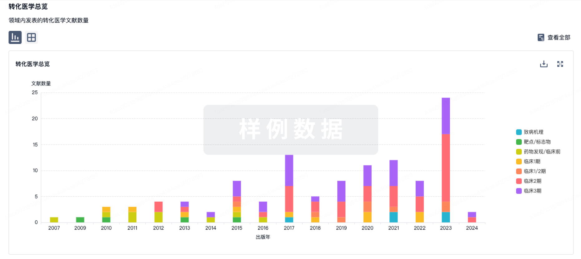
药物交易
使用我们的药物交易数据加速您的研究。
登录
或
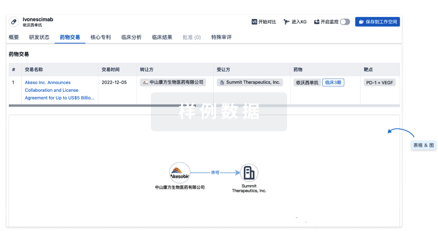
核心专利
使用我们的核心专利数据促进您的研究。
登录
或

临床分析
紧跟全球注册中心的最新临床试验。
登录
或
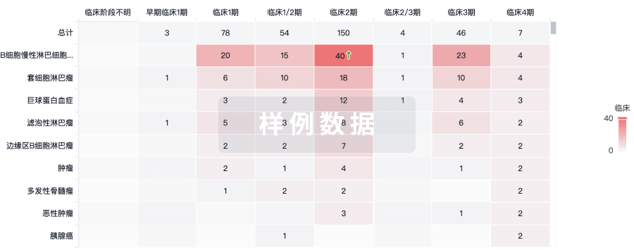
批准
利用最新的监管批准信息加速您的研究。
登录
或
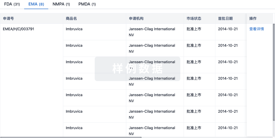
特殊审评
只需点击几下即可了解关键药物信息。
登录
或
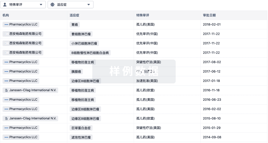
提出任何生物医药研究问题
让每一步生物医药决策都精准制胜
基于十亿级生物数据与全球情报的精准 AI,赋能早期药物发现
立即开始免费试用!
智慧芽新药情报库是智慧芽专为生命科学人士构建的基于AI的创新药情报平台,助您全方位提升您的研发与决策效率。
立即接入生物医药数据包
智慧芽新药库数据也通过智慧芽开放平台,以MCP或API服务形式对外开放。立即使用我们的MCP服务定制您的LLM智能体。
生物序列数据库
生物药研发创新
免费使用
化学结构数据库
小分子化药研发创新
免费使用