预约演示
更新于:2026-09-30

Swiss Federal Institute of Technology
更新于:2026-09-30
概览
标签
神经系统疾病
其他疾病
肿瘤
小分子化药
化学药
ASO
疾病领域得分
一眼洞穿机构专注的疾病领域

技术平台
公司药物应用最多的技术
靶点
公司最常开发的靶点
关联
靶点 |
作用机制 |
在研机构 |
原研机构 |
在研适应症 |
非在研适应症 |
最高研发阶段 |
首次获批国家/地区 |
首次获批日期 |

靶点 |
作用机制 |
在研机构 |
原研机构 |
在研适应症 |
非在研适应症 |
最高研发阶段 |
首次获批国家/地区 |
首次获批日期 |
靶点 |
作用机制 |
在研机构 |
原研机构 |
在研适应症 |
非在研适应症 |
最高研发阶段 |
首次获批国家/地区 |
首次获批日期 |
NCT07837752
Exercise and Prolonged Fasting to Improve Interscapular Brown Fat Mitochondrial Function and Enhance Metabolic Flexibility in Postmenopausal Women (EMPOWER-BAT)

NCT07704606
Measurement of Metabolites in the Breath and Blood in Patients With Altered Metabolism and Healthy Individuals
NCT07701603
Effects of Blood Gas Perturbations on Neuromuscular Function of the Diaphragm and Lower Limbs at Rest and During Exercise.
100 项与 Swiss Federal Institute of Technology 相关的临床结果
登录后查看更多信息
登录后查看更多信息
2026-12-31SCIENCE AND TECHNOLOGY OF ADVANCED MATERIALS
Sputtering yield for metal halide perovskite devices patterning
Article
作者: Proniakova, Daria ; Yakunin, Sergii ; Liu, Xuqi ; Tsarev, Sergey ; Kovalenko, Maksym V. ; Wu, Erfu ; Shorubalko, Ivan
Metal halide perovskites (MHPs) are emerging semiconductors with unique optoelectronic properties promising for highly rewardable applications. Water and polar solvents instability hinders the introduction of MHPs into CMOS technology infrastructure and is the main challenge for patterning and integration into electronic systems. Recently, dry etching in combination with standard lithography was demonstrated as a viable technology to address the problem. In this work, we investigate the dry etching of MHPs using argon (Ar) ion milling. Simulated etch rates using Ziegler's model are validated with experimental measurements. Assuming a linear sum of elemental sputtering yields results in total sputtering yield values for complex MHPs (CsPbIBr2, CsPbBr2Cl) that agree well with experimental data. Interestingly, ignoring the organic part of the hybrid halide perovskite MAPbI2Br gives a valid estimation of the sputtering yield. At a typical processing ion energy of 700 eV, Ar milling achieves rates of approximately 1-2 nm/s across various perovskite compositions. Photodetectors (PDs) fabricated under optimized etching conditions retain typical photoresponse, demonstrating the device functionality can be preserved after etching.
2026-10-01Nature Reviews Clinical Oncology
CRISPR in clinical oncology: translational advances from molecular diagnostics to therapeutics
Review
作者: Grigg, Samuel ; Blombery, Piers ; Shembrey, Carolyn ; Corn, Jacob E ; Fareh, Mohamed ; Seymour, John F ; Casan, Joshua M L
Cancer care is increasingly driven by molecular classification, yet many key oncogenic drivers remain undruggable, and intrinsic or acquired resistance to treatment frequently limits durable clinical benefit. CRISPR-Cas technologies provide a modular, programmable platform to interrogate and directly manipulate cancer biology via sequence-specific targeting of DNA or RNA and have advanced from experimental tools to the early stages of clinical translation. In this Review, we outline how CRISPR-enabled functional genomics approaches can reveal unexpected cancer dependencies and resistance mechanisms. We discuss emerging applications of CRISPR-based diagnostics in oncology that convert precise nucleic acid sequence recognition into rapid mutation detection. We also discuss applications of CRISPR in therapeutic strategies ranging from ex vivo immune cell engineering to nascent in vivo interventions that directly target tumour-related sequences such as fusion junctions or single-nucleotide variants. Finally, we highlight technological and regulatory challenges, including effective delivery of the editing machinery to cells in vivo, safety and platform-level regulatory frameworks, that will determine the clinical utility of CRISPR-based diagnostics and therapies in oncology.
2026-08-14PHYSICS IN MEDICINE AND BIOLOGY
Monte Carlo-based prediction models for nanodroplet-mediated proton range verification
Article
作者: Collado-Lara, Gonzalo ; Carlier, Bram ; Heymans, Sophie V ; Sterpin, Edmond ; Himmelreich, Uwe ; van der Heyden, Brent ; Piot, Wout ; Giammanco, Andrea ; Bomhals, Alec
Abstract:
Objective.
Radiation-sensitive nanodroplets were recently proposed for
in vivo
proton range verification. Specifically, the ultrasound contrast generated by radiation-induced droplet vaporization enables visualization of radiation deposition. Here, we developed and validated an
in silico
model to predict the nanodroplet radiation response from the original treatment plan, ultimately enabling contrast-mediated treatment verification.
Approach.
By combining the theory of radiation-induced nucleation of superheated emulsions with Monte Carlo simulations, we determined the effective fluence of ionizing particles contributing to the nanodroplet vaporization response. This fluence we assumed to be linearly proportional to the number of vaporization events. The resulting model performance was compared to experimental observations in phantoms and healthy rats
. Main results.
The modeled effective fluence closely mimicked the vaporization distributions in phantoms, with a sub-millimeter range retrieval performance at body temperature. We also demonstrated the possibility to apply the model directly to preclinical computed tomography scans, yielding promising qualitative agreement with the radiation-induced contrast.
Significance.
The presented prediction model forms an essential step towards clinical implementation of nanodroplet-mediated proton range verification. Moreover, the simplicity and generalizability of the model enables its use for a wide range of radiotherapy applications.
100 项与 Swiss Federal Institute of Technology 相关的药物交易
登录后查看更多信息
100 项与 Swiss Federal Institute of Technology 相关的转化医学
登录后查看更多信息
组织架构
使用我们的机构树数据加速您的研究。
登录
或
管线布局
2026年10月11日管线快照
管线布局中药物为当前组织机构及其子机构作为药物机构进行统计,早期临床1期并入临床1期,临床1/2期并入临床2期,临床2/3期并入临床3期
药物发现
5
16
临床前
临床1期
1
15
其他
登录后查看更多信息
当前项目
登录后查看更多信息
药物交易
使用我们的药物交易数据加速您的研究。
登录
或
转化医学
使用我们的转化医学数据加速您的研究。
登录
或
营收
使用 Synapse 探索超过 36 万个组织的财务状况。
登录
或
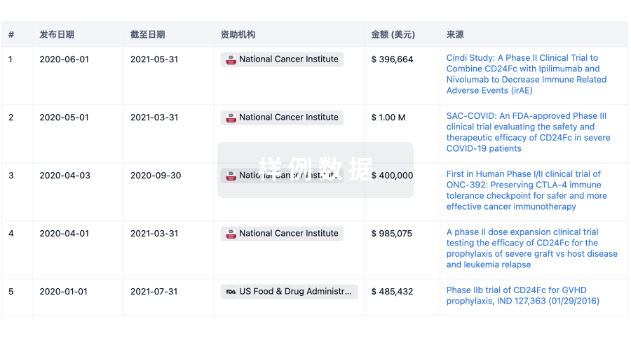
科研基金(NIH)
访问超过 200 万项资助和基金信息,以提升您的研究之旅。
登录
或
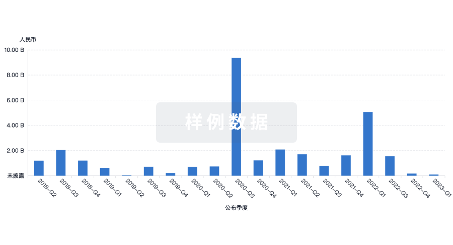
投资
深入了解从初创企业到成熟企业的最新公司投资动态。
登录
或
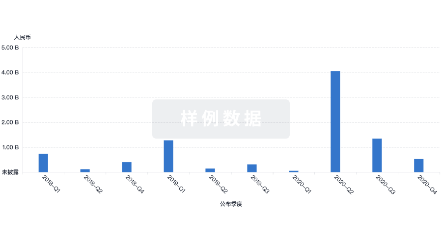
融资
发掘融资趋势以验证和推进您的投资机会。
登录
或
提出任何生物医药研究问题
让每一步生物医药决策都精准制胜
基于十亿级生物数据与全球情报的精准 AI,赋能早期药物发现
立即开始免费试用!
智慧芽新药情报库是智慧芽专为生命科学人士构建的基于AI的创新药情报平台,助您全方位提升您的研发与决策效率。
立即接入生物医药数据包
智慧芽新药库数据也通过智慧芽开放平台,以MCP或API服务形式对外开放。立即使用我们的MCP服务定制您的LLM智能体。
生物序列数据库
生物药研发创新
免费使用
化学结构数据库
小分子化药研发创新
免费使用