预约演示
更新于:2025-10-25
Ajmaline
西萝芙木碱
更新于:2025-10-25
概要
基本信息
结构/序列
分子式C20H26N2O2 |
InChIKeyCJDRUOGAGYHKKD-IWMNNKGHSA-N |
CAS号4360-12-7 |
关联
6
项与 西萝芙木碱 相关的临床试验NCT04580992
Defining the Electrocardiographic Effect of Propofol on the Ajmaline Provocation Drug Challenge: A Prospective Trial

EUCTR2018-000752-18-NL
Ajmaline provocation in asymptomatic PLN and PKP2 mutation carriers for early detection of Arrhythmogenic Cardiomyopathy - Ajmaline provocation in ACM
NCT03491475
Dynamicity of Echocardiography During Ajmaline Test
100 项与 西萝芙木碱 相关的临床结果
登录后查看更多信息
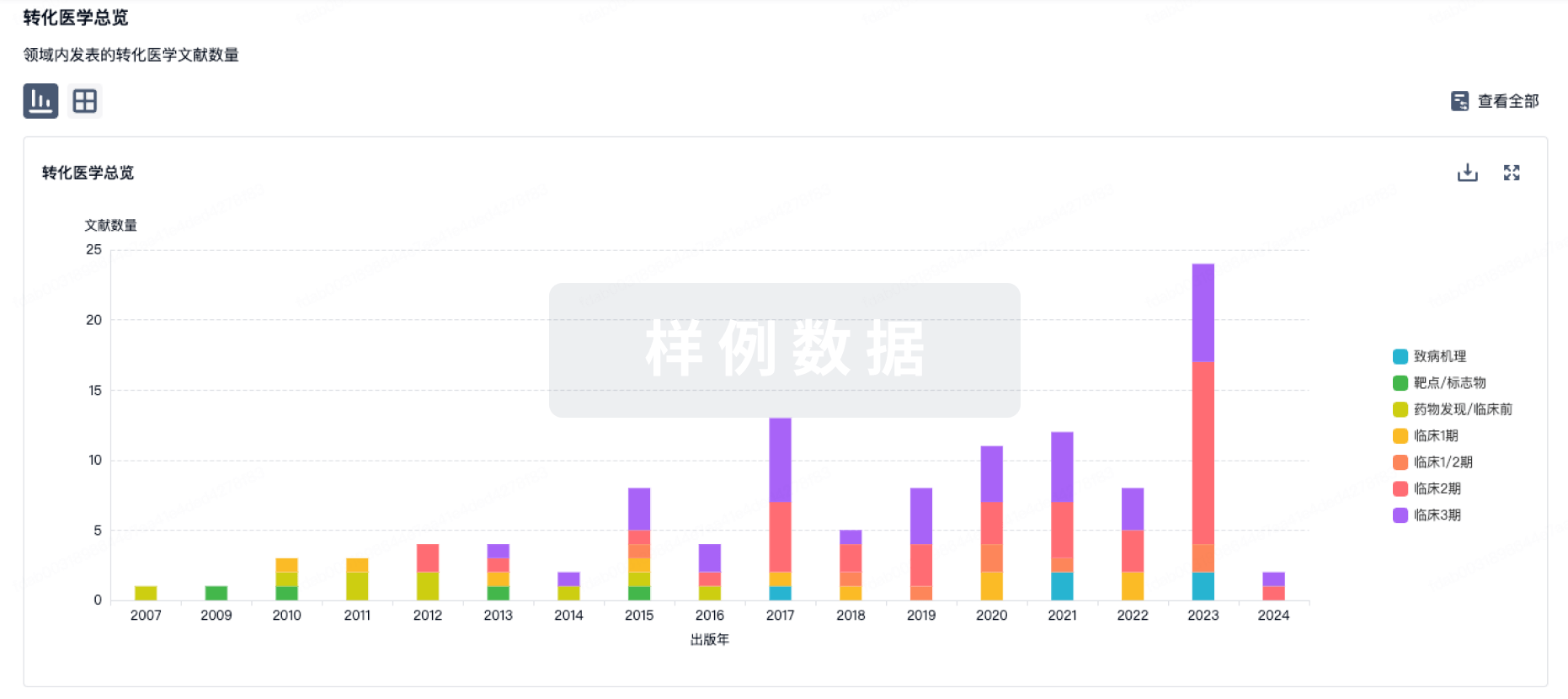
100 项与 西萝芙木碱 相关的转化医学
登录后查看更多信息
100 项与 西萝芙木碱 相关的专利(医药)
登录后查看更多信息
1,330
项与 西萝芙木碱 相关的文献(医药)2025-10-07EUROPACE
Genetic variants associated with ventricular arrhythmias during ajmaline test in Brugada syndrome
Article
作者: Vlaeminck, Jelle ; Uyttebroeck, Sophie ; Vergara, Pasquale ; Helsen, Christine ; Ströker, Erwin ; Audiat, Charles ; de Asmundis, Carlo ; Pappaert, Gudrun ; Brugada, Pedro ; Van Dooren, Sonia ; Della Rocca, Domenico Giovanni ; Pannone, Luigi ; Sieira, Juan ; De Schutter, Elke ; Almorad, Alexandre ; Giron, Philippe ; Sarkozy, Andrea ; Overeinder, Ingrid ; Sorgente, Antonio ; Bala, Gezim ; Gharaviri, Ali ; La Meir, Mark ; Chierchia, Gian Battista ; Eltsov, Ivan ; Pölsler, Laura
2025-10-01PLANTA MEDICA
Alkaloid Composition and Antiarrhythmic Activity of the Extracts from Rauvolfia serpentina Tissue Culture
Article
作者: Konvalyuk, Iryna ; Bieda, Oleksandr ; Soloviev, Anatoliy ; Mozhylevska, Ludmyla ; Khromov, Aleksandr ; Yarmoluk, Sergiy ; Kunakh, Viktor ; Andreev, Igor ; Dobrelia, Natalia
Abstract:
Rauvolfia serpentina produces a number of indole alkaloids and has long been used in the traditional treatment of arrhythmia. Given the shortage of natural resources, the K-27M strain of R. serpentina tissue culture was established. The aim was to evaluate the antiarrhythmic activity of extracts with different compositions and ratios of indole alkaloids derived from the cell biomass of the K-27M strain. Chemical analysis was conducted using HPLC-MS. Adrenaline-induced (rats) and ischemia-reperfusion arrhythmia (isolated guinea pig hearts) models were used to study the antiarrhythmic activity of the extracts. Extracts were obtained from dry (extracts 1, 2, and 3) and fresh biomass (fractions 1 and 2 of extract 4). Extract 1 contained ajmaline and acetylajmaline (the total indole alkaloid content (TIAC) was 2.2% of the dry biomass); extract 2–ajmaline, acetylajmaline, and raucaffricine (TIAC 6.4%); extract 3–ajmaline and raucaffricine (TIAC 29.0%). Fraction 1 of
extract 4 was dominated by vomilenine, methylajmalicine, ajmalicine, and raufloridine (TIAC 65.0%), and fraction 2 of extract 4 contained acetylajmaline (TIAC 47.4%). Extracts 1 and 2 containing negligible amounts of indole alkaloids showed a weak proarrhythmic effect. Fractions 1 and 2 of extract 4 had a pronounced antiarrhythmic effect in the adrenaline-induced arrhythmia model. In addition, fraction 2 of extract 4 had an antiarrhythmic effect in the ischemia-reperfusion arrhythmia model. The level of this activity depended on the composition and ratio of alkaloids in the extract. Thus, the K-27M strain of R. serpentina tissue culture is a promising source of indole alkaloids with antiarrhythmic activity.
2025-08-01HEART RHYTHM
Catheter ablation to prevent malignant ventricular arrhythmias in symptomatic Brugada syndrome: A systematic review and meta-analysis
Article
作者: Marcon, Lorenzo ; Almorad, Alexandre ; Sarkozy, Andrea ; Doundoulakis, Ioannis ; Giordano, Federica ; Brugada, Pedro ; Pelargonio, Gemma ; Ballacci, Federico ; de Asmundis, Carlo ; Imazio, Massimo ; Scacciavillani, Roberto ; Della Rocca, Domenico Giovanni ; Vetta, Giampaolo ; Narducci, Maria Lucia ; Sieira, Juan ; Chierchia, Gian Battista ; Pannone, Luigi
BACKGROUND:
Brugada syndrome (BrS) is a cardiac channelopathy predisposing individuals to malignant ventricular arrhythmias (VAs) and sudden cardiac death. Substrate modification with catheter ablation (CA) has emerged as an interesting option to prevent recurrence of VA.
OBJECTIVE:
This systematic review and meta-analysis aimed to assess the efficacy and safety of CA in patients with high-risk symptomatic BrS.
METHODS:
We conducted a systematic review and meta-analysis of studies comparing CA with standard therapy in patients with symptomatic BrS. We systematically searched PubMed and Embase databases from inception to June 15, 2025. The primary endpoint was recurrence of malignant VA or appropriate implantable cardioverter-defibrillator therapy during follow-up. Random-effects models were used to calculate pooled risk ratios with 95% confidence intervals (CIs).
RESULTS:
Five studies with 584 patients were included in the meta-analysis (331 ablation and 253 control). CA was associated with a 78% reduction in the primary endpoint compared with standard therapy (risk ratio 0.22; 95% CI 0.05-0.99; P = .049), with an estimated number needed to treat of 2.5. The time-to-event analysis confirmed the robustness of the findings (hazard ratio 0.17; 95% CI 0.04-0.73; P = .017). Procedural success rates were high: electrocardiogram normalization after Ajmaline challenge in 98.1% (95% CI 83.9-100.0), elimination of abnormal electrograms in 100% (95% CI 99.6-100.0), and noninducibility achieved in 94.9% of patients. Complication rate was low at 2.4% (95% CI 0.0-9.0).
CONCLUSION:
These findings support CA to prevent recurrence of VA in patients with symptomatic BrS on top of standard therapy, with high procedural success rates and an acceptable safety profile.
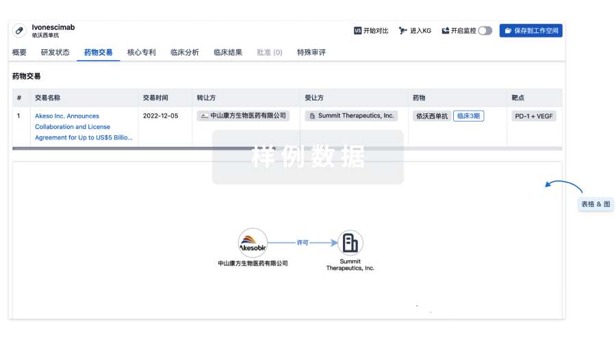
100 项与 西萝芙木碱 相关的药物交易
登录后查看更多信息
研发状态
10 条最早获批的记录, 后查看更多信息
登录
| 适应症 | 国家/地区 | 公司 | 日期 |
|---|---|---|---|
| 心律失常 | - | - | - |
登录后查看更多信息
临床结果
临床结果
适应症
分期
评价
查看全部结果
登录后查看更多信息
转化医学
使用我们的转化医学数据加速您的研究。
登录
或
药物交易
使用我们的药物交易数据加速您的研究。
登录
或
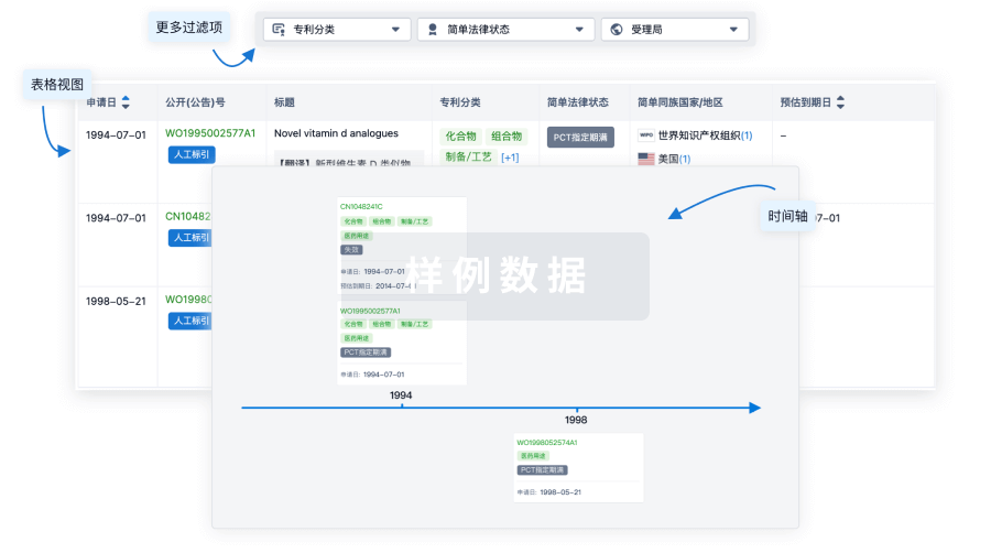
核心专利
使用我们的核心专利数据促进您的研究。
登录
或
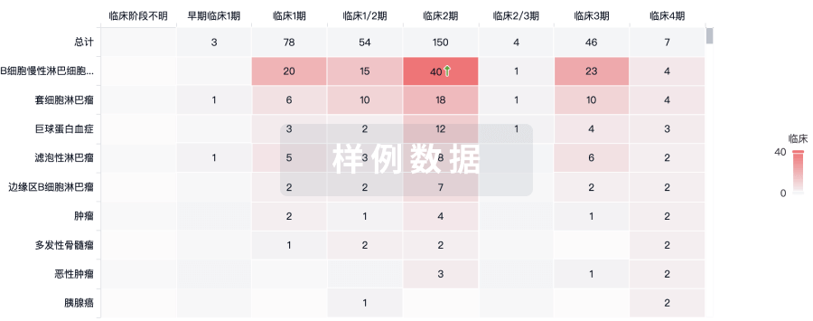
临床分析
紧跟全球注册中心的最新临床试验。
登录
或
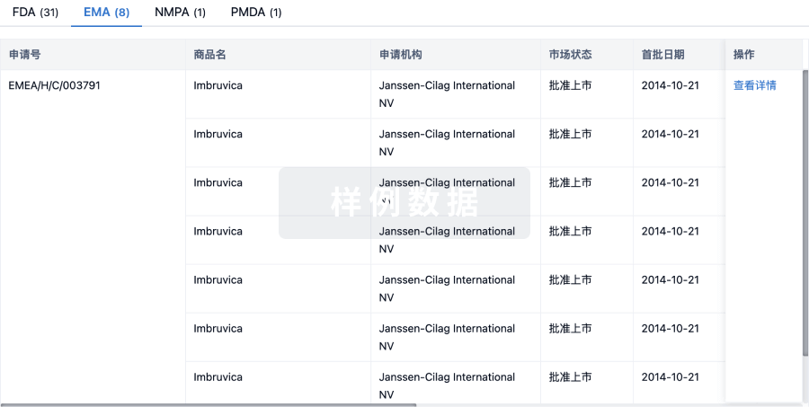
批准
利用最新的监管批准信息加速您的研究。
登录
或
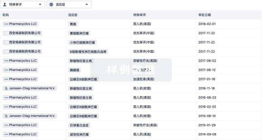
特殊审评
只需点击几下即可了解关键药物信息。
登录
或
生物医药百科问答
全新生物医药AI Agent 覆盖科研全链路,让突破性发现快人一步
立即开始免费试用!
智慧芽新药情报库是智慧芽专为生命科学人士构建的基于AI的创新药情报平台,助您全方位提升您的研发与决策效率。
立即开始数据试用!
智慧芽新药库数据也通过智慧芽数据服务平台,以API或者数据包形式对外开放,助您更加充分利用智慧芽新药情报信息。
生物序列数据库
生物药研发创新
免费使用
化学结构数据库
小分子化药研发创新
免费使用