预约演示
更新于:2026-07-18
111In-panitumumab
更新于:2026-07-18
概要
基本信息
药物类型 抗体偶联核素、诊断用放射药物 |
别名 Indium In 111 Panitumumab |
靶点 |
作用方式 拮抗剂 |
作用机制 EGFR拮抗剂(表皮生长因子受体erbB1拮抗剂) |
治疗领域 |
在研适应症 |
非在研适应症- |
非在研机构- |
权益机构- |
最高研发阶段临床1期 |
首次获批日期- |
最高研发阶段(中国)- |
特殊审评- |
登录后查看时间轴
结构/序列
使用我们的ADC技术数据为新药研发加速。
登录
或

Sequence Code 103929L

当前序列信息引自: *****
Sequence Code 194636H
当前序列信息引自: *****
关联
3
项与 111In-panitumumab 相关的临床试验NCT05945875
Official Title Targeted Dual Modality Imaging (TDMI) for Detection and Removal of Head and Neck Cancer
NCT05901545
Evaluating 111In Panitumumab for Nodal Staging in Head and Neck Cancer
NCT04840472
Pilot Evaluating 111In-Panitumumab for Nodal Staging in Head and Neck Cancer

100 项与 111In-panitumumab 相关的临床结果
登录后查看更多信息
100 项与 111In-panitumumab 相关的转化医学
登录后查看更多信息
100 项与 111In-panitumumab 相关的专利(医药)
登录后查看更多信息
2
项与 111In-panitumumab 相关的文献(医药)2026-01-14Theranostics
Use of Dual-Modality Antibody Imaging for Assessment of Lymph Node Metastases in Head and Neck Cancer
Article
作者: Topf, Michael C ; Rosenthal, Eben L ; McAdoo, Ashtyn G ; Hoang, Joseph ; Grice, Jared ; Smith, Gary ; Akhund, Ramsha ; Zinn, Kurt R ; Rosenberg, Adam J ; Meeks, Nicole ; Lee, Yu-Jin ; Hom, Marisa E
Surgical management of head and neck squamous cell carcinoma (HNSCC) patients often requires resection of the deep cervical lymph nodes. Fluorescence guided surgery (FGS) is a growing area of oncologic surgery that has shown promise for this purpose, however, it is limited by the signal penetration through the neck. To address this, we sought to evaluate the efficacy of the anti-EGFR antibody [111In]panitumumab to detect metastatic lymph nodes both on preoperative imaging and intraoperatively. We hypothesized that the addition of a radiolabeled antibody would be safe and effective in detecting malignant tissue when given alone or together with the optical agent panitumumab-IRDye800CW (pan800). Methods: Seventeen patients were enrolled and received 5 mCi of [111In]panitumumab with nine patients (53%) receiving [111In]panitumumab and pan800. SPECT/CT scans were performed prior to surgical resection. Intraoperatively, patients underwent gamma tracing and the patients who received pan800 also underwent optical fluorescence imaging. Resected specimens were compared to final histopathology to determine the sensitivity and specificity of the two tracers. Results: No adverse events related to the [111In]panitumumab were reported. SPECT/CT performed 3 days post-injection showed higher tumor-to-blood pool ratio compared to earlier scans (p = 0.04), and when compared to pathological assessment, could detect disease greater than 1 cm in diameter. Intraoperatively, resection of the primary tumor resulted in a significant drop in gamma counts (p < 0.001). In vivo detection of metastatic lymph nodes using the radiotracer was inconsistent and attributed to high background counts as patients who underwent surgery before 48 hours post-injection had significantly higher precordial counts than patients who underwent surgery after 48 hours, even when corrected for time, weight, and dose (p = 0.04). Ex vivo, metastatic lymph nodes had gamma counts almost twice that of benign (p < 0.0001). The separately dosed pan800 and [111In]panitumumab showed strong co-localization within metastatic lymph nodes (R = 0.87). Conclusion: Intravenous infusion of [111In]panitumumab in HNSCC patients is safe, whether administered alone or with pan800. Consistent with other studies, ex vivo application of the radiotracer differentiated tumor containing tissue from benign. However, intraoperative use of the radiotracer to detect metastatic lymph nodes was limited by background signal at early timepoints and signal decay at later timepoints.
2013-04-01Radiology1区 · 医学
Orthotopic Pleural Mesothelioma in Mice: SPECT/CT and MR Imaging with HER1- and HER2-targeted Radiolabeled Antibodies
1区 · 医学
Article
作者: Martin W. Brechbiel ; Marcelino Bernardo ; Peter L. Choyke ; Diane E. Milenic ; Tapan K. Nayak
PURPOSE:
To evaluate the potential of anti-human epidermal growth factor receptor (HER)1- and anti-HER2-targeted radiolabeled antibodies and magnetic resonance (MR) imaging for imaging of orthotopic malignant pleural mesothelioma (MPM) in mouse models.
MATERIALS AND METHODS:
Animal studies with 165 mice were performed in accordance with National Institutes of Health guidelines for the humane use of animals, and all procedures were approved by the institutional Animal Care and Use Committee. Flow cytometry studies were performed to evaluate HER1 and HER2 expression in NCI-H226 and MSTO-211H mesothelioma cells. Biodistribution and single photon emission computed tomography (SPECT)/computed tomography (CT) imaging studies were performed in mice (four or five per group, depending on tumor growth) bearing subcutaneous and orthotopic MPM tumors by using HER1- and HER2-targeted indium 111 ((111)In)- and iodine 125 ((125)I)-labeled panitumumab and trastuzumab, respectively. Longitudinal MR imaging over 5 weeks was performed in three mice bearing orthotopic tumors to monitor tumor growth and metastases. SPECT/CT/MR imaging studies were performed at the final time point in the orthotopic models (n = 3). The standard unpaired Student t test was used to compare groups.
RESULTS:
Orthotopic tumors and pleural effusions were clearly visualized at MR imaging 3 weeks after tumor cell inoculation. At 2 days after injection, the mean (111)In-panitumumab uptake of 29.6% injected dose (ID) per gram ± 2.2 (standard error of the mean) was significantly greater than the (111)In-trastuzumab uptake of 13.6% ID/g ± 1.0 and the (125)I-panitumumab uptake of 7.4% ID/g ± 1.2 (P = .0006 and P = .0001, respectively). MR imaging fusion with SPECT/CT provided more accurate information about (111)In-panitumumab localization in the tumor, as the tumor was poorly visualized at CT alone.
CONCLUSION:
This study demonstrates the utility of radiolabeled anti-HER1 antibodies in the imaging of MPM in preclinical models.
SUPPLEMENTAL MATERIAL:
http://radiology.rsna.org/lookup/suppl/doi:10.1148/radiol.12121021/-/DC1.
100 项与 111In-panitumumab 相关的药物交易
登录后查看更多信息
研发状态
10 条进展最快的记录, 后查看更多信息
登录
| 适应症 | 最高研发状态 | 国家/地区 | 公司 | 日期 |
|---|---|---|---|---|
| 复发性头颈部鳞状细胞癌 | 临床1期 | 美国 | 2023-08-22 |
登录后查看更多信息
临床结果
临床结果
适应症
分期
评价
查看全部结果
登录后查看更多信息
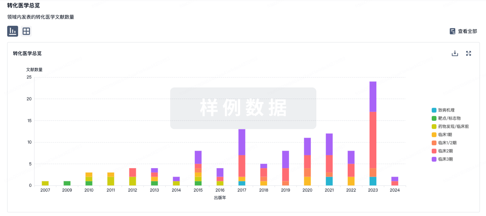
转化医学
使用我们的转化医学数据加速您的研究。
登录
或
药物交易
使用我们的药物交易数据加速您的研究。
登录
或

核心专利
使用我们的核心专利数据促进您的研究。
登录
或

临床分析
紧跟全球注册中心的最新临床试验。
登录
或
批准
利用最新的监管批准信息加速您的研究。
登录
或
生物类似药
生物类似药在不同国家/地区的竞争态势。请注意临床1/2期并入临床2期,临床2/3期并入临床3期
登录
或

特殊审评
只需点击几下即可了解关键药物信息。
登录
或
芽仔
全新生物医药AI Agent 覆盖科研全链路,让突破性发现快人一步
立即开始免费试用!
智慧芽新药情报库是智慧芽专为生命科学人士构建的基于AI的创新药情报平台,助您全方位提升您的研发与决策效率。
立即开始数据试用!
智慧芽新药库数据也通过智慧芽数据服务平台,以API或者数据包形式对外开放,助您更加充分利用智慧芽新药情报信息。
生物序列数据库
生物药研发创新
免费使用
化学结构数据库
小分子化药研发创新
免费使用