预约演示
更新于:2026-02-26
SR-18292
更新于:2026-02-26
概要
基本信息
权益机构- |
最高研发阶段药物发现 |
首次获批日期- |
最高研发阶段(中国)- |
特殊审评- |


关联
100 项与 SR-18292 相关的临床结果
登录后查看更多信息
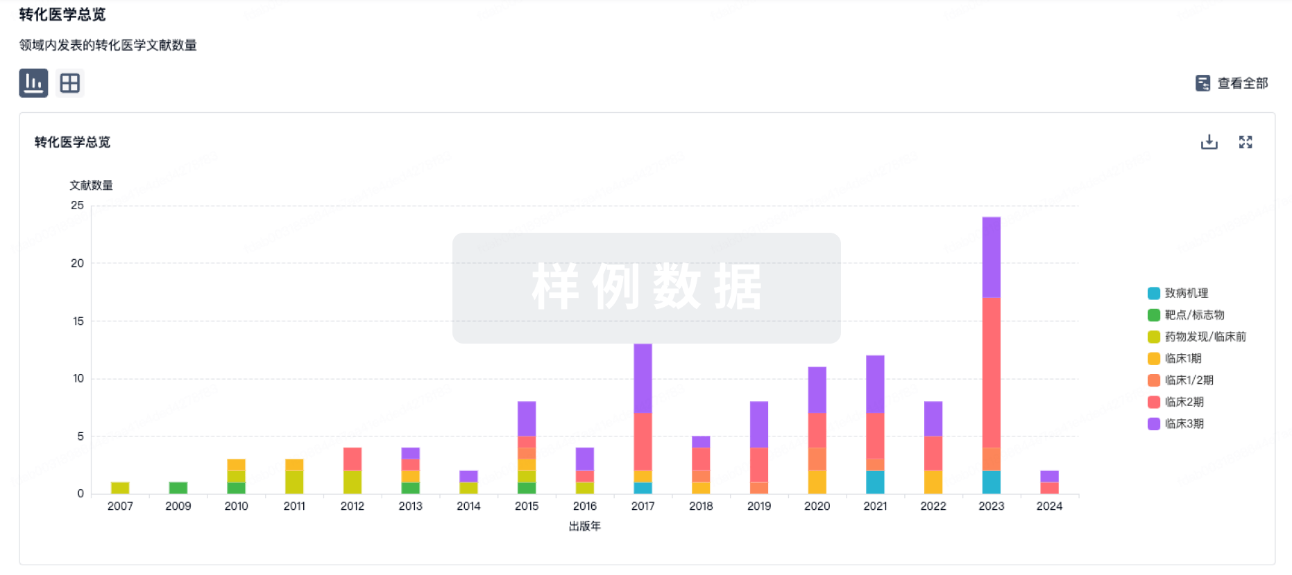
100 项与 SR-18292 相关的转化医学
登录后查看更多信息
100 项与 SR-18292 相关的专利(医药)
登录后查看更多信息
38
项与 SR-18292 相关的文献(医药)2026-04-01·BIOCHEMICAL AND BIOPHYSICAL RESEARCH COMMUNICATIONS
MK-3903 alleviates myocardial ischemia/reperfusion injury and mitochondrial dysfunction associated with AMKP-PGC-1α signaling
Article
作者: Gao, Jing ; Jiang, Wen-Xi ; Li, Pang-Bo ; Li, Hui-Hua
Myocardial ischemia/reperfusion (I/R) injury frequently occurs in acute coronary artery disease after timely reperfusion to rescue the ischemic heart. AMP-activated protein kinase (AMPK) is a key sensor that regulates metabolic metabolism and mitochondrial function and protects against myocardial I/R injury. Thus, pharmacologically activating AMPK modulation of AMPK has been suggested as a potential approach for attenuating myocardial I/R injury. MK-3903 is a potent and selective activator of AMPK, but its importance and mechanism of action in modulating this disease remain unclear. I/R was modeled in wild-type mice pretreated with MK-3903 (30 mg/kg). In addition, hypoxia/reoxygenation (H/R) was modeled using neonatal rat cardiomyocytes (NRCMs), and these cells were treated with MK-3903 and SR-18292 (a PGC-1α inhibitor). Our results revealed that in I/R model mice, the administration of MK-3903 dramatically alleviated myocardial dysfunction; reduced the infarct size, myocyte apoptosis, oxidative stress and inflammation; along with increased AMPK-PGC-1α signaling, improved imitochondrial biogenesis and the balance of mitochondrial dynamics. Conversely, the treatment of NRCMs with SR-18292 markedly diminished the cardioprotective effects of MK-3903 following H/R in vitro. In conclusion, these data demonstrate that MK-3903 may attenuate myocardial I/R injury and mitochondrial dysfunction possibly by activating AMPK-PGC-1α signaling and highlight its potential as a candidate for further investigation in ischemic heart injury.
2025-09-01·Zhongguo Zhong yao za zhi = Zhongguo zhongyao zazhi = China journal of Chinese materia medica
[Cajanolactone A ameliorates hepatocyte steatosis by regulating mitochondrial quality control via PGC-1α].
Article
作者: Yang, Rui-Yi ; Guo, Yu-Jia ; Hu, Ying-Jie ; Gan, Li-Zhen ; Cao, You-You
To explore the effects and mechanisms of cajanolactone A(CLA) on hepatocyte steatosis, with a focus on mitochondrial quality control(MQC) regulated by peroxisome proliferator-activated receptor-γ-coactivator-1α(PGC-1α). Human liver HHL-5 cells were induced with fatty acids(oleic acid-palmitic acid=2∶1) to develop steatosis, followed by exposure to different concentrations(2, 4, and 8 μmol·L~(-1)) of CLA. Lovastatin(LOV), the PGC-1α agonist ZLN005, and the PGC-1α inhibitor SR18292 served as control groups. Lipid accumulation was assessed by BODIPY staining, and flow cytometry. The levels of triglycerides(TG), total cholesterol(TC), and non-esterified fatty acids(NEFA) were measured by corresponding kits. The mitochondrial DNA(mtDNA) levels were determined by qPCR. The number and morphology of mitochondria(Mt) were observed by transmission electron microscopy. The mitochondrial quality was detected by Mt-specific fluorescent probe labeling. The functions of Mt were evaluated by JC-1 mitochondrial membrane potential and the ATP assay. The expression levels of PGC-1α and its associated transcription factors, including peroxisome proliferator-activated receptor α(PPARA), nuclear respiratory factor 1(NRF1), nuclear respiratory factor 2(NRF2), mitochondrial transcription factor A(TFAM), mitofusion 2(MFN2), optic atrophy 1(OPA1), autophagy receptor protein p62, beclin 1, and microtubule-associated protein 1 light chain 3β(LC3B), were quantified by RT-qPCR and Western blot. The results showed that CLA significantly reduced lipid accumulation, promoted lipolysis, increased Mt quantity, and improved the mitochondrial morphology, structure, and function in hepatocytes with steatosis. Furthermore, CLA up-regulated the expression of PGC-1α, PPARA, NRF1, NRF2, TFAM, MFN2, OPA1, p62, beclin 1, and LC3B. In conclusion, CLA may ameliorate hepatic steatosis by regulating the PGC-1α pathway and maintaining mitochondrial homeostasis.
2025-06-01·Science China-Life Sciences
Integrative analysis based on CRISPR screen identifies apilimod as a potential therapeutic agent for cisplatin-induced acute kidney injury treatment
Article
作者: Shen, Lu ; Zhang, Yutong ; Du, Huihui ; Lv, Cai ; Cao, Zhongyu ; Yang, Fan ; Tan, Jie ; Wang, Zhenting ; Zhou, Wei ; Chu, Yunpeng ; Qin, Shengying ; Zhang, Yingtian ; Huai, Cong ; Bao, Wei ; Chen, Luan ; Li, Shiyi ; Wei, Muyun ; Lin, Yunxiao
Acute kidney injury (AKI), a life-threatening side effect of cisplatin therapy, significantly limits the drug's therapeutic potential. In this study, we conducted a genome-wide CRISPR/Cas9 knockout screen in human renal tubular epithelial cells, integrating the results with transcriptome analyses and the Connectivity Map (CMap) database. Apilimod and elacridar emerged as the top two candidates of mitigating cisplatin-induced nephrotoxicity, with apilimod demonstrating superior efficacy in drug matrix experiments. Apilimod reduced cisplatin-induced apoptosis, inflammation and reactive oxygen species (ROS) generation. Transcriptome analyses suggested that apilimod may protect against cisplatin-induced nephrotoxicity via modulating lipid metabolism. In vitro experiments revealed that apilimod significantly ameliorated cisplatin-induced lipotoxicity by enhancing lipid clearance and upregulating PGC1α-mediated fatty acid oxidation. Mechanism experiments showed that apilimod induces the nuclear translocation of TFEB through the inhibition of its target, PIKfyve, thereby enhancing PGC1α expression and ameliorating lipotoxicity. These protective effects of apilimod were simulated by siRNA-mediated PIKfyve knockdown and diminished by the PGC1α inhibitor SR-18292 and siRNA targeting TFEB, confirming the role of the PIKfyve/TFEB/PGC1α signaling axis in apilimod's renoprotective effects. In vivo, apilimod alleviated apoptosis, inflammation, and lipid accumulation in a cisplatin-induced AKI mouse model. Additionally, apilimod treatment did not compromise the antitumor effect of cisplatin in cancer cells or tumor-bearing mice. Overall, our study suggests that apilimod could be a promising therapeutic agent for the treatment of cisplatin-induced AKI and revealed its underlying molecular mechanism.
2
项与 SR-18292 相关的新闻(医药)2024-08-12
·生物谷
研究表明,将一种小分子与羟基脲结合使用,可以通过不同的机制提高胎儿血红蛋白的生成。这可能为那些对羟基脲单独治疗效果不佳的镰状细胞病患者提供一种重要的新治疗选择。
镰状细胞病虽然罕见,但却是最常见的遗传性血液疾病。根据美国疾病控制预防中心(CDC)的数据,美国有超过 10 万人患有这种疾病,其中 90% 以上是黑人。尽管一种名为羟基脲(hydroxyurea)的药物可以减轻疼痛并减少住院次数,但并非所有成年人都对这种治疗方法反应良好。
在一项新的研究中,来自波士顿大学波士顿医学中心的研究人员发现了一种新的小分子,可以减少镰状红细胞并改善症状。这一发现为开发更有效的疗法提供了概念验证。相关研究结果发表在2024年8月2日的Science Advances期刊上,论文标题为“PGC-1α agonism induces fetal hemoglobin and exerts antisickling effects in sickle cell disease”。
论文通讯作者、波士顿医学中心镰状细胞病卓越研究中心研究员 Shuaiying Cui 博士说,“我们发现了一种很有希望的方法,可以为传统治疗方法无效的镰状细胞病患者带来希望。”
镰状细胞病患者的红细胞会变成“镰状”或新月形,这是因为一种基因突变影响血红蛋白的功能,而血红蛋白是将氧气从肺部输送到其他组织的蛋白。这种突变会使血红蛋白分子粘在一起,阻碍血液流动,导致患者剧痛发作。
然而,胎儿血红蛋白(人们通常在出生后就停止制造)可以减少镰状红细胞的数量。羟基脲通过提高胎儿血红蛋白发挥作用,但会导致毒副作用,而且并非对所有人都有帮助。
在这项研究中,研究者测试了一种靶向PGC-1 α蛋白的小分子对胎儿血红蛋白生成和镰状红细胞的影响,其中PGC-1 α是一种在脂肪组织中发现的蛋白,在红细胞的成熟和存活中起作用。他们发现这种称为SR-18292的小分子能增加人类造血干细胞中的胎儿血红蛋白生成,并减少镰状细胞病小鼠体内畸形红细胞的数量。这表明SR-18292本身就能改善镰状细胞病的病理。不过,当与羟基脲联合使用时,SR-18292 对胎儿血红蛋白的生成产生了更大的影响。
Cui说,“我们的研究表明,将一种小分子与羟基脲结合使用,可以通过不同的机制提高胎儿血红蛋白的生成。这可能为那些对羟基脲单独治疗效果不佳的镰状细胞病患者提供一种重要的新治疗选择。”
图片来自Science Advances, 2024, doi:10.1126/sciadv.adn8750
为了确定SR-18292是否会影响控制胎儿血红蛋白生成的基因,研究者对接受该分子处理的人类造血干细胞进行了单细胞RNA测序。他们发现,许多基因在接受SR-18292处理后表达不同,其中包括BCL11A的下调,其中BCL11A通常抑制胎儿血红蛋白的产生,是第一种CRISPR基因编辑疗法治疗镰状细胞病的靶基因。
美国食品药品管理局(FDA)最近批准了治疗镰状细胞病的疗法,波士顿医学中心正在为患者提供这些疗法,但这些疗法成本高昂,而且由于程序复杂,还不能在全球范围内推广治疗。Cui希望这项新的研究是为治疗资源匮乏的社区的患者开发一种治疗方法的第一步。
Cui 说,“这一突破标志着波士顿医学中心在为所有患者寻求更有效的镰状细胞病治疗方法的道路上迈出了重要一步。有朝一日,我们希望我们的药物能应用于全球各地可能无法获得现有基因疗法的镰状细胞病患者。”(生物谷Bioon.com)
参考资料:
Yanan Sun et al. PGC-1α Agonism Induces Fetal Hemoglobin and Exerts Anti-Sickling Effects in Sickle Cell Disease. Science Advances, 2024, doi:10.1126/sciadv.adn8750.
临床1期
2024-04-02
·奇点网
*仅供医学专业人士阅读参考ω-3多不饱和脂肪酸,特别是二十二碳六烯酸(DHA),被证明对人类心血管健康、认知功能等具有广泛的健康益处。在抗肿瘤方面,动物研究表明,DHA可以通过直接诱导肿瘤细胞死亡和抑制肿瘤转移来发挥抗肿瘤作用,同时目前也有多项临床研究在评估DHA补充对于癌症患者的有效性。不过迄今为止,还没有足够研究描述抗肿瘤机制中DHA对于免疫细胞的调节作用。陆军军医大学邓有才教授团队的最新研究成果填补了这一空白,于近日发表在《癌症免疫学研究》杂志上[1]。他们发现,自然杀伤细胞(NK)对DHA的吸收较好,DHA可以特异性通过改善NK细胞的代谢和提高线粒体活性来增强其细胞毒性,从而对NK细胞的抗肿瘤能力发挥免疫调节作用。论文首页截图目前为止,关于DHA免疫调节作用的观点尚有分歧,于是研究团队展开此次研究。他们着重考察了DHA饮食对不同类型免疫细胞的影响。结果发现,尽管富含DHA的饮食会轻微减少野生型小鼠的NK细胞数量,但是对于黑色素瘤小鼠而言,DHA饮食增强了其NK细胞的细胞毒性,肿瘤微环境中能够产生杀伤因子IFN-γ的NK细胞数量增多,而对T细胞、B细胞等其它免疫细胞的数量和活性影响较小。进一步的scRNA-seq等技术分析表明,与常规饮食相比,DHA大幅增强了NK细胞的线粒体氧化磷酸化信号通路和细胞毒性相关通路的活性。DHA对不同类型免疫细胞的信号通路的影响此外还揭示,DHA如此关照于NK细胞而非其它免疫细胞,是因为NK细胞能够更好地吸收DHA,这可能归因于与DHA转运相关的特定蛋白(SLC27A3、FABP3和SLC27A1等)在NK细胞中的表达水平明显高于T细胞、B细胞。小鼠实验中,小鼠分别接受三周的常规饮食和DHA饮食喂养,之后两组均注射具有转移性的黑色素瘤细胞。结果显示,较对照组相比,DHA饮食组小鼠的肺部肿瘤结节明显较少;当耗尽NK细胞时,DHA饮食无法再减少小鼠的肿瘤负荷。以上结果意味着,DHA的抗肿瘤效果不仅可以通过直接作用于肿瘤细胞来发挥,还可以依赖于NK细胞的效应功能。结合体外实验,研究团队揭示DHA调控NK细胞的机制:DHA通过增加NK细胞中蛋白质PGC-1α的水平来促进NK细胞的线粒体活性,使线粒体数量和膜电位增强、氧化磷酸化活动增强,从而提高其抗肿瘤效应。DHA以浓度依赖性方式促进NK细胞产生IFN-γ,而PGC-1α抑制剂(SR-18292)可以逆转DHA引起的IFN-γ产生、线粒体活性的增加。给DHA饮食的黑色素瘤小鼠模型注射PGC-1α抑制剂时,小鼠的肺部肿瘤结节数量增加,说明PGC-1α信号的缺失会消除DHA对肿瘤定植的抑制作用。PGC-1α抑制剂逆转DHA的抗肿瘤效果总之这项研究结论强调,以往的观点大多聚焦于DHA对肿瘤细胞的直接抑制作用,而忽视DHA的免疫调节作用。研究团队发现,DHA作为一种营养补充剂,富含DHA的饮食能够特异性地提高肿瘤微环境中的NK细胞抗肿瘤活性以抑制肿瘤生长,并表明DHA优先激活NK细胞,是因为NK细胞可以更好地吸收DHA,这些为癌症预防和治疗提供了新的视角。参考文献:[1]https://aacrjournals.org/cancerimmunolres/article/doi/10.1158/2326-6066.CIR-23-0359/741956/The-3-polyunsaturated-fatty-acid-docosahexaenoic本文作者丨张艾迪
细胞疗法免疫疗法临床研究
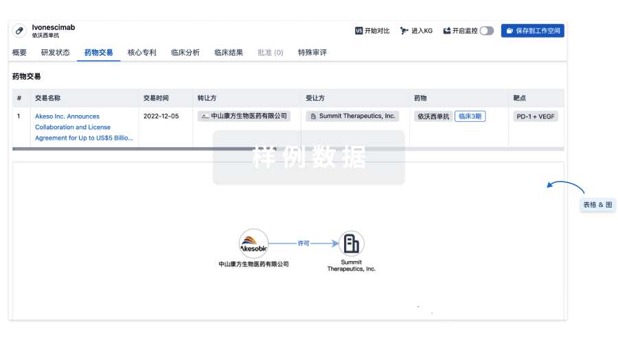
100 项与 SR-18292 相关的药物交易
登录后查看更多信息
研发状态
10 条进展最快的记录, 后查看更多信息
登录
| 适应症 | 最高研发状态 | 国家/地区 | 公司 | 日期 |
|---|---|---|---|---|
| 2型糖尿病 | 临床前 | 美国 | - | |
| 2型糖尿病 | 临床前 | 美国 | - | |
| 镰状细胞血症 | 药物发现 | 美国 | 2024-08-12 |
登录后查看更多信息
临床结果
临床结果
适应症
分期
评价
查看全部结果
| 研究 | 分期 | 人群特征 | 评价人数 | 分组 | 结果 | 评价 | 发布日期 |
|---|
No Data | |||||||
登录后查看更多信息
转化医学
使用我们的转化医学数据加速您的研究。
登录
或
药物交易
使用我们的药物交易数据加速您的研究。
登录
或
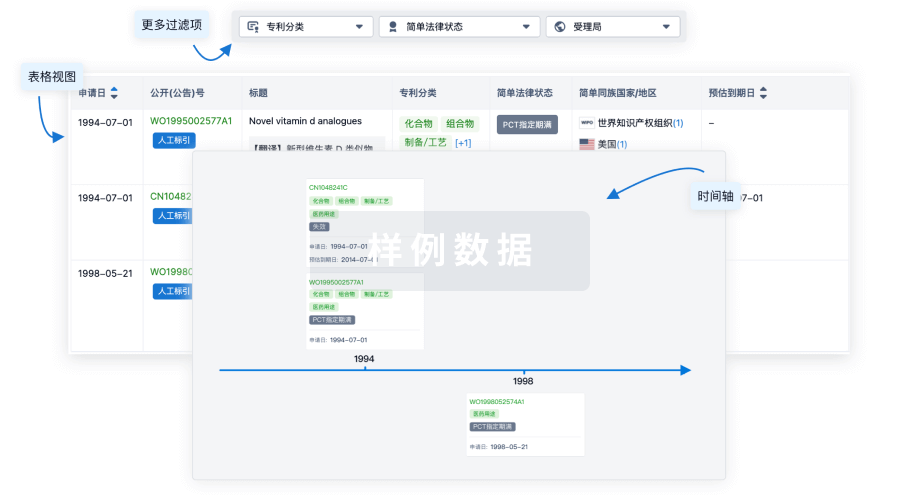
核心专利
使用我们的核心专利数据促进您的研究。
登录
或
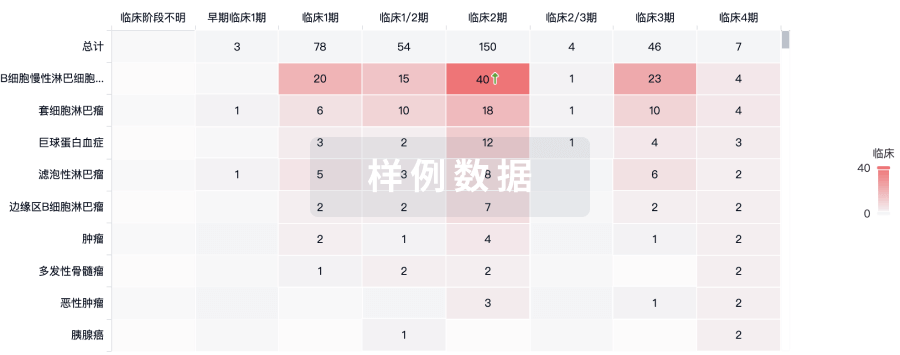
临床分析
紧跟全球注册中心的最新临床试验。
登录
或
批准
利用最新的监管批准信息加速您的研究。
登录
或
特殊审评
只需点击几下即可了解关键药物信息。
登录
或
生物医药百科问答
全新生物医药AI Agent 覆盖科研全链路,让突破性发现快人一步
立即开始免费试用!
智慧芽新药情报库是智慧芽专为生命科学人士构建的基于AI的创新药情报平台,助您全方位提升您的研发与决策效率。
立即开始数据试用!
智慧芽新药库数据也通过智慧芽数据服务平台,以API或者数据包形式对外开放,助您更加充分利用智慧芽新药情报信息。
生物序列数据库
生物药研发创新
免费使用
化学结构数据库
小分子化药研发创新
免费使用