预约演示
更新于:2026-08-29
Porcine Fibrin (Guangzhou Baitejing Pharmaceutical Technology)
猪源纤维蛋白贴(广州市拜特凇医药科技)
更新于:2026-08-29
概要
基本信息
非在研机构- |
权益机构- |
最高研发阶段临床1/2期 |
首次获批日期- |
最高研发阶段(中国)临床1/2期 |
特殊审评- |

登录后查看时间轴
关联
2
项与 猪源纤维蛋白贴(广州市拜特凇医药科技) 相关的临床试验CTR20213208
DBT用于肝脏/脾脏/胰腺切除手术时止血作用的I/II期探索性临床试验
初步评价DBT用于肝脏/脾脏/胰腺切除术止血时的有效性和安全性
开始日期2021-12-30 |
申办/合作机构 广州市拜特凇医药科技有限公司 [+1] |
ChiCTR-OOC-16008927
Change in health-related quality of life & immunity among menopausal women using Danggui Buxue Tang
开始日期1990-01-01 |
100 项与 猪源纤维蛋白贴(广州市拜特凇医药科技) 相关的临床结果
登录后查看更多信息
100 项与 猪源纤维蛋白贴(广州市拜特凇医药科技) 相关的转化医学
登录后查看更多信息
100 项与 猪源纤维蛋白贴(广州市拜特凇医药科技) 相关的专利(医药)
登录后查看更多信息
750
项与 猪源纤维蛋白贴(广州市拜特凇医药科技) 相关的文献(医药)2026-09-01·ENVIRONMENTAL POLLUTION
Long-term trends and patterns of legacy and emerging cosmetic UV filters in German waters
Article
作者: Koschorreck, Jan ; Duffek, Anja ; Winter, Eric ; Backhaus, Thomas ; Brendler, Luise
Organic UV filters contained in sunscreens and other cosmetics can potentially end up in the aquatic environment. Various studies have demonstrated that UV filters are widespread in water bodies, yet there is a lack of comprehensive understanding of their spatial and temporal distribution in Germany. The objective of this study is to investigate the occurrence of this group of substances in suspended particulate matter (SPM) and water samples, and to explore potential links to market changes reported in the literature. We developed an analytical target method in which SPM was extracted using accelerated solvent extraction (ASE), the extracts were purified, and then analyzed using liquid chromatography-tandem mass spectrometry (LC-MS/MS) with a quadrupole time-of-flight (Q-TOF) mass analyzer. The method detected 10 of 12 organic UV filters in SPM samples from the German Environmental Specimen Bank (ESB). Avobenzone (BMDBM) and iscotrizinol (DBT) showed average concentrations of 105 ng/g dry weight (dw) and 34 ng/g dw, respectively, in 2022. DBT showed a comprehensive significant upward trend at all Rhine stations monitored between 2005 and 2022. Diethylaminohydroxybenzoylhexyl benzoate (DHHB) also showed a clear and statistically significant upward trend at these stations, with a notable increase since 2017 at the Koblenz station. High-resolution mass spectrometry (HRMS) non-target screening (NTS) data confirmed the presence of additional polar organic UV filters in SPM and water samples, specifically camphor benzalkonium methosulfate (CBM), 4-aminobenzoic acid (PABA), ensulizole (PBSA), and sulisobenzone (BP-4). We recommend using a combination of target and NTS methods to achieve a comprehensive assessment of water pollution.
2026-07-07·ANALYTICAL CHEMISTRY
Ferroptosis Revealed by a Bifunctional Nanopipette Sensing the Intracellular Iron-Reactive Oxygen under Dibenzothiophene Exposure
Article
作者: Xiao, Shan ; Meng, Xianyang ; Du, Yuying ; Yu, Yanmin ; Lu, Liping ; Wang, Xiayan
Ferroptosis, as an iron-dependent form of regulated cell death, is characterized by disrupted iron homeostasis and excessive lipid peroxidation. However, current methods usually rely on population analysis of a single biomarker and are unable to obtain the real-time dynamic interplay of key intracellular species. Here, we developed a bifunctional nanopipette that can synchronously determine Fe2+ and H2O2 at the single-cell level by integrating scanning electrochemical microscopy (SECM) and scanning ion conductance microscopy (SICM). This nanopipette features bifunctions, whose inner surface was modified with 1,10-phenanthroline-4-carboxylic acid (Phen-COOH) for capturing Fe2+ detection using SICM, while the outer surface was coated with Prussian blue (PB) for catalyzing H2O2 using SECM. The two interfaces were characterized by scanning electron microscopy (SEM) and X-ray photoelectron spectroscopy (XPS). Their high sensitivity (Fe2+, 0.65 nA/μM; H2O2, - 0.09 nA/μM) and good selectivity are attributed to the high catalysis of PB and the capture ability of phenanthroline. In addition, the morphology of HepG2 cells was imaged in situ in real time. Ferroptosis effects on HepG2 cells under dibenzothiophene (DBT) exposure were investigated using this SECM-SICM platform. DBT exposure induced a coordinated increase in intracellular Fe2+ and H2O2, accompanied by reduced mitochondrial activity, increased malondialdehyde accumulation, and enhanced lactate dehydrogenase release, which indicate that DBT triggers ferroptosis of HepG2 cells through an iron-dependent oxidative mechanism. This work provides a single-cell analytical strategy for dissecting ferroptosis-related pathways and evaluating the toxicological effects of environmental pollutants.
2026-07-01·Journal of Pediatric Nursing-Nursing Care of Children & Families
Nurse-led Buteyko versus diaphragmatic breathing retraining in hospitalized children with asthma: A three-group quasi-experimental study
Article
作者: Osman, Atiat ; Ramadan, Osama Mohamed Elsayed ; Mahdy, Noura Mohammed ; Katooa, Nouran Essam ; Abunar, Areej ; Mohamed, Aziza Ibrahim Abd El Kader ; Mohamed, Fatma Abdellah ; Hafiz, Alaa ; Elsharkawya, Nadia Bassuoni
BACKGROUND:
Pediatric asthma may involve dysfunctional breathing patterns and autonomic dysregulation not directly addressed by pharmacotherapy. Comparative evidence on breathing retraining in hospitalized children remains limited.
PURPOSE:
To compare nurse-led Buteyko Breathing Technique (BBT) and Diaphragmatic Breathing Technique (DBT) versus routine care on heart rate (HR), respiratory rate (RR), systolic blood pressure (SBP), peripheral oxygen saturation (SpO₂), Control Pause (CP) time, and Modified Becker's Score (MBS) in hospitalized children with bronchial asthma.
DESIGN AND METHODS:
A three-group quasi-experimental pretest-posttest study enrolled 90 hospitalized children aged 6-12 years in BBT, DBT, or routine care groups (30/group) at the inpatient pediatric department of a university hospital in Upper Egypt, preceded by a small pilot study with nine children (three per group) not included in the main analysis, which confirmed feasibility and informed sample-size refinement. Two nurse-delivered sessions (30-45 min each) were initiated 24-48 h after stabilization with prescribed pharmacotherapy.
RESULTS:
Both intervention groups were associated with significant reductions in HR and RR versus controls on adjusted analyses: HR (F = 6.72, p = .002, partial η2 = 0.136) and RR (F = 9.18, p < .001, partial η2 = 0.178). BBT was associated with the largest CP improvement; normalization (≥ 40 s) occurred in 60.0% of BBT versus 0% in DBT and controls (F = 12.05, p < .001, partial η2 = 0.221). SpO₂ normalized in 56.7% of BBT, 23.3% of DBT, and 0% of controls. Adjusted SBP differences were not significant (p = .250).
CONCLUSIONS:
Nurse-led breathing retraining was associated with short-term physiological improvement during pediatric asthma hospitalization, with BBT showing a greater association with improvement in CP and oxygenation. Given the non-randomized design, baseline severity imbalance, brief dose, and absence of follow-up, findings are preliminary and require confirmation in rigorously designed randomized controlled trials.
1
项与 猪源纤维蛋白贴(广州市拜特凇医药科技) 相关的新闻(医药)2025-11-11
·生物通
在现代风湿免疫疾病的治疗中,生物制剂和靶向合成疾病修饰抗风湿药物(b/tsDMARDs)已成为重要手段。这些药物通过针对特定的炎症通路,如肿瘤坏死因子(TNF)、白细胞介素(IL)等,显著改善了患者的临床预后。然而,尽管有多种药物可供选择,仍有一部分患者对单一生物制剂治疗反应不足,难以达到治疗目标。因此,双生物制剂治疗(Dual Biologic Therapy, DBT)作为一种可能的策略,被提出以克服治疗抵抗。DBT通过同时抑制多个炎症通路,理论上可以增强治疗效果,但也伴随着更高的感染风险,尤其是在某些特定的药物组合中。在类风湿关节炎(Rheumatoid Arthritis, RA)领域,DBT的研究表明,虽然其在部分难治性病例中显示出一定的疗效提升,但与单药治疗相比,感染率显著增加。例如,一项2004年的研究显示,将阿那白滞素(anakinra)与依那西普(etanercept)联合使用时,尽管有轻微的疗效提升,但严重感染的发生率明显上升。另一项2006年的研究则发现,虽然ACR50/70的响应率有所提高,但严重感染和不良事件的发生率也显著增加。尽管这些早期研究引发了对DBT安全性的担忧,但后续的一些研究探索了不同的药物组合,例如利妥昔单抗(rituximab)与JAK抑制剂的联合使用。然而,即使在这些研究中,DBT的疗效提升并不明显,且感染风险依然存在,尤其是在高剂量组合中。这表明,在RA患者中,DBT可能更适合那些对其他治疗方案无效的难治性病例,但其使用仍需谨慎评估风险与收益。在银屑病关节炎(Psoriatic Arthritis, PsA)方面,DBT的应用更为广泛,尤其是在涉及多领域(如关节炎、皮肤病变、肠病)的复杂病例中。例如,一项研究显示,将阿普斯特(apremilast)与TNF抑制剂或IL-23抑制剂联合使用,能够显著改善患者的皮肤和关节症状,同时感染风险相对较低。此外,一些案例报告指出,使用维多珠单抗(vedolizumab)与TNF抑制剂的联合治疗,在控制肠道和关节炎症方面具有积极作用,且未观察到严重的感染事件。然而,这些研究大多基于回顾性分析或个案报告,缺乏大规模的随机对照试验(RCTs)支持。因此,虽然DBT在PsA中的应用前景较好,但其长期疗效和安全性仍需进一步验证。对于轴性脊柱关节炎(Axial Spondyloarthritis, axSpA),DBT的研究还处于初步阶段。一些小规模的回顾性研究和个案报告显示,联合使用不同机制的生物制剂可能带来一定的临床改善,尤其是在单一药物治疗无效的情况下。例如,一名患者在使用依那西普和英夫利昔单抗的联合治疗后,出现了显著的临床缓解,且未发生严重感染。此外,另一项研究中,维多珠单抗与戈利木单抗(golimumab)的联合治疗也有效控制了患者的肠道和关节症状。尽管这些案例提供了初步的证据,但目前的证据仍有限,且缺乏标准化的评估体系,使得DBT在axSpA中的应用仍需更多高质量的研究支持。DBT的潜在优势在于其能够同时作用于多个炎症通路,从而更全面地控制疾病活动。然而,这一策略的实施也面临诸多挑战。首先,现有的研究数据存在局限性,大多数研究基于小样本量或单中心经验,缺乏广泛的适用性和长期随访结果。其次,不同地区的感染风险、治疗可及性和药物监测能力存在显著差异,这使得DBT在全球范围内的应用受到地理因素的影响。例如,在资源丰富的地区,可能更容易开展多中心研究和长期随访,而在资源有限的地区,DBT的应用可能受限于缺乏足够的医疗资源和专业知识。此外,DBT的疗效并不总是优于单药治疗。在RA和PsA的研究中,尽管部分病例显示了联合治疗的优势,但整体上并没有一致的证据表明DBT在所有情况下都更有效。这可能与患者个体差异、疾病异质性以及药物组合的选择有关。因此,在临床实践中,DBT的使用应当基于对患者具体情况的全面评估,尤其是在那些对现有治疗方案反应不佳的情况下。为了更好地指导DBT的应用,未来的研究方向应包括以下几个方面。首先,开发能够预测治疗反应的生物标志物,这有助于精准选择适合DBT的患者群体,减少不必要的治疗风险。例如,在RA中,代谢组学分析已经发现了一些能够预测特定生物制剂疗效的代谢物标志物,这些标志物的进一步研究和验证可能为个性化治疗提供重要依据。其次,探索诱导免疫耐受的新策略,这可能为解决治疗抵抗问题提供新的思路。例如,通过靶向CD4? T细胞或调节性T细胞(Tregs)的功能,可以尝试在不损害整体免疫系统的情况下,实现对自身免疫反应的控制。此外,随着CAR-T细胞疗法的发展,这一技术在风湿免疫疾病中的应用也引起了广泛关注。CAR-T细胞通过工程改造,能够特异性地攻击致病性免疫细胞,减少对正常组织的损伤,为难治性疾病的治疗提供了新的可能性。总体而言,DBT作为一种潜在的治疗策略,在某些特定的风湿免疫疾病中可能带来一定的临床益处。然而,由于现有的研究证据尚不充分,且感染风险较高,其应用应当谨慎。未来的研究需要更多高质量的临床试验,以明确DBT在不同疾病类型和患者群体中的疗效和安全性。同时,结合生物标志物和个体化治疗策略,可能有助于提高DBT的适用性和成功率。此外,考虑到不同地区的医疗资源差异,还需要推动针对特定环境和条件的DBT研究,以确保其在全球范围内的安全和有效应用。最终,DBT的推广应建立在充分的科学证据和临床经验基础上,确保其在真正需要的患者中发挥最大作用,同时最小化潜在的风险。
引进/卖出临床研究并购临床结果优先审批
100 项与 猪源纤维蛋白贴(广州市拜特凇医药科技) 相关的药物交易
登录后查看更多信息
研发状态
10 条进展最快的记录, 后查看更多信息
登录
| 适应症 | 最高研发状态 | 国家/地区 | 公司 | 日期 |
|---|---|---|---|---|
| 术后出血 | 临床2期 | 中国 | 2021-12-30 | |
| 术后出血 | 临床2期 | 中国 | 2021-12-30 |
登录后查看更多信息
临床结果
临床结果
适应症
分期
评价
查看全部结果
| 研究 | 分期 | 人群特征 | 评价人数 | 分组 | 结果 | 评价 | 发布日期 |
|---|
No Data | |||||||
登录后查看更多信息
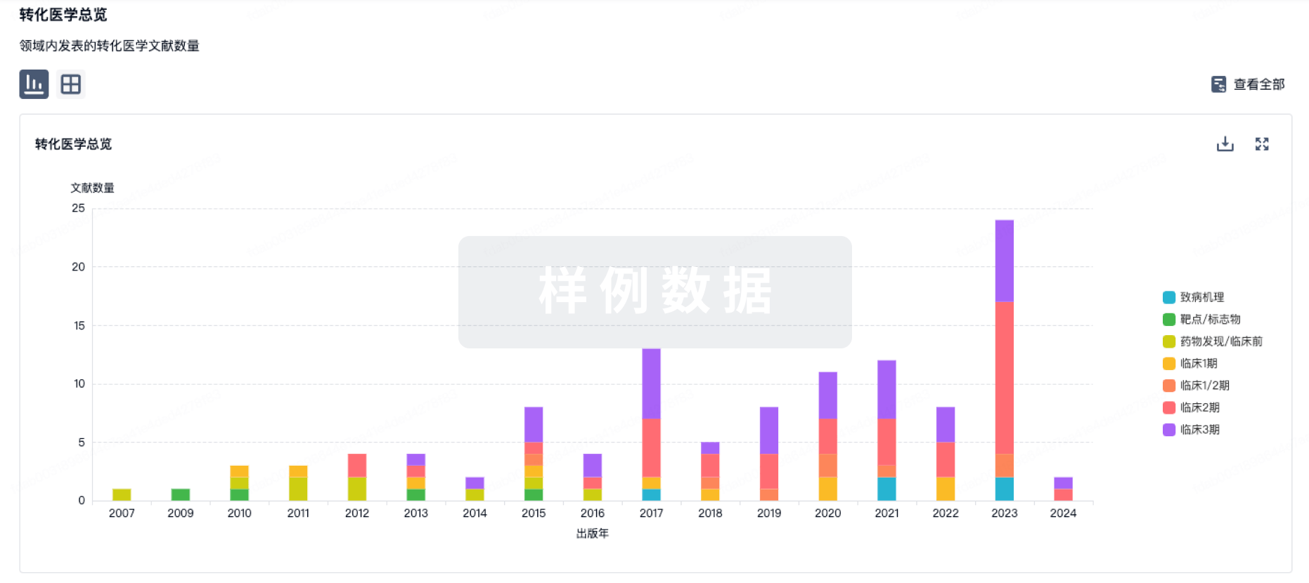
转化医学
使用我们的转化医学数据加速您的研究。
登录
或
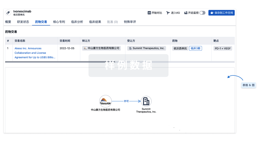
药物交易
使用我们的药物交易数据加速您的研究。
登录
或
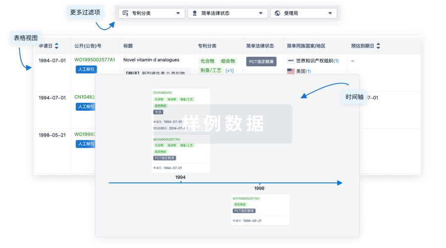
核心专利
使用我们的核心专利数据促进您的研究。
登录
或
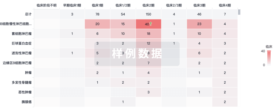
临床分析
紧跟全球注册中心的最新临床试验。
登录
或
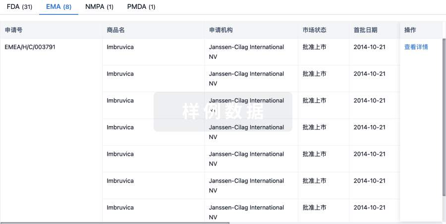
批准
利用最新的监管批准信息加速您的研究。
登录
或
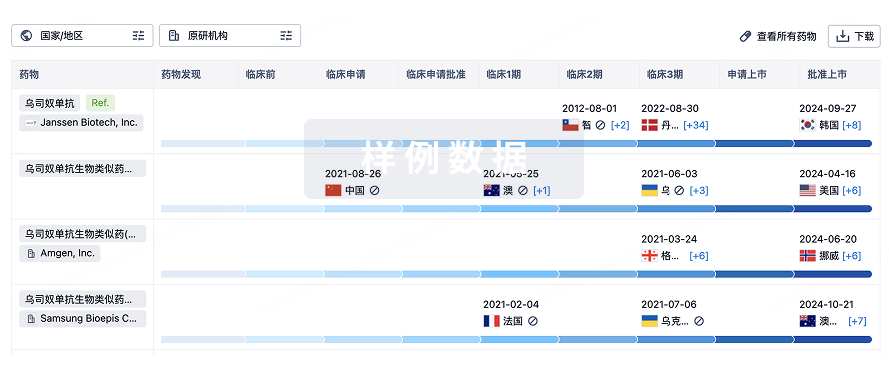
生物类似药
生物类似药在不同国家/地区的竞争态势。请注意临床1/2期并入临床2期,临床2/3期并入临床3期
登录
或
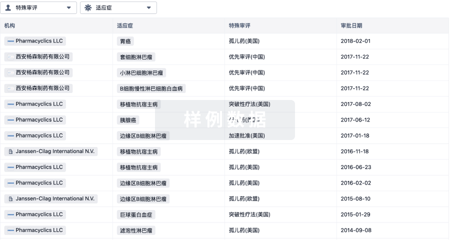
特殊审评
只需点击几下即可了解关键药物信息。
登录
或
提出任何生物医药研究问题
让每一步生物医药决策都精准制胜
基于十亿级生物数据与全球情报的精准 AI,赋能早期药物发现
立即开始免费试用!
智慧芽新药情报库是智慧芽专为生命科学人士构建的基于AI的创新药情报平台,助您全方位提升您的研发与决策效率。
立即接入生物医药数据包
智慧芽新药库数据也通过智慧芽开放平台,以MCP或API服务形式对外开放。立即使用我们的MCP服务定制您的LLM智能体。
生物序列数据库
生物药研发创新
免费使用
化学结构数据库
小分子化药研发创新
免费使用