预约演示
更新于:2026-06-27
T-01(Hangzhou Cheetah Cell Therapeutics)
更新于:2026-06-27
概要
基本信息
原研机构 |
非在研机构- |
权益机构- |
最高研发阶段临床阶段不明 |
首次获批日期- |
最高研发阶段(中国)临床阶段不明 |
特殊审评- |
结构/序列
Sequence Code 1332736687

关联
1
项与 T-01(Hangzhou Cheetah Cell Therapeutics) 相关的临床试验NCT05776355
NKG2D CAR-NK Cell Therapy for Patients With Platinum-Resistant Recurrent Ovarian Cancer
This trial will explore the maximum tolerated dose (MTD)of NKG2D CAR-NK cells in the treatment of platinum-resistant, relapsed epithelial ovarian cancer in a dose-escalation manner, and observe the clinical safety and efficacy.
开始日期2023-03-01 |
申办/合作机构 |
100 项与 T-01(Hangzhou Cheetah Cell Therapeutics) 相关的临床结果
登录后查看更多信息
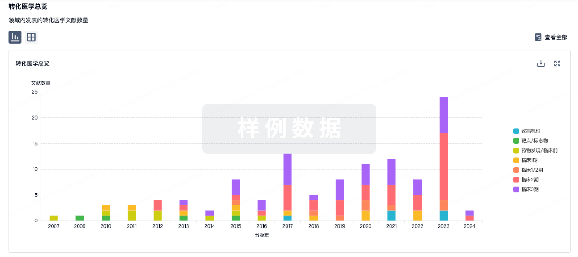
100 项与 T-01(Hangzhou Cheetah Cell Therapeutics) 相关的转化医学
登录后查看更多信息
100 项与 T-01(Hangzhou Cheetah Cell Therapeutics) 相关的专利(医药)
登录后查看更多信息
29
项与 T-01(Hangzhou Cheetah Cell Therapeutics) 相关的文献(医药)2025-08-01·BIOORGANIC CHEMISTRY
Tilmicosin derivatives as topoisomerase I/II inhibitors: Rational design, synthesis, and antibacterial evaluation
Article
作者: Wang, Qin ; Gu, Chang-Chun ; Jia, Zi-Han ; Xia, Ya-Mu ; Cao, Meng-Nan ; Gao, Wei-Wei ; Yu, Ri-Lei
The antibacterial mechanisms of macrolides primarily focus on ribosome inhibition, while their potential interactions with topoisomerases (Topo) remain to be thoroughly explored. In this study, novel tilmicosin (TIM) derivatives were designed and screened with strong binding affinities to Topo I/II by using molecular docking. Then, four TIM derivatives (T-1, T-2, T-16, and T-17) were selected as potential candidates for subsequent synthesis. In vitro antibacterial activities were evaluated, among them, T-1 and T-16 exhibited superior antibacterial effects against most bacteria, with a particularly notable MIC of 1 μg/mL against E. coli ATCC 8739. The inhibitory activities of T-1 (IC50 = 17 μM) and T-16 (IC50 = 15 μM) against Topo II were significantly higher than that of TIM. The inhibitory effects of T-1/T-16 may stem from optimized binding to the residue Asp or Met via hydrogen bonding. Gel electrophoresis analysis demonstrated that T-1 and T-16 effectively induce unwinding of supercoiled pUC19 plasmid, further confirming their interference with bacterial Topo II activity, thereby leading to disruption of DNA metabolism. The antibacterial mechanisms were also investigated by molecular dynamics (MD), which forms a closed-loop verification with the above-described inhibitory effect against Topo II and unwinding ability towards pUC19 plasmid. HPLC combined with electrochemical analysis indicated that T-1 and T-16 possessed better stability in acidic aqueous solutions and a longer metabolic half-life in mice compared to TIM. To further evaluate the in vivo anti-infective efficacy, different mice infection models were established. The results suggested that T-1 and T-16 not only exhibit excellent in vivo antibacterial activity and biocompatibility but also effectively treat infections.
2025-07-01·BIOORGANIC & MEDICINAL CHEMISTRY LETTERS
Development and evaluation of biphenyl-based small-molecule radiotracers for PET imaging of PD-L1 in tumor
Article
作者: Lin, Jianguo ; Zhu, Junyi ; Qiu, Ling ; Zhou, Yuxuan ; Hu, Xin ; Cai, Shuyue ; Wang, Qianhui ; Zhang, Nan ; Lv, Gaochao ; Xie, Quan
Accurate identification of programmed cell death ligand 1 (PD-L1) expression is crucial for anti-tumor immunotherapy. However, the heterogeneity of PD-L1 expression in tumors makes it challenging to detect by immunohistochemistry. In this study, we developed two novel PD-L1 small-molecule PET tracers, [18F]LGT-1 and [18F]LGT-2, to enable the non-invasive and precise measurement of PD-L1 expression in tumors through PET imaging. The radiochemical yields for [18F]LGT-1 and [18F]LGT-2 were 12.54±2.73% and 10.54±2.21%, respectively, with both tracers exhibiting approximately 98% radiochemical purity and molar activities of 12.23±2.84 GBq/μmol and 11.41±1.47 GBq/μmol. Both tracers demonstrated good stability in PBS (pH 7.4) and mouse serum after 2 hours of incubation. In cellular uptake assays, [18F]LGT-1 achieved a maximum uptake of 5.47±0.03 %AD at 4 hours, which could be significantly inhibited by the non-radioactive compound LGT-1. In contrast, [18F]LGT-2 exhibited high non-specific uptake in tumor cells. PET imaging revealed that [18F]LGT-1 quickly accumulated in tumors within 5 minutes, achieving an uptake of 1.48±0.15 %ID/mL, and maintained a stable level for 60 minutes, while [18F]LGT-2 showed minimal tumor uptake. Additionally, [18F]LGT-1 had significantly lower liver uptake compared to [18F]LGT-2. Despite the high uptake in non-target tissues for [18F]LGT-1, which complicates its application, this study provides new insights for developing novel PD-L1 small-molecule tracers, with further optimization of the tracers currently in progress.
2024-11-01·Heliyon
Production performance, serum lipid profile and gut health in Indian native Kadaknath chickens fed diet incorporated with liquorice root powder
Article
作者: Deo, Chandra ; Gowthaman, V. ; Tiwari, A.K. ; V, Gowthaman ; Sharma, Divya ; Biswas, Avishek ; A K, Tiwari
The principal constituent of liquorice root (Glycyrrhiza glabra) is glycyrrhizin, a triterpene saponin that is approximately many times sweeter than sucrose, the main active component. This study aimed to investigate the dietary liquorice root powder (LRP) on production performance, serum biochemical, gut health and carcass characteristics of Kadaknath (KN) birds as replacement of antibiotic growth promoter. Day-old Kadaknath chicks (n = 240) with uniform body weight were selected randomly and divided into six different treatments, each one with five replicates and eight birds per replicate, and raised in battery brooder cages for 15 weeks. Corn soya based basal diet (T1) was prepared. In addition to the basal diet, five experimental diets were created with varying amounts of LRP i.e., T2: T1+ 0.1 % LRP, T3: T1+ 0.3 % LRP, T4: T1+ 0.5 % LRP, T5: T1+ 0.7 % LRP, and T6: T1+ 0.0335 % Chlortetracycline (CTC). Body weight gain and feed intake significantly (P ≤ 0.05) increased in T3 group on 0-5 wks and 5-9 wks of age. Significant (P ≤ 0.01) reduction in the feed intake was noted in the T5 group which was fed with maximum level (0.7 %) of inclusion of LRP. Dietary inclusion of liquorice in higher doses resulted in a significant (P ≤ 0.05) decrease in serum lipids such as triglyceride, LDL, and total cholesterol concentrations and a significant increase in the HDL cholesterol. Decrease in the coliform count of caecum significantly (P ≤ 0.05), but dose-dependent lactobacilli proliferation was seen in the caecum of treated birds (P ≤ 0.01). Supplementation of liquorice root powder in kadaknath birds resulted in significant increase (P ≤ 0.05) in the villus length and VH: CD ratio. Thus it may be concluded that dietary supplementation of liquorice root powder improved the bird's growth performance, serum lipid profile and gut health of Kadaknath birds.
10
项与 T-01(Hangzhou Cheetah Cell Therapeutics) 相关的新闻(医药)2026-06-03
2026年6月3日,中国苏州,康宁杰瑞生物制药(股票代码:9966.HK)宣布,公司自主研发的EGFR/HER3双特异性抗体双载荷偶联药物(ADC)JSKN021治疗晚期恶性实体瘤的Ⅰ期临床研究(研究编号:JSKN021-101)已完成首例患者给药。JSKN021是康宁杰瑞第6款进入临床研究阶段的ADC,也是公司首款进入临床阶段的双载荷ADC。
表皮生长因子受体EGFR与HER3是细胞增殖信号通路的关键调控因子,在肺癌、头颈癌、乳腺癌、卵巢癌、食管癌、胃癌、结直肠癌等多种实体瘤中高表达,且与患者不良预后密切相关。HER3激酶活性有限,主要通过与EGFR、HER2形成异二聚体激活致癌信号;而EGFR基因变异也会引发信号通路异常活化,驱动肿瘤进展。流行病学研究进一步证实,HER3介导的信号激活是EGFR酪氨酸激酶抑制剂(TKI)耐药发生的关键诱因。基于EGFR/HER3双靶点设计的ADC药物,能够有效克服单靶点ADC的抗原异质性局限,已成为实体瘤治疗极具潜力的创新策略。
JSKN021是一种靶向EGFR/HER3的“2-in-1”双抗双载荷ADC药物。该药物采用糖基定点偶联技术将两种不同细胞毒性载荷定点、定量偶联至抗体的Fc区域,工艺过程简单、稳定、高效,在临床前研究中展现出康宁杰瑞自主技术平台特有的高稳定性与高安全性。在2025年美国癌症研究协会(AACR)年会上公布的临床前研究数据显示,JSKN021能有效抑制HER3阳性、EGFR阳性或双阳性肿瘤细胞的生长。此外,在多种CDX模型中,JSKN021的肿瘤抑制效果显著优于单载荷ADC。
JSKN021-101是一项在中国晚期恶性实体瘤受试者中开展的开放、多中心、Ⅰ期临床研究,分为剂量递增和剂量优化两个阶段,旨在评估JSKN021的安全性、耐受性、药代动力学(PK)及抗肿瘤活性,并确定最佳生物学剂量(OBD)和/或推荐Ⅱ期剂量(RP2D)。
关于JSKN021
JSKN021是一种全球首创的双抗双载荷抗体偶联药物(ADC),由靶向EGFR和HER3的双特异性抗体与新型拓扑异构酶Ⅰ抑制剂(T01)及微管蛋白抑制剂MMAE偶联而成。通过精确调控双抗结合亲和力设计,JSKN021在有效应对肿瘤异质性的同时最大限度降低靶向脱瘤毒性。该分子具有增强的稳定性和更高的均一性,通过结合T01(DAR为4)与MMAE(DAR为2)双重载荷机制,可有效克服单载荷治疗策略出现的无应答及耐药性问题。目前JSKN021正在中国开展用于治疗晚期恶性实体瘤的Ⅰ期临床研究。
康宁杰瑞(股票代码:9966.HK)是一家以创新驱动、聚焦肿瘤治疗领域的生物制药公司,依托自主研发的单域抗体、双特异性抗体、糖基定点偶联、连接子载荷、双载荷偶联及高浓度皮下制剂等核心技术平台,构建了具有差异化创新和国际竞争力的产品矩阵,覆盖抗体偶联药物(ADC)、双抗及单域抗体等前沿领域。
公司已有2款产品获批上市,恩沃利单抗注射液KN035(商品名:恩维达®,全球首个皮下注射PD-(L)1抑制剂),在肿瘤治疗的便捷性和可及性上做出了重大突破;安尼妥单抗注射液KN026(商品名:恩尼妥®,中国首个获批上市的国产自研HER2双抗药物),重塑HER2阳性胃癌二线治疗新标准;6款双抗ADC、双载荷ADC药物已进入临床阶段,并且正快速推进下一代ADC新药管线。目前公司已与石药集团、ArriVent、Glenmark等合作方达成多项针对产品或技术平台的战略合作。
“康达病患,瑞济万家”。康宁杰瑞致力于解决未满足的临床需求,开发高效、安全、具有全球竞争优势的抗肿瘤药物,让患者长期高质量生存,以“中国智造”的抗癌方案惠及全球患者。
欢迎访问公司网站:www.alphamabonc.com
前瞻性陈述
本新闻稿包含与我们未来业务、财务表现和涉及康宁杰瑞未来事件相关的声明,这些声明或构成前瞻性陈述。此类陈述包括预测和估计及关联的基本假设、有关潜在可能性的计划和期望的陈述,以及有关未来活动、运营及表现的陈述。这些陈述或可用诸如"预期"、"期待"、"预计"、"打算"、"计划"、"相信"、"寻求"、"估计"、"将"或类似含义的词语来标识。此类陈述基于康宁杰瑞管理和业务运营的当前某些假设,且受包括但不限于政治、经济、法律环境和商业环境等多种风险和不确定性的影响,康宁杰瑞的实际经营结果、表现或业绩可能较相关前瞻性陈述中明确或暗含的描述呈现(正面或负面)变化。除适用法律的要求外,无论出于新信息、未来事件还是其他原因,康宁杰瑞均无义务公开更新任何前瞻性陈述,且不为无法实现该等前瞻性陈述而承担责任。
医药信息声明
康宁杰瑞不推荐任何已获批或正在研发的药品/适应症临床使用,本文所包含的任何信息不应被看作是任何药物的申请、推广或广告。
2026-04-20
·商图药讯
4月16日,IGC2026 北京・第十一届免疫基因及细胞治疗大会在北京盛大启幕!六大高规格平行论坛重磅开启,百余项前沿议题深度研讨,汇聚 100 + 顶尖行业专家领衔开讲,聚焦体内细胞治疗、免疫细胞、干细胞与外泌体、基因治疗、mRNA 核酸药物及先进治疗药品质量研究与控制法规全赛道;紧跟818号令新政导向,深度解码双轨制监管与临床转化路径,覆盖 CGT 全产业链创新方向;3000 +行业精英齐聚一堂,70+优质展商同台亮相,全场座无虚席、热度拉满,共绘细胞与基因治疗产业高质量发展新蓝图。
本篇体内细胞治疗专场首日报道,带您直击第十一届IGC北京站首日不容错过的精彩内容!
现场精彩演讲内容
IGC2026 4月16日·北京
IGC
演讲话题:体内制备CAR-T细胞治疗复发/难治多发性骨髓瘤及更多
演讲嘉宾:胡豫,武汉协和医院前任院长,华中科技大学血液病学研究所所长
胡所长介绍体内 BCMA CAR‑T治疗复发难治多发性骨髓瘤的研究。先对比传统 CAR‑T 流程复杂、可及性差、髓外疗效不佳等局限,讲解病毒载体、LNP‑RNA两大体内制备技术平台。重点展示团队ESO‑T01临床前与 Ⅰ 期临床数据,其缓解率高、对髓外病灶有效,存在双相 CRS反应且可控。同时介绍体内 CAR‑T 在自身免疫病、实体瘤的拓展应用,指出当前递送、持久性等挑战,展望其在多疾病的应用前景。
演讲话题:体内CAR-T的技术路径选择:病毒or mRNA-LNP
演讲嘉宾:孙召朋,石药集团北京研究二院,疫苗研究所/细胞与基因治疗研究所所长
孙博士围绕体内 CAR‑T 技术路径选择,对比病毒与 mRNA‑LNP 路线。先讲行业背景,MNC 密集布局,国内研发同步。分析药学层面,慢病毒需改造包膜、质控难;LNP 靶向偶联与免疫原性是挑战。临床数据显示,慢病毒短期疗效佳,需验证长期效果;LNP 更适配自身免疫病。石药布局两款核心产品,SYS6055 为国内首个注册临床体内 CAR‑T。最后指出行业挑战,两条路线互补,应按适应症选择,未来聚焦递送优化、适应症拓展。
演讲话题:从载体骨架到工艺:体内 CAR-T 慢病毒载体的系统性设计与开发策略
演讲嘉宾:宣春玲,蓬勃生物慢病毒载体工艺开发部总监
宣总围绕体内 CAR-T 慢病毒载体(tLVV)展开,对比体外与体内 CAR-T 在生产、临床、成本及可及性的优势,介绍主流递送系统。重点讲解 tLVV 载体设计,包括靶向、融合、启动子等关键元件,推出 LentiBone III‑TCS 骨架解决 CAR 脱靶风险。通过 DOE 优化质粒配比与上下游工艺,将回收率大幅提升。同时解读国内外体内基因治疗法规,明确质量控制与生产要求,提供从载体设计到工艺开发再到合规申报的全流程解决方案。
演讲话题:In Vivo CAR-T 细胞疗法动物模型的选择与评价策略研究
演讲嘉宾:仇晓雷,上海南方模式生物科技有限公司,肿瘤管线负责人
仇总介绍体内 CAR‑T 细胞疗法的动物模型选择与评价。先讲解体内 CAR‑T 概念、病毒 / LNP 两大递送路线及国内外临床与 BD 进展。重点分享南模生物临床前评价体系,推荐Hu‑PBMC、Hu‑HSC 人源化免疫重建小鼠模型,可用于 CD19、BCMA、GPC3 等靶点及双靶点体内 CAR‑T 药效、安全性评价。展示多瘤种模型资源,为体内 CAR‑T 临床前研发提供标准化动物模型与评价方案支撑。
演讲话题:治疗肿瘤与自免的体内细胞治疗创新研发和临床开发
演讲嘉宾:张永克,驯鹿生物首席科学官兼高级副总裁
张总介绍了驯鹿公司的发展历程和技术平台,重点讨论了其基于慢病毒载体的CAR-T细胞治疗技术。张总解释了该技术相比传统方法的优势,包括更低的生产成本、不需要清零过程以及更好的患者olerability。张总还详细介绍了公司正在开发的CD20-CAR和CD19-CAR产品线,并分享了临床前研究数据,显示该技术仅对T细胞进行特异性转染,且在体外和动物模型中表现出良好的安全性和活性。
演讲话题:In vivo CAR T-cell therapy for autoimmune diseases自身免疫疾病的体内CAR T细胞疗法
演讲嘉宾:陈竹,中国科学技术大学附属第一医院(安徽省立医院),风湿免疫科行政主任,主任医师
陈主任围绕体内 CAR‑T 治疗自身免疫病的临床进展。先回顾 CAR‑T 发展及在难治性 SLE、RA 等疾病的传统体外治疗成果,指出体外制备复杂、成本高、需清淋等局限。重点分享团队全球首个体内 CAR‑T 治疗 SLE 临床研究,采用 LNP‑mRNA 递送 CD19 CAR,无需清淋、快速起效、安全性好,患者症状显著改善。同时阐述病毒与 LNP 两大技术路线挑战,展望靶向 IGHV4‑34 的精准治疗方向。
演讲话题:tpLNP递送体内CAR-T mRNA疾病治疗突破与创新
演讲嘉宾:郭磊,北京百替生物,创始人
郭总介绍百替生物tpLNP 免疫细胞靶向体内 CAR‑T 核酸药物递送系统。传统体外 CAR‑T 工艺复杂、毒性高、实体瘤效果差,体内 mRNA CAR‑T 更具优势。团队开发tpLNP 新型聚合物载体,解决传统 LNP 肝靶向、免疫原性等问题,实现脾脏精准富集,高效转染 T 细胞。经 AI 优化的 CAR mRNA 与 tpLNP 组合,在低剂量下展现更强肿瘤杀伤,安全性良好。管线覆盖淋巴瘤、自免、脏器纤维化、实体瘤,计划 2026Q4 启动临床,为体内 CAR‑T 提供高效递送方案。
演讲话题:体从体内CAR-T到新一代抗体偶联药物:CMC核心挑战的破局之路
演讲嘉宾:方子辉,宜诺生物创始人、CEO
方总分享宜诺生物从体内 CAR‑T 到新一代抗体偶联 LNP 的 CMC 破局方案。先介绍 ADC、AOC、Ab‑tLNP 市场趋势,指出体内 CAR‑T 已成热门赛道,LNP 路线安全性佳但肝靶向与偶联均一性是瓶颈。重点讲解抗体定点偶联 LNP 技术,优化工艺实现高偶联效率、低抗体残留,适配全类型抗体,具备 50L GMP 放大与 6 个月稳定性。建立纳米流式等完整质控体系,明确 IND 申报思路,提供从 mRNA、LNP 到偶联的全流程 CMC 解决方案,支撑体内 CAR‑T 与核酸药物高效转化。
演讲话题:tLNP 介导的体内CAR-T产品的质量研究
演讲嘉宾:陆航,嘉译生物医药,创始人兼首席执行官
陆总系统讲解 tLNP 介导体内 CAR‑T 产品的质量研究框架。先梳理自体 CAR‑T 上市产品与审评要点,明确体内 CAR‑T 属于基因治疗 / 细胞治疗双属性产品。对比慢病毒与 LNP 递送优劣,介绍 AI 优化 CAR 设计与 tLNP 平台化方案。重点解读国内外 ATMP、基因 / 细胞治疗相关法规与指导原则,建立mRNA 原液、偶联抗体、tLNP 制剂三级质量评价体系,涵盖理化、纯度、含量、活性、安全性等全项检测方法,明确生物学效力(Potency)评价标准与非临床 / CMC / 临床评价路径。
演讲话题:基于mRNA工程的体内CAR-X破局之道
演讲嘉宾:罗凤燕,瑞吉生物,研发总监
罗总介绍瑞吉生物基于mRNA 工程的体内 CAR‑X 技术突破。公司拥有 mRNA 全要素平台,布局 20 余项专利,获 CDE/FDA/TGA 认可。针对传统体外 CAR‑T 成本高、周期长、毒性大等痛点,开发LNP‑mRNA 体内递送系统,靶向 CD8+ T 细胞效率超 80%,可瞬时表达、无需清淋、可重复给药,安全性更高。同时搭建 AI 抗体设计平台,覆盖 CAR‑T、CAR‑NK、CAR‑M,布局血液瘤、自免、纤维化等多适应症。平台具备全球专利、临床验证与自免适配优势,提供一体化 mRNA 药物开发解决方案。
演讲话题:基于环形RNA的体内CAR T开发
演讲嘉宾:杨赟,环码生物董事长兼CTO
杨总介绍环码生物基于环形 RNA(circRNA)的体内 CAR‑T 开发。circRNA 比 mRNA 更稳定、免疫原性低、无需碱基修饰 / 加帽加尾,是下一代 RNA 平台。公司拥有 AI 序列设计、免疫靶向 LNP、GMP 生产三大核心平台,筛选出高脾脏靶向脂质,优化 circRNA‑CAR 序列。核心管线CC2401(CD19 靶点)用于自免疾病,临床前显示低剂量即可长效表达、深度清除 B 细胞,优于 mRNA‑LNP。产品现货、可重复给药、无预处理、非病毒整合,已完成 IIT,推进临床 Ⅰ 期,布局自免与肿瘤领域。
演讲话题:基于circRNA-电穿孔策略构建CAR-T疗法与肿瘤疫苗突破
演讲嘉宾:邱满堂,北京大学人民医院研究员、麒明生物创始人
邱总介绍基于环状 RNA(circRNA)的创新肿瘤治疗策略。circRNA 比 mRNA 更稳定、表达更持久,是下一代 RNA 技术。团队研发NeoAna 成环体系,实现低免疫原性、高纯度与规模化生产。开展四大应用:1)circRNA 编码DLL3 CAR‑T,高效杀伤小细胞肺癌;2)LNP 递送 circRNA 体内 CAR‑T;3)circRNA 肿瘤疫苗,靶向 NY‑ESO‑1 等靶点,联合 PD‑1 显著抑瘤;4)AI 设计个性化新抗原疫苗。相关成果已获多项专利与论文发表,推动 circRNA 在 CAR‑T 与肿瘤疫苗的临床转化。
圆桌讨论:in vivo CAR-T 病毒与非病毒递送技术下的靶向性、药学、工艺开发的难点与策略
1.无论是病毒载体还是LNP,体内转导的核心难点都在于特异性靶向T细胞。各位在实践中选择了哪些靶向策略?企业该如何根据靶点与适应症选择技术路线
2. 传统CAR-T可在回输前进行严格质控,但in vivo产品的“终产品”在患者体内生成。各位认为,应建立哪些新的质控指标?药学研究与监管标准如何同步?
3.病毒vs非病毒:两条路线的工艺难点有何不同?各位在实际开发中遇到过哪些具体工艺瓶颈?如何解决的
4. in vivo CAR-T处于早期阶段,但MNC已开始密集扫货,从投资和BD角度看,当前融资环境下投资机构的估值逻辑有哪些
主持人:竺添,引正基因CEO&联合创始人
郑玉芬,约印医疗基金,董事长
梁高峰,河南科技大学基础医学与法医学院教授、国家重点研发计划首席、河南省微纳技术与转化医学创新团队带头人
杨赟,环码生物董事长兼CTO
郭磊,北京百替生物,创始人
以上内容由组委会整理编辑而成,未全经本人审阅,
如有偏差或疏漏之处,请以实际为准。
现场盛况 精彩集锦
IGC2026 4月16日·北京
IGC
扫描上方二维码
查看更多现场精彩照片!
随着818号令、828号令相继落地,博鳌乐城、广州南沙等先行试点区也在如火如荼陆续批准临床应用,CGT行业的监管框架日趋清晰。
IGC品牌年会将在下半年 前沿创新药产业发展速度最快的珠江三角洲区域中打造精品细分技术论坛,聚焦体内及免疫细胞治疗、干细胞与类器官、基因治疗的创新研发,覆盖早研、CMC研究、以及临床开发热点。
期待您的建言献策,扫二维码完成问卷的填写,将有机会获取价值四千元含餐IGC 2026-广州站 大会入场门票!
下一届IGC 火热预定中!
期待下届再会!
扫描左侧二维码
咨询合作事宜
2026-03-26
2026年3月26日,中国苏州,康宁杰瑞生物制药(股票代码:9966.HK)公布了截至2025年12月31日的2025年度业绩和近期业务进展。
亮点概览
● KN026(安尼妥单抗)在二三线HER2阳性胃癌中取得PFS与OS双强阳性,该数据在2025年ESMO大会上以LBA口头报告形式重磅发布。首个NDA获NMPA受理并纳入优先审评审批,顺利通过药品注册核查及GMP符合性检查,上市在即。一线HER2阳性乳腺癌及新辅助治疗的两项Ⅲ期临床研究已完成入组,辅助治疗乳腺癌Ⅲ期临床已启动。
● JSKN003(普康安尼妥单抗)凭借“高效低毒”优势,于2025年获得CDE两项突破性疗法认定及FDA突破性疗法、孤儿药、快速通道三项国际认定;多项研究优异的疗效和安全性数据在ASCO和ESMO大会上发布;针对二线HER2阳性乳腺癌、HER2低表达乳腺癌、铂耐药卵巢癌、HER2阳性结直肠癌的四项Ⅲ期临床研究全面推进,其中二线HER2阳性乳腺癌Ⅲ期已完成入组。
● JSKN016Ⅰ、Ⅱ期研究累计入组超460例,完成剂量扩展和剂量选择,肺癌和乳腺癌在后线确认疗效后,进行了一线联合化疗/免疫的探索,三阴性乳腺癌Ⅲ期临床已启动。同时高浓度皮下制剂已在澳大利亚获批开展临床。
● JSKN033完成二线宫颈癌POC数据积累与剂量优化确认,二线子宫内膜癌POC队列入组中,同时推进肺癌Ⅱ期探索及启动联合化疗一线治疗宫颈癌Ⅱ期临床。通过ADC/IO组合,继续强化泛瘤种和全人群开发。
● 基于模块化、可迭代的技术平台,创新分子持续产出并高效推进临床。JSKN022已进入第6个爬坡剂量,显示初步疗效和良好的安全性;JSKN027获批Ⅰ期临床并完成首例患者给药;JSKN021临床前数据在AACR年会发布,IND申请已获CDE受理。
财务概览
● 2025年度实现营业收入566.24百万元人民币,其中归属于本公司的产品收入130.13百万元人民币。
● 2025年度研发开支572.16百万元人民币,同比增长41.57%。
● 2025年度年内亏损113.95百万元人民币。
● 财务状况安全稳健,截至2025年12月31日现金储备为1350.32百万元人民币。
业务摘要
一、产品管线
公司依托自主研发的单域抗体、双特异性抗体、糖基定点偶联、连接子载荷、双载荷偶联及高浓度皮下制剂等核心技术平台,构建了具有差异化创新和国际竞争力的产品矩阵,覆盖抗体偶联药物(ADC)、双抗及单域抗体等前沿领域:其中全球首个皮下注射PD-(L)1抑制剂恩沃利单抗注射液(研发代号:KN035,商品名:恩维达®)已获批上市,HER2双特异性抗体安尼妥单抗注射液(研发代号:KN026)针对二线及以上HER2阳性胃癌的新药上市申请(NDA)已获国家药品监督管理局(NMPA)受理,2个双抗ADC产品处于Ⅲ期临床研究阶段,另有多个双抗ADC和双载荷ADC正在快速推进临床研究。
KN035(恩沃利单抗,恩维达®)
一种创新抗肿瘤免疫治疗药物,是全球首个获批上市的皮下注射PD-(L)1抑制剂和肿瘤领域单域抗体。患者无需进行静脉滴注,可以在30秒内完成给药,在便利性和依从性方面具有显著优势,尤其适用于体弱、高龄及有静脉输注反应的患者。恩沃利单抗已获美国食品药品监督管理局(FDA)授予三项孤儿药资格(ODD),用于治疗晚期胆道癌、软组织肉瘤及胃癌和胃食管结合部癌;获NMPA授予突破性疗法认定,用于治疗高肿瘤突变载荷(TMB-H)不可切除或转移性实体瘤。
报告期内主要进展
● 2025年6月,恩沃利单抗3项单药或联合用药的Ⅱ期临床研究数据在2025年美国临床肿瘤学会(ASCO)年会上以壁报形式发布,同时另有8项研究数据在线发表。
● 2025年8月,恩沃利单抗治疗晚期肺癌的真实世界研究结果在Thoracic Cancer全文发表。
● 2025年10月,恩沃利单抗6项联合用药的Ⅱ期临床研究数据在2025年欧洲肿瘤内科学会(ESMO)大会上以壁报形式发布,优异疗效跨越胆道癌、宫颈癌、肺癌等多个实体瘤领域。
● 2025年12月,恩沃利单抗联合化疗一线治疗广泛期小细胞肺癌(ES-SCLC)的Ⅱ期临床研究结果在BMC Medicine全文发表。
● 2025年12月,恩沃利单抗获FDA授予孤儿药资格,用于治疗胃癌和胃食管结合部癌。
报告期后进展
● 2026年1月,恩沃利单抗联合吉西他滨和奥沙利铂(GEMOX)方案,用于一线治疗不可切除或转移性胆道癌的NDA获NMPA受理。
KN026(安尼妥单抗)
HER2异二聚体双抗,可同时结合HER2的两个非重叠表位,阻断HER2信号,能通过抗体诱导的受体聚集,增强ADCC和CDC效应,同时下调细胞表面HER2受体。KN026已获FDA授予孤儿药资格,用于治疗HER2阳性或HER2低表达胃癌;获NMPA授予突破性疗法认定,用于一线标准治疗失败的HER2阳性胃癌(包括胃食管结合部腺癌)。
报告期内主要进展
● 2025年1月,KN026联合多西他赛一线治疗HER2阳性复发或转移性乳腺癌的Ⅱ期临床研究结果在Cancer Communications全文发表。
● 2025年3月,KN026联合KN046治疗除乳腺癌之外的HER2阳性实体瘤的Ⅱ期研究结果在Signal Transduction and Targeted Therapy全文发表。
● 2025年4月,KN026联合化疗二线及以上治疗HER2阳性胃癌(包括胃食管结合部癌)的Ⅱ/Ⅲ期临床研究完成首次期中分析,达到无进展生存期(PFS)和总生存期(OS)的主要终点。是首个在二线胃癌适应症中获得阳性结果的HER2双抗药物。
● 2025年4月,KN026联合白蛋白结合型多西他赛HB1801一线治疗HER2阳性复发转移性乳腺癌的Ⅲ期临床研究完成全部患者入组。
● 2025年6月,KN026联合KN046治疗HER2阳性乳腺癌的Ⅱ期研究结果在Clinical Cancer Research全文发表。
● 2025年8月,KN026联合白蛋白结合型多西他赛HB1801用于HER2阳性早期或局部晚期乳腺癌的新辅助治疗Ⅲ期临床研究完成全部患者入组。
● 2025年9月,KN026联合化疗用于至少接受过一种系统性治疗失败的HER2阳性局部晚期、复发或转移性胃癌/胃食管结合部腺癌的NDA获NMPA受理,并已获优先审评审批资格。
● 2025年10月,KN026 Ⅲ期临床研究期中分析结果以LBA口头报告形式在2025年ESMO大会上发布。KN026在曲妥珠单抗经治的二三线HER2阳性胃癌/胃食管结合部癌中取得PFS和OS双强阳性,有望改变二线及以上胃癌治疗的临床指南。
● 2025年11月,KN026针对HER2阳性胃癌/胃食管结合部腺癌的Ⅱ期临床研究结果在Cancer Communications全文发表。
● 2025年12月,KN026顺利通过药品注册核查(药学)及GMP符合性检查。
● KN026联合化疗(联合或不联合恩朗苏拜单抗)一线治疗HER2阳性局部晚期或转移性胃癌/胃食管结合部腺癌的Ⅱ期临床研究已启动,目前正在顺利进行中。
报告期后进展
● 2026年1月,KN026针对HER2阳性胃癌/胃食管结合部腺癌患者的Ⅲ期临床研究结果发表于肿瘤学领域顶级期刊Annals of Oncology。
● 2026年3月,KN026联合白蛋白结合型多西他赛HB1801和化疗辅助治疗HER2阳性乳腺癌的Ⅲ期临床研究完成首例患者给药。
2026年预期里程碑(2026年Q2及以后)
● KN026二线及以上治疗HER2阳性胃癌/胃食管结合部癌适应症在国内获批上市销售。
● KN026新辅助治疗HER2阳性乳腺癌的Ⅲ期临床研究数据发布并申报上市。
● KN026一线治疗HER2阳性乳腺癌的Ⅲ期临床研究数据发布并申报上市。
JSKN003(普康安尼妥单抗)
通过将KN026 Fc上糖基定点偶联获得DAR4的均一稳定的ADC。JSKN003能够结合肿瘤细胞HER2的两个表位,通过增强细胞内吞释放拓扑异构酶Ⅰ抑制剂,发挥肿瘤杀伤作用。较同类ADC药物具有更好的血清稳定性、更低的血液学毒性、更强的肿瘤抑制和旁观者杀伤效应,显著扩大了治疗窗。JSKN003已获FDA授予快速通道资格认定,用于治疗晚期或转移性铂耐药复发性上皮性卵巢癌、原发性腹膜癌或输卵管癌(PROC)(全人群);已获FDA授予突破性疗法认定,用于治疗贝伐珠单抗经治的HER2有表达PROC;已获NMPA授予两项突破性疗法认定,用于治疗PROC,及奥沙利铂、氟尿嘧啶和伊立替康治疗失败的HER2阳性晚期结直肠癌。此外,JSKN003已获FDA授予孤儿药资格,用于治疗胃癌及胃食管结合部癌。
报告期内主要进展
● 2025年2月,获CDE批准开展JSKN003对比恩美曲妥珠单抗(T-DM1)二线及以上治疗HER2阳性晚期乳腺癌的Ⅲ期临床研究,并于当月完成首例患者给药。
● 2025年2月,JSKN003对比研究者选择化疗治疗PROC的Ⅲ期临床研究完成首例患者给药,目前正在顺利进行中。
● 2025年3月,JSKN003获CDE突破性疗法认定,用于治疗PROC,且不限HER2表达水平。
● 2025年6月,JSKN003在澳大利亚Ⅰ期临床研究和中国Ⅰ/Ⅱ期临床研究中治疗非原发性铂难治的铂耐药卵巢癌、多线治疗后进展的HER2阳性乳腺癌以及晚期HER2高表达(IHC 3+)胃肠道肿瘤的疗效与安全性的三项汇总分析结果在2025年ASCO年会上发布。
● 2025年6月,JSKN003的一项临床前研究结果在RSC Chemical Biology全文发表。
● 2025年7月,JSKN003获FDA授予孤儿药资格,用于治疗胃癌及胃食管结合部癌。
● 2025年7月,JSKN003获FDA批准在美国开展一项治疗PROC的Ⅱ期临床研究,且不限HER2表达水平。
● 2025年9月,JSKN003对比恩美曲妥珠单抗(T-DM1)治疗HER2阳性晚期乳腺癌的Ⅲ期临床研究完成全部患者入组。
● 2025年10月,JSKN003获CDE突破性疗法认定,用于治疗既往经奥沙利铂、氟尿嘧啶和伊立替康治疗失败的HER2阳性晚期结直肠癌。
● 2025年10月,JSKN003治疗原发性铂难治卵巢癌、HER2阳性转移性结直肠癌的2项临床研究结果及1项对比研究者选择化疗治疗铂耐药卵巢癌的Ⅲ期临床研究设计在2025年ESMO大会上发布。JSKN003优异的疗效和安全性数据展现了“高效低毒”的差异化优势,和“不限癌种、不限HER2表达”的广谱特性,预示着其广阔的临床应用前景。
● 2025年10月,JSKN003获CDE批准开展对比研究者选择的方案(瑞戈非尼/呋喹替尼/曲氟尿苷替匹嘧啶)治疗经奥沙利铂、氟尿嘧啶和伊立替康治疗失败的HER2阳性晚期结直肠癌的Ⅲ期临床研究,并于2026年2月完成首例患者给药。
● 2025年10月,JSKN003获FDA快速通道资格认定,用于治疗不限HER2表达水平的PROC。
● 2025年12月,JSKN003获FDA突破性疗法认定,用于治疗贝伐珠单抗经治的晚期或转移性HER2有表达PROC。
● JSKN003治疗不可切除局部晚期或转移性HER2低表达乳腺癌的Ⅲ期临床研究顺利进行中。
● JSKN003联合KN026、免疫(IO)及化疗一线和围手术期治疗HER2阳性胃癌/胃食管结合部癌的Ⅱ期临床研究正在顺利进行中。
2026年预期里程碑(2026年Q2及以后)
● JSKN003二线及以上治疗HER2阳性乳腺癌的Ⅲ期临床研究数据读出并提交pre-BLA。
● JSKN003治疗PROC的Ⅲ期临床研究完成全部患者入组。
● JSKN003后线治疗HER2低表达乳腺癌的Ⅲ期临床研究完成全部患者入组。
JSKN016
利用单域抗体和双抗平台开发的靶向TROP2和HER3双抗,通过糖基定点偶联获得DAR4的均一稳定的ADC。JSKN016与肿瘤细胞表面的受体结合后,阻断相应信号通路,通过增强细胞内吞释放拓扑异构酶Ⅰ抑制剂,发挥肿瘤杀伤作用。
报告期内主要进展
● JSKN016Ⅰ、Ⅱ期临床研究累计入组超460例,完成了剂量扩展和剂量选择。
● JSKN016单药后线治疗HR阳性乳腺癌的队列扩展已完成入组。
● JSKN016联合卡培他滨治疗CDK4/6经治未经化疗的HR阳性乳腺癌Ⅱ期临床研究正在顺利进行中。
● 2025年12月,获CDE批准开展联合口服SERD治疗CDK4/6经治HR阳性、HER2阴性乳腺癌的Ⅱ期临床研究,并于2026年2月完成首例患者给药。
● JSKN016单药后线治疗驱动基因阳性(EGFR突变/少见突变)非小细胞肺癌的Ⅱ期临床研究已完成疗效确认。
● JSKN016联合伏美替尼一线及二线治疗EGFR突变非小细胞肺癌的Ⅱ期临床研究顺利进行中。
● JSKN016联合依沃西单抗和卡铂一线治疗驱动基因阴性非小细胞肺癌的Ⅱ期临床研究顺利进行中。
报告期后进展
● 2026年3月,JSKN016对比研究者选择方案治疗经至少二线系统性治疗失败的不可手术切除的局部晚期、复发或转移性三阴性乳腺癌的Ⅲ期临床研究完成首例患者给药。
● 2026年3月,获澳大利亚Bellberry临床研究伦理委员会同意开展JSKN016高浓度皮下制剂治疗晚期实体瘤的Ⅰ期临床研究。
2026年预期里程碑(2026年Q2及以后)
● JSKN016联合用药治疗CDK4/6经治HR阳性乳腺癌临床研究完成美国Pre-IND沟通。
● JSKN016皮下制剂澳洲Ⅰ期临床研究完成首例患者给药。
● JSKN016单药治疗三阴性乳腺癌与HR阳性乳腺癌数据发布。
● JSKN016非小细胞肺癌单药和联合用药临床数据发布。
● JSNK016联合用药一线治疗非小细胞肺癌Ⅲ期临床研究启动。
JSKN033
一种ADC(JSKN003)和PD-L1(恩沃利单抗) 组成的高浓度皮下注射复方制剂,结合免疫治疗和ADC的优势,并通过皮下给药进一步提升安全性和可及性。
报告期内主要进展
● JSKN033 Ⅰ、Ⅱ期临床研究累计入组约130例。
● 2025年12月,联合铂类化疗(联合或不联合贝伐珠单抗)用于一线治疗晚期宫颈癌的Ⅱ期临床研究申请获CDE受理,并于2026年3月正式获批。
● JSKN033单药二线及以上治疗宫颈癌的两个剂量组已完成入组,完成POC数据的积累以及剂量优化确认。
● JSKN033单药二线及以上治疗子宫内膜癌的POC队列入组中。
● JSKN033治疗HER2突变/有表达的非小细胞肺癌Ⅱ期临床研究顺利进行中。
2026年预期里程碑(2026年Q2及以后)
● JSKN033联合用药一线治疗宫颈癌Ⅱ期临床研究启动。
● JSKN033启动后线治疗宫颈癌(全人群)注册临床研究。
JSKN022
利用抗体定向进化,获得针对PD-L1和整合素αvβ6的双功能单域抗体,通过糖基定点偶联获得DAR4的均一稳定的ADC。JSKN022与肿瘤细胞表面的PD-L1和/或整合素αvβ6结合后,通过内吞,释放拓扑异构酶I抑制剂,杀伤肿瘤细胞。JSKN022阻断PD-L1和PD-1,激活肿瘤免疫反应。同时阻断αvβ6整合素,抑制TGFB1/3的生成,改变肿瘤免疫微环境。
报告期内主要进展
● 2025年4月,JSKN022临床前研究数据在2025年美国癌症研究协会(AACR)年会上发布。数据显示,JSKN022在体外和体内模型中,对整合素αvβ6和/或PD-L1表达阳性的肿瘤细胞具有显著的抗肿瘤活性。
● 2025年10月,获CDE批准开展治疗晚期恶性实体瘤的Ⅰ期临床研究,并于当月完成首例患者给药。目前已进入第6个爬坡剂量,显示初步疗效和良好的安全性。
2026年预期里程碑(2026年Q2及以后)
● JSKN022完成剂量递增和剂量扩展。
JSKN027
一种全球首创的可同时靶向PD-L1和VEGFR2的双抗ADC,通过糖基定点偶联技术将可裂解连接子及拓扑异构酶Ⅰ抑制剂载荷精准偶联至抗体的Fc区域,在保持良好安全性的同时实现有效的抗肿瘤活性。JSKN027的抗肿瘤作用基于其独特的三重协同机制:除具备ADC典型的靶向杀伤及旁观者效应外,还可以通过阻断VEGF/VEGFR2信号通路抑制肿瘤血管生成,并通过阻断PD-1/PD-L1免疫检查点信号通路解除免疫抑制。以上综合机制有助于增强抗肿瘤治疗的整体疗效,并克服前线治疗耐药。
报告期后进展
● 2026年3月,获CDE批准开展治疗晚期恶性实体瘤的Ⅰ期临床研究,并于当月完成首例患者给药。
2026年预期里程碑(2026年Q2及以后)
● JSKN027完成剂量递增和剂量扩展。
JSKN021
一种全球首创的双抗双载荷ADC,由靶向EGFR和HER3的双特异性抗体与新型拓扑异构酶Ⅰ抑制剂(T01)及微管蛋白抑制剂MMAE偶联而成。通过精确调控双抗结合亲和力设计,JSKN021在有效应对肿瘤异质性的同时最大限度降低靶向脱瘤毒性。该分子具有增强的稳定性和更高的均一性,通过结合T01(DAR为4)与MMAE(DAR为2)双重载荷机制,可有效克服单载荷治疗策略出现的无应答及耐药性问题。
报告期内主要进展
● 2025年4月,JSKN021临床前研究数据在2025年AACR年会上发布。数据显示,JSKN021能有效抑制HER3阳性、EGFR阳性或双阳性肿瘤细胞的生长,在多种CDX模型中的肿瘤抑制效果显著优于单载荷ADC。
报告期后进展
● 2026年3月,JSKN021用于治疗晚期恶性实体瘤的Ⅰ期临床研究申请获CDE受理。
2026年预期里程碑(2026年Q2及以后)
● JSKN021完成剂量递增。
KN046
一种双特异性(BsAb)免疫检查点抑制剂,由 CTLA-4与PD-L1单域抗体融合组成,可靶向富集于PD-L1表达的肿瘤微环境。
报告期内主要进展
● 2025年2月,KN046联合仑伐替尼治疗晚期不可切除或转移性肝细胞癌的Ⅱ期临床研究结果在Nature Communications全文发表。
二、生产基地
公司产业化基地按照NMPA、FDA及欧洲药品管理局(EMA)的GMP标准建设,现有产能满足临床至商业化阶段的规模化生产需求。新建的ADC原液和制剂车间已正式投产,进一步夯实了ADC平台的产业化基础。2025年顺利通过津巴布韦MCAZ GMP符合性检查;2026年初相继通过沙特SFDA cGMP、澳大利亚TGA GMP符合性检查,质量体系持续获得国际权威监管机构认可。
三、其他摘要
● 2025年9月,公司在2025中国医药决策者峰会(2025 CHDC)与中国创新药十年成就巡礼活动中登“中国创新药十年荣耀”榜单,获评“行业引领Biotech公司”。
● 2025年11月,公司在第十七届中国医药企业家科学家投资家大会(2025启思会)上第七次获授予“中国医药创新企业100强”。
● 2025年12月,公司在格隆汇“年度卓越公司评选”中获“年度创新力奖”。
有关上述内容的更多信息,请参阅公司在香港联交所及公司官网上发布的2025年度业绩公告。
关于康宁杰瑞
康宁杰瑞(股票代码:9966.HK)是一家以创新驱动、聚焦肿瘤治疗领域的生物制药公司,依托自主研发的单域抗体、双特异性抗体、糖基定点偶联、连接子载荷、双载荷偶联及高浓度皮下制剂等核心技术平台,构建了具有差异化创新和国际竞争力的产品矩阵,覆盖抗体偶联药物(ADC)、双抗及单域抗体等前沿领域。
公司已有1款产品获批上市,恩沃利单抗注射液KN035(商品名:恩维达®,全球首个皮下注射PD-(L)1抑制剂),在肿瘤治疗的便捷性和可及性上做出了重大突破;HER2双抗KN026(安尼妥单抗注射液)针对二线及以上HER2阳性胃癌的上市申请已获国家药品监督管理局(NMPA)受理;5款双抗ADC药物已进入临床阶段,并且正快速推进以双载荷ADC为代表的下一代ADC新药管线。目前公司已与石药集团、ArriVent、Glenmark等合作方达成多项针对产品或技术平台的战略合作。
“康达病患,瑞济万家”。康宁杰瑞致力于解决未满足的临床需求,开发高效、安全、具有全球竞争优势的抗肿瘤药物,让患者长期高质量生存,以“中国智造”的抗癌方案惠及全球患者。
康宁杰瑞前瞻性陈述
本新闻稿包含与我们未来业务、财务表现和涉及康宁杰瑞未来事件相关的声明,这些声明或构成前瞻性陈述。此类陈述包括预测和估计及关联的基本假设、有关潜在可能性的计划和期望的陈述,以及有关未来活动、运营及表现的陈述。这些陈述或可用诸如"预期"、"期待"、"预计"、"打算"、"计划"、"相信"、"寻求"、"估计"、"将"或类似含义的词语来标识。此类陈述基于康宁杰瑞管理和业务运营的当前某些假设,且受包括但不限于政治、经济、法律环境和商业环境等多种风险和不确定性的影响,康宁杰瑞的实际经营结果、表现或业绩可能较相关前瞻性陈述中明确或暗含的描述呈现(正面或负面)变化。除适用法律的要求外,无论出于新信息、未来事件还是其他原因,康宁杰瑞均无义务公开更新任何前瞻性陈述,且不为无法实现该等前瞻性陈述而承担责任。
医药信息声明
康宁杰瑞不推荐任何已获批或正在研发的药品/适应症临床使用,本文所包含的任何信息不应被看作是任何药物的申请、推广或广告。
优先审批AACR会议临床3期申请上市孤儿药
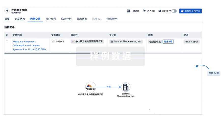
100 项与 T-01(Hangzhou Cheetah Cell Therapeutics) 相关的药物交易
登录后查看更多信息
研发状态
10 条进展最快的记录, 后查看更多信息
登录
| 适应症 | 最高研发状态 | 国家/地区 | 公司 | 日期 |
|---|---|---|---|---|
| 卵巢癌 | 临床阶段不明 | 中国 | 2023-03-01 | |
| 铂耐药性卵巢癌 | 临床阶段不明 | 中国 | 2023-03-01 |
登录后查看更多信息
临床结果
临床结果
适应症
分期
评价
查看全部结果
| 研究 | 分期 | 人群特征 | 评价人数 | 分组 | 结果 | 评价 | 发布日期 |
|---|
No Data | |||||||
登录后查看更多信息
转化医学
使用我们的转化医学数据加速您的研究。
登录
或
药物交易
使用我们的药物交易数据加速您的研究。
登录
或
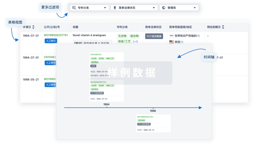
核心专利
使用我们的核心专利数据促进您的研究。
登录
或
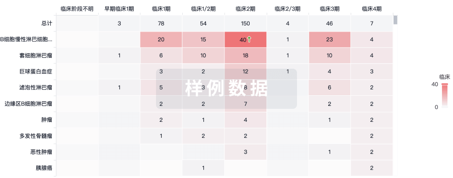
临床分析
紧跟全球注册中心的最新临床试验。
登录
或
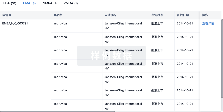
批准
利用最新的监管批准信息加速您的研究。
登录
或
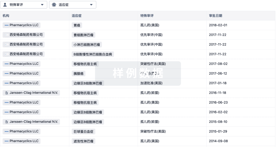
特殊审评
只需点击几下即可了解关键药物信息。
登录
或
生物医药百科问答
全新生物医药AI Agent 覆盖科研全链路,让突破性发现快人一步
立即开始免费试用!
智慧芽新药情报库是智慧芽专为生命科学人士构建的基于AI的创新药情报平台,助您全方位提升您的研发与决策效率。
立即开始数据试用!
智慧芽新药库数据也通过智慧芽数据服务平台,以API或者数据包形式对外开放,助您更加充分利用智慧芽新药情报信息。
生物序列数据库
生物药研发创新
免费使用
化学结构数据库
小分子化药研发创新
免费使用