预约演示
更新于:2025-10-30
MSC-Exos
更新于:2025-10-30
概要
基本信息
非在研机构- |
权益机构- |
最高研发阶段临床前 |
首次获批日期- |
最高研发阶段(中国)- |
特殊审评- |

关联
2
项与 MSC-Exos 相关的临床试验ChiCTR2500103555
Multi-center clinical trial study on the safety and effectiveness of stem cell-derived extracellular vesicles for wound repair and skin appendage regeneration

ChiCTR2100042948
the safety and the efficacy study of allogenic adipose MSC-Exos in the treatment of anti-MDA5 positive dermatonyositis- associated interstitial lung disease
100 项与 MSC-Exos 相关的临床结果
登录后查看更多信息
100 项与 MSC-Exos 相关的转化医学
登录后查看更多信息
100 项与 MSC-Exos 相关的专利(医药)
登录后查看更多信息
104
项与 MSC-Exos 相关的文献(医药)2026-02-01TISSUE & CELL
MSC-derived exosomes promote chondrocyte proliferation and inhibit apoptosis by suppressing inflammation in osteoarthritis
Article
作者: Cheng, Wendan ; Ji, Chaochao
BACKGROUND:
Osteoarthritis (OA) is marked by reduced chondrocyte proliferation, increased apoptosis, and an inflammatory microenvironment. Mesenchymal stem cell-derived exosomes (MSC-Exos) are promising modulators of chondrocyte function; however, their precise roles remain unclear. This study explores whether MSC-Exos protect chondrocytes from IL-1β-induced injury via these effects, in vitro and in a rat OA model.
METHODS:
BMSC-derived exosomes (BMSC-Exos) were isolated via ultracentrifugation and characterized (TEM, NTA, Western blotting). Chondrocyte injury was induced by IL-1β (10 ng/mL). Effects of BMSC-Exos on proliferation (CCK-8), apoptosis (Annexin V/PI), migration (wound healing/Transwell), inflammatory cytokines (ELISA), and NF-κB p65 phosphorylation (Western blotting) were evaluated. In vivo, OA was induced by intra-articular sodium iodoacetate in rats, and MSC-Exos were administered weekly. Histological staining (H&E and Safranin O-Fast Green) was performed to assess cartilage repair, immunohistochemical (IHC) analysis for IL-1β, TNF-α, IL-6, type II collagen and aggrecan was conducted, and analysis of p65 phosphorylation in cartilage tissue was conducted.
RESULTS:
BMSC-Exos significantly increased chondrocyte proliferation, reduced apoptosis, suppressed TNF-α, IL-6, and IL-1β expression, and inhibited p65 phosphorylation in vitro. In vivo, BMSC-Exos attenuated cartilage erosion, subchondral bone exposure, synovial hyperplasia and inflammatory cell infiltration. IHC analysis demonstrated that BMSC-Exos treatment significantly reduced the expression of IL-1β, TNF-α, IL-6, while restoring the levels of cartilage matrix components in cartilage compared to the OA model group. Reduced phospho-p65 levels correlated with these histological and molecular improvements.
CONCLUSION:
MSC-Exos protect chondrocytes from inflammatory injury via promoting proliferation, inhibiting apoptosis, reducing inflammation, and suppressing NF-κB, with therapeutic potential for OA.
2025-11-01CORNEA
Therapeutic Efficacy and Mechanisms of Mesenchymal Stem Cells and Their Exosomes in the Treatment of Ocular Graft-Versus-Host Disease
Review
作者: Ye, Chenxi ; Hong, Jing ; Liu, Shuwan
Purpose::
To provide a comprehensive overview of the clinical manifestations, pathophysiology, and current treatments of ocular graft-versus-host disease (oGVHD), and to explore the therapeutic potential of mesenchymal stem cells (MSCs) and their exosomes (MSC-Exos) as novel treatments.
Methods::
A literature review was conducted using major databases (PubMed, Web of Science, Scopus) to identify studies published between 2000 and 2024. The focus was on clinical trials, case series, and experimental studies related to oGVHD. Data were synthesized to summarize current knowledge.
Results::
oGVHD affects 40% to 60% of Hematopoietic Stem Cell Transplantation recipients, causing symptoms such as dryness, irritation, and vision impairment. Current treatments often fail to provide long-term relief and may cause significant side effects. MSCs and MSC-Exos offer a promising alternative because of their immunosuppressive and regenerative properties. Preclinical and clinical studies have demonstrated their safety and efficacy in improving oGVHD symptoms and promoting corneal health.
Conclusions::
MSCs and MSC-Exos show promise in treating oGVHD, with potential to improve symptoms and promote tissue repair. Further research is needed to establish their long-term safety and efficacy.
2025-10-01Drug Delivery and Translational Research
Mouse mesenchymal stem cell-derived exosomal miR-205-5p modulates LPS-induced macrophage polarization and alleviates lung injury by regulating the USP7/FOXM1 axis
Article
作者: Li, Bo ; Feng, Yinglu ; Yao, Shanglong ; Li, Haopeng ; Tang, Min
Exosomal microRNAs produced from mesenchymal stem cells (MSCs) are crucial in the management of acute lung injury (ALI). In this work, mMSCs separated from bone marrow were used to extract exosomes (MSC-Exos). MSC-Exos treatment attenuated pathological changes and scores, and edema in ALI mice. Also, MSC-Exos administration modulated the concentrations of inflammatory factors as well as the macrophage polarization both in vivo and in vitro. Upregulation of miR-205-5p in MSC-Exos regulated the macrophage polarization and the contents of inflammatory factors in animal and cell models. MiR-205-5p targeted USP7, and negatively modulated the expression of USP7. USP7 interacted with FOXM1, and reduced the ubiquitination degradation of FOXM1. MSC-derived exosomal miR-205-5p modulated ubiquitination of FOXM1 by targeting USP7. The ameliorative effect of MSC-Exos on the macrophage polarization and the inflammatory factors release was reversed with the overexpression of USP7 in animal and cell models. Collectively, MSC-derived exosomal miR-205-5p regulated lipopolysaccharide (LPS)-induced macrophage polarization and alleviated lung injury by the USP7/FOXM1 axis, which developed a potential target for the treatment of ALI.
100 项与 MSC-Exos 相关的药物交易
登录后查看更多信息
研发状态
10 条进展最快的记录, 后查看更多信息
登录
| 适应症 | 最高研发状态 | 国家/地区 | 公司 | 日期 |
|---|---|---|---|---|
| 系统性硬皮病 | 临床前 | 意大利 | 2025-07-03 |
登录后查看更多信息
临床结果
临床结果
适应症
分期
评价
查看全部结果
登录后查看更多信息
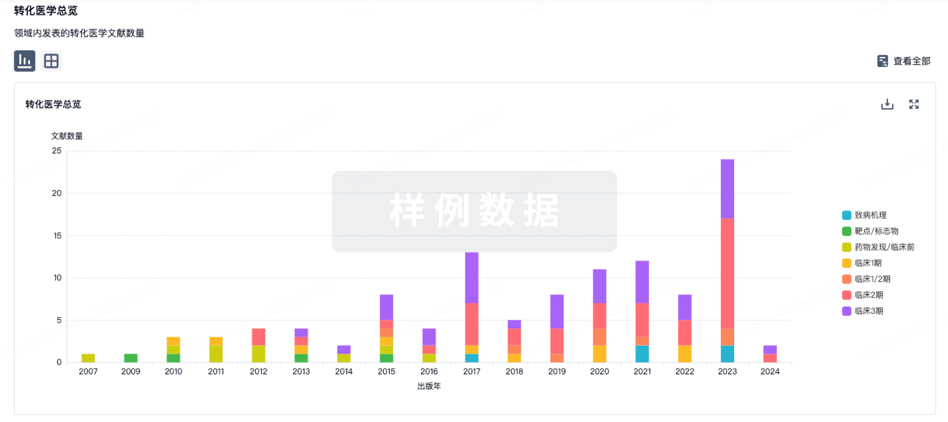
转化医学
使用我们的转化医学数据加速您的研究。
登录
或
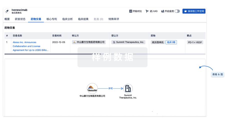
药物交易
使用我们的药物交易数据加速您的研究。
登录
或
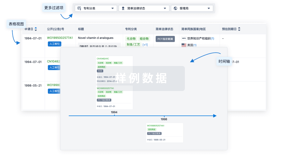
核心专利
使用我们的核心专利数据促进您的研究。
登录
或
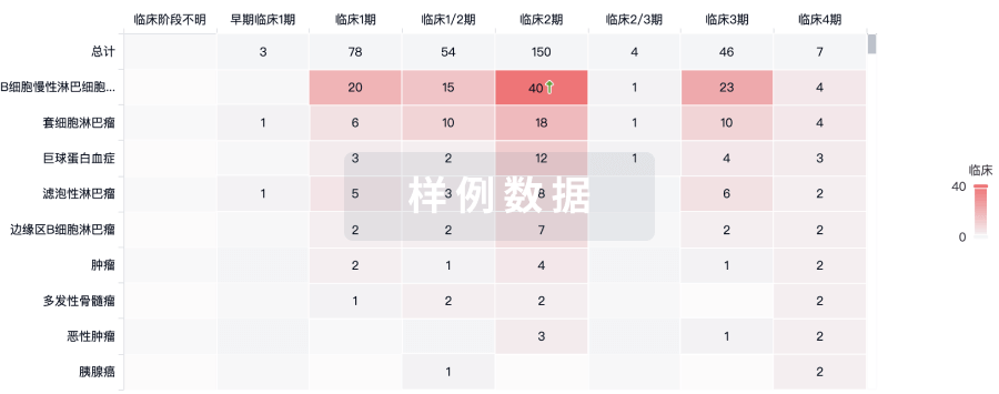
临床分析
紧跟全球注册中心的最新临床试验。
登录
或
批准
利用最新的监管批准信息加速您的研究。
登录
或
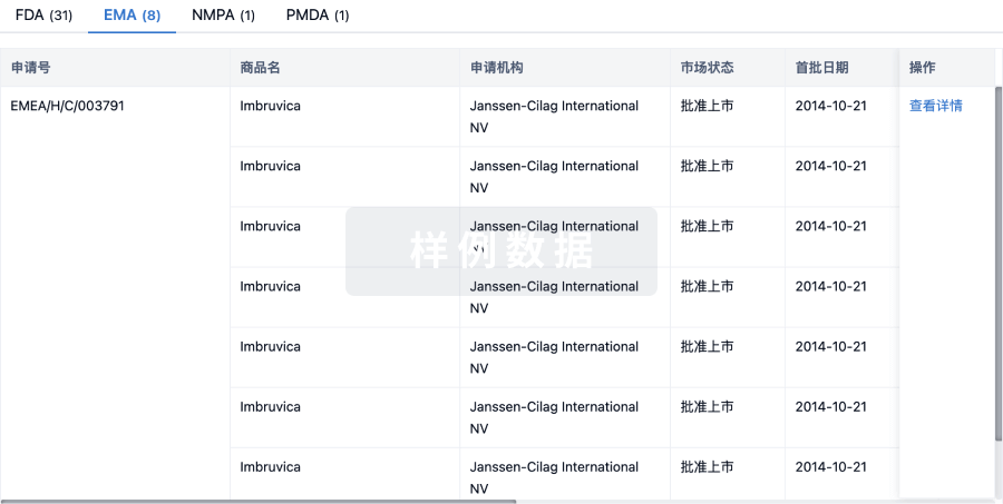
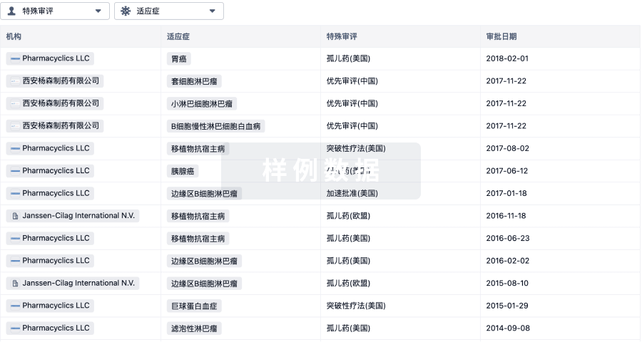
特殊审评
只需点击几下即可了解关键药物信息。
登录
或
提出任何生物医药研究问题
让每一步生物医药决策都精准制胜
基于十亿级生物数据与全球情报的精准 AI,赋能早期药物发现
立即开始免费试用!
智慧芽新药情报库是智慧芽专为生命科学人士构建的基于AI的创新药情报平台,助您全方位提升您的研发与决策效率。
立即接入生物医药数据包
智慧芽新药库数据也通过智慧芽开放平台,以MCP或API服务形式对外开放。立即使用我们的MCP服务定制您的LLM智能体。
生物序列数据库
生物药研发创新
免费使用
化学结构数据库
小分子化药研发创新
免费使用