预约演示
更新于:2025-05-07
Swiss Institute of Allergy & Asthma Research
私营公司|Switzerland
私营公司|Switzerland
更新于:2025-05-07
概览
关联
3
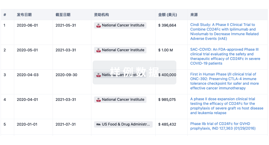
项与 Swiss Institute of Allergy & Asthma Research 相关的临床试验NCT06269315
Assessment of Skin Barrier Disruption Due to Daily Use of Cleaning Products and Hand Disinfectants
The goal of this clinical trial is to assess the effects of household detergents and hand disinfectants on human skin barrier integrity among healthy adult volunteers.
The main questions it aims to answer are:
How does exposure to household detergents and hand disinfectants impact skin barrier function?
Are there differences in skin barrier disruption between various types of cleaning agents and disinfectants?
Participants will:
Undergo patch testing with controlled amounts of household detergents and hand disinfectants on a small area of their skin.
Be monitored for adverse reactions during and after exposure. Have their skin barrier integrity assessed using electrical impedance spectroscopy at multiple time points post-exposure.
If there is a comparison group:
Researchers will compare the effects of different types of cleaning agents, hand disinfectants, and their ingredients on skin barrier integrity to determine variations in their impact.
开始日期2024-04-01 |
申办/合作机构 |
NCT06244524
Exploring the Influence of Elevated Indoor CO2 Levels on Allergic Skin Disorders
The goal of this clinical trial is to investigate the impact of elevated indoor CO2 levels on skin barrier function and inflammation in healthy adults. The main questions it aims to answer are:
How do increased indoor CO2 levels contribute to type 2 inflammation and barrier dysfunction in human skin? What is the demonstrable impact of high CO2 exposure on the human skin barrier and transcriptome? Participants will be exposed to controlled levels of CO2 in either well-ventilated or non-ventilated (closed) bedrooms.
We will evaluate epithelial barrier function by electrical impedance spectroscopy (EIS), collect skin biopsy samples, and investigate the change induced by high indoor CO2 exposure. Healthy adults meeting the inclusion criteria will be included and those with chronic skin conditions, allergies, or recent systemic therapy will be excluded.
Researchers will compare participants exposed to elevated CO2 levels in closed bedrooms with those in well-ventilated bedrooms to determine if skin barrier integrity and transcriptome variations are observed.
开始日期2024-03-01 |
NCT03581747
Assessment of Skin Epithelial Barrier Defects in Patients With Allergic Skin Disorders by Electrical Impedance Spectroscopy
The primary function of epithelial tissues is to form a barrier between the body and the external environment, in order to protect the internal tissues from environmental stresses, by minimizing water loss and preventing the entry of pathogens, pollutants and allergens. Allergic disorders, such as atopic dermatitis, have been associated to an impaired epithelial barrier function. Indeed, defects in the epithelial barriers allow tissue-damaging factors to enter the tissue and thus activate the immune response. This study aims to establish a method to assess the epithelial barrier function in vivo by electrical impedance (EI) spectroscopy, a new technique for the characterisation of epithelial tissue. By this technique, a harmless electrical signal is sent through the skin and the response of the tissue is analysed, which is influenced by several cellular properties, such as shape, orientation and size. In order to validate this technique, skin of mice was treated with some molecules able to destroy the epithelial barrier. The investigators observed that, after damaging the barrier, a decrease of the EI can be detected, consistent with the type and degree of the damage.
Based on this result, the investigators believe that this technique is a good candidate as an in vivo method to determine skin barrier defects, which might be used in the future as an early diagnostic tool for the prediction of the risk to develop atopic dermatitis in young subjects, allowing the possibility to apply in time possible preventive measures. In addition, this technique might be suitable for the evaluation of a given therapy during the hospitalisation. To confirm this hypothesis, in the present study patients with atopic dermatitis will be recruited. EI measurements will be performed in both lesional and non-lesional skin and values will be compared in order to detect any difference in the electrical response due to the inflammatory state. In addition, in order to evaluate whether these patients have an appreciable defect in their skin electrical behaviour, the investigators will compare non-lesional and lesional skin of patients with skin of healthy volunteers. Peripheral venous blood and skin biopsies will be collected, in oder to characterise several immune cell populations, to detect specific skin barrier mutations and to measure serum cytokines and immunoglobulins. These and some other parameters and will be analysed in order to identify a possible correlation with the EI.
Based on this result, the investigators believe that this technique is a good candidate as an in vivo method to determine skin barrier defects, which might be used in the future as an early diagnostic tool for the prediction of the risk to develop atopic dermatitis in young subjects, allowing the possibility to apply in time possible preventive measures. In addition, this technique might be suitable for the evaluation of a given therapy during the hospitalisation. To confirm this hypothesis, in the present study patients with atopic dermatitis will be recruited. EI measurements will be performed in both lesional and non-lesional skin and values will be compared in order to detect any difference in the electrical response due to the inflammatory state. In addition, in order to evaluate whether these patients have an appreciable defect in their skin electrical behaviour, the investigators will compare non-lesional and lesional skin of patients with skin of healthy volunteers. Peripheral venous blood and skin biopsies will be collected, in oder to characterise several immune cell populations, to detect specific skin barrier mutations and to measure serum cytokines and immunoglobulins. These and some other parameters and will be analysed in order to identify a possible correlation with the EI.
开始日期2018-08-01 |
100 项与 Swiss Institute of Allergy & Asthma Research 相关的临床结果
登录后查看更多信息
0 项与 Swiss Institute of Allergy & Asthma Research 相关的专利(医药)
登录后查看更多信息
767
项与 Swiss Institute of Allergy & Asthma Research 相关的文献(医药)2025-02-01·Clinical & Experimental Allergy
Allergen Immunotherapy for the Prevention and Treatment of Asthma
Review
作者: Shamji, Mohamed H. ; Akdis, Cezmi ; Floch, Véronique Bordas‐Le ; Bozek, Andrzej ; Taillé, Camille ; Guilleminault, Laurent ; Canonica, Walter G. ; Pfaar, Oliver ; Mascarell, Laurent ; Batard, Thierry
2025-01-01·Allergy
Allergen‐specific B cell responses in oral immunotherapy‐induced desensitization, remission, and natural outgrowth in cow's milk allergy
Article
作者: Deniz, Gunnur ; Boonpiyathad, Tadech ; Satitsuksanoa, Pattraporn ; Jansen, Kirstin ; Boyd, Scott D. ; Barletta, Elena ; Babayev, Huseyn ; van de Veen, Willem ; Sokolowska, Milena ; Wirz, Oliver ; Yücel, Esra Özek ; Lopez, Juan‐Felipe ; Nadeau, Kari ; Chang, Iris ; Tahralı, İlhan ; Spits, Hergen ; Akdis, Mübeccel ; Mirer, David ; Akdis, Cezmi A. ; Schneider, Stephan R. ; Globinska, Anna ; Tan, Ge ; Kıykım, Ayca
2024-10-28·European Heart Journal
Integrated plasma proteomics and myocardial tissue transcriptomics reveal elevated fibrosis markers in arrhythmogenic cardiomyopathy patients
作者: Akdis, D A ; Winnik, S H ; Wilzeck, V ; Tan, G ; Brunckhorst, C ; Saguner, A M ; Akdis, C A ; Duru, F ; Costa, S ; Matter, C
5
项与 Swiss Institute of Allergy & Asthma Research 相关的新闻(医药)2024-07-16
Novel concepts highlight a holistic approach to overall health and physical performance of athletes (professional sporters and physically active persons), linked with their nutrition and exposure to pollution and toxic substances affecting the integrity of their barriers of skin and mucosal body surfaces and healthy microbiome.
DAVOS, Switzerland, July 16, 2024 /PRNewswire/ -- The health of athletes and physically active people is in the focus of the current research in the Swiss Institute of Allergy and Asthma Research (SIAF) in collaboration with the Sports Medicine Department of the Davos Hospital (Swiss Research Institute for Sports Medicine - SRISM) on the effects of environmental pollutants and nutrition on the barrier function of the skin, respiratory tract and digestive tract. Epithelial barrier defects play a crucial role in defense against external influences such as viruses, bacteria, allergens and environmental pollutants. An intact barrier function is therefore essential for health and well-being.
While modern living conditions expose us to a variety of toxic and harmful substances, athletes are faced with particular challenges due to their increased risk of exposure to these substances. Various cleaning products, air pollutants, small plastic particles (micro and nanoplastic) and food additives can damage the barrier function of skin and mucous membranes, which can lead to a disturbed composition of the microbiome (skin and intestinal bacterial flora) and chronic inflammatory reactions.
The situation is particularly challenging for top athletes. The cumulative exposure to environmental toxins, cleaning agents, high levels of air pollution and special nutritional conditions can affect their overall health and sports performance. Reduced resistance to infection; increased hygiene conditions; increased burden of air pollution affect the health and performance of the athletes depending on their intensity of exercise, type of indoor, outdoor and water sports. Despite the well-known health benefits of physical activity, athletes must therefore also pay attention to a healthy diet and the protection of their epithelial barriers and healthy microbiome in order to minimize long-term health consequences and keep up with their top performance.
In addition, our studies reviewed here demonstrate how food preferences, in particular the consumption of processed foods, can impair the epithelial barrier function. The use of emulsifiers and other additives in sports nutrition can lead to a disruption of the epithelial barriers and microbiome, which is associated with an increased risk of several diseases, such as allergies, autoimmunity and neuropsychiatric conditions.
Together these novel concepts highlight a holistic approach to overall health and physical performance of athletes, linked with their nutrition and exposure to pollution and toxic substances affecting the integrity of their barriers of skin and mucosal body surfaces and healthy microbiome.
Link for free download:
Correspondence: Prof. Dr. Cezmi A. Akdis, MD
Director
Swiss Institute for Allergy and Asthma Research (SIAF)
Herman-Burchard-Strasse 9
CH-7265 Davos Wolfgang
Phone: +41 81 410 08 48
e-mail: [email protected]
Dr. Walter Kistler
Head of Department, Davos Sports & Health
Spital Davos AG
Promenade 4
CH-7270 Davos Platz
Phone: +41 81 414 84 84
e-mail: [email protected]
Web:
This information was brought to you by Cision
The following files are available for download:
2023-10-10
Nomenclature of allergic diseases and hypersensitivity
reactions: Adapted to modern needs: An EAACI position paper published in 'Allergy'
DAVOS, Switzerland, Oct. 10, 2023 /PRNewswire/ -- The revision of the current allergic disease nomenclature based on symptoms and organ dysfunction has been long-awaited at the time of modern patient-tailored treatments and precision medicine. The new classification is based on disease mechanisms, thus facilitating targeted and personalised disease management.
EAACI is the world leader in allergy science and education. The world's key opinion leaders gathered around the initiative of the European Academy of Allergy and Clinical Immunology (EAACI) to present a new classification of allergic disorders based on the mechanisms of diseases. Published online today in Allergy, the position paper reveals the new way allergic diseases are perceived.
We expect the new classification to profoundly change the healthcare professionals' approach to managing allergic diseases since it provides key solutions to a personalised approach
. It is focused on the roles of immune-competent cells, changes in the tissues, the role of microbial infections, and genetic and epigenetic influences, all affecting the protective epithelial barrier of the skin, respiratory tract and gut.
The exponential growth of precision diagnostic tools, including omic technology, molecular diagnostics, imaging, sophisticated genetic and epigenetic editing, nano-technologies, etc., compels us to introduce a more nuanced concept, moving the field towards precision and personalised medicine. The general consensus and fast dissemination of the new nomenclature of allergic diseases are crucial to developing the entire field of management of immune-mediated diseases.
The cultural change brought by the new nomenclature will lead to novel concepts of diagnostic tools, improving therapies, and disease management and will guide future research into more innovative strategies for patient care. This will include new pinpoint targeted immune-based therapies, especially with substances made from living organisms, called biologicals, allergen immunotherapy, as well as strategies to alter the composition of the microbiome in humans among many others.
The value of an idea lies in the use of it. We hope that the new nomenclature for allergic diseases developed by EAACI will help healthcare professionals and patients find a better way to manage and even cure allergic diseases.
Contact:
Prof. Marek Jutel MD, Head of the Department of Clinical Immunology, Wroclaw Medical University, Poland. [email protected], phone +48 71 784 17 40
Prof. Ioana Agache MD, Faculty of Medicine, Transylvania University, Brasov, Romania. [email protected], phone +40 727 849 321
Prof. Dr. Cezmi A. Akdis, Director, Swiss Institute of Allergy and Asthma Research, Davos, Switzerland. [email protected], phone: ++41 78 738 82 84
Direct link to download free access article:
SOURCE SIAF
免疫疗法
2023-09-29
STOCKHOLM, Sept. 29, 2023 /PRNewswire/ --
SciBase Holding AB ("SciBase") (STO:SCIB)
, a leading developer of augmented intelligence-based solutions for skin disorders announced today the publication of a collaborative scientific project with the Swiss Institute of Allergy and Asthma Research (SIAF) in Davos, Switzerland. The study is the first to use Nevisense to measure skin barrier function in human excised skin samples, demonstrating Nevisense as an effective laboratory research device.
Skin barrier research comprises one of the fastest-growing fields within the dermatology community, including the development of new therapeutics, testing cosmetics, and beauty products, testing other consumer goods, and the scientific investigation of diseases related to the skin barrier.
Nevisense and its underlying Electrical Impedance Spectroscopy (EIS) technology were used to assess the effect of commercially available detergents on the skin barrier of mice and human skin samples. The top-line results of the study indicated that:
EIS is a reliable biomarker/indicator of the skin barrier function in both ex vivo (lab research) and in vivo (human studies) models, with a higher sensitivity than trans-epidermal water loss (TEWL).
EIS showed a significant correlation with protein biomarkers that are associated with inflammatory pathways.
EIS further correlated with the expression of skin barrier-related genes.
EIS is a fast and reliable tool for investigating skin barrier function in both ex vivo and in vivo models, with possible applications in dermatological and cosmetic studies.
"We are very encouraged to see that EIS measurements could identify skin barrier impairment at such an early stage and with high sensitivity. EIS provides an increasingly useful tool for such evaluations" said Professor Cezmi Akdis at SIAF (Swiss Institute of Asthma and Allergy Research).
"This publication represents a rewarding collaborative effort between several leading organizations in the skin barrier research space. We are proud of the technology underpinning the success of this project. Skin barrier research has grown rapidly to include diseases like eczema, psoriasis, allergic disorders, and many others. We believe that Nevisense has the potential to become the state-of-the-art research tool within the skin barrier space, and now we can expand this to include ex vivo, in vivo, and human studies," says Pia Renaudin, appointed Chief Executive Officer of SciBase.
The full results of the study have now been published in the scientific journal Allergy Household laundry detergents disrupt barrier integrity and induce inflammation in mouse and human skin - Rinaldi - Allergy - Wiley Online Library. The study was carried out jointly from SIAF (CH), SciBase (SE), and Genoskin (FR), amongst others.
For more information, please contact:
Tord Lendau, Chairman of the Board
Tel: +46 70 810 01 67
Email: [email protected]
Certified Advisor (CA):
Vator Securities
Tel: +46 8 580 065 99
Email: [email protected]
About SciBase and Nevisense
SciBase is a global medical technology company headquartered in Stockholm, Sweden, that has developed a unique point of care platform for the non-invasive detection of skin cancer and other skin conditions. SciBase is a pioneer within augmented intelligence, combining artificial intelligence with Electrical Impedance Spectroscopy (EIS) to provide objective information that assists dermatologists and others in clinical decision-making. SciBase's products include Nevisense and Nevisense Go and to date the platform addresses the areas of melanoma detection, non-melanoma skin cancer detection and skin barrier assessment. Nevisense is the only FDA-approved device for the detection of melanoma and the only MDR-approved technology for skin cancer detection in Europe. SciBase's technology is based on more than 20 years of academic research at the Karolinska Institute in Stockholm, Sweden. For more information please visit
. All press-releases and financial reports can be found here :
The following files are available for download:
SOURCE SciBase
临床结果
100 项与 Swiss Institute of Allergy & Asthma Research 相关的药物交易
登录后查看更多信息
100 项与 Swiss Institute of Allergy & Asthma Research 相关的转化医学
登录后查看更多信息
组织架构
使用我们的机构树数据加速您的研究。
登录
或
管线布局
2026年07月12日管线快照
无数据报导
登录后保持更新
药物交易
使用我们的药物交易数据加速您的研究。
登录
或
转化医学
使用我们的转化医学数据加速您的研究。
登录
或
营收
使用 Synapse 探索超过 36 万个组织的财务状况。
登录
或
科研基金(NIH)
访问超过 200 万项资助和基金信息,以提升您的研究之旅。
登录
或
投资
深入了解从初创企业到成熟企业的最新公司投资动态。
登录
或
融资
发掘融资趋势以验证和推进您的投资机会。
登录
或
生物医药百科问答
全新生物医药AI Agent 覆盖科研全链路,让突破性发现快人一步
立即开始免费试用!
智慧芽新药情报库是智慧芽专为生命科学人士构建的基于AI的创新药情报平台,助您全方位提升您的研发与决策效率。
立即开始数据试用!
智慧芽新药库数据也通过智慧芽数据服务平台,以API或者数据包形式对外开放,助您更加充分利用智慧芽新药情报信息。
生物序列数据库
生物药研发创新
免费使用
化学结构数据库
小分子化药研发创新
免费使用