预约演示
更新于:2026-05-11

Osaka University Hospital
更新于:2026-05-11
概览
关联
JPRN-UMIN000055677
Impact of mechanical ventilation strategy with electrical impedance tomography in patients at high-risk of postoperative pulmonary complications: A randomized clinical study - Impact of mechanical ventilation strategy with electrical impedance tomography in patients at high-risk of postoperative pulmonary complications: A randomized clinical study
JPRN-UMIN000055306
Swallowing Assessment with Virtual Reality - Swallowing Assessment with VR
JPRN-UMIN000049252
Evaluation of the effectiveness of VR teaching materials in retraining pharmacists after in situ simulation of emergency response-Multicenter randomized controlled trial. - Evaluation of the effectiveness of VR teaching materials in retraining pharmacists after in situ simulation of emergency response.
100 项与 Osaka University Hospital 相关的临床结果
登录后查看更多信息
登录后查看更多信息
2026-05-01NUCLEAR MEDICINE AND BIOLOGY
Enhanced tumor retention of the novel LAT1-targeting PET probe [18F]FAMT-OMe: A comparative study with [18F]FAMT in glioma mouse models
Article
作者: Watabe, Tadashi ; Kobayashi, Takanori ; Kirihata, Mitsunori ; Isohashi, Kayako ; Kanai, Yoshikatsu ; Kato, Hiroki ; Sampunta, Thosapol ; Tomiyama, Noriyuki ; Kaneda-Nakashima, Kazuko ; Kurimoto, Kenta ; Naka, Sadahiro ; Ohta, Yoichiro ; Tatsumi, Mitsuaki
INTRODUCTION:
[18F]fluoro-L-α-methyltyrosine ([18F]FAMT) has been reported as a positron-emission tomography (PET) probe that has high specificity for L-type amino acid transporter 1 (LAT1), which is overexpressed in various malignant tumors. However, [18F]FAMT showed rapid washout from the tumor and high retention in the kidney. This study aimed to develop and evaluate a novel LAT1-targeting PET probe, [18F]FAMT-OMe, and compare its performance with [18F]FAMT in glioma xenograft mice.
METHODS:
[18F]-FAMT-OMe was synthesized via nucleophilic substitution. The uptake of [18F]FAMT-OMe and [18F]FAMT was compared in in vitro studies using C6 glioma and U-87MG cells. PET scans were performed on C6 glioma- and U-87MG tumor-bearing mice (n = 20 each) following intravenous administration of either [18F]FAMT-OMe or [18F]FAMT. After PET/computed tomography (CT) imaging, the organs were weighed and the radioactivity present was measured using a gamma counter.
RESULTS:
In vitro analyses demonstrated higher uptake of [18F]FAMT-OMe compared with [18F]FAMT in both C6 and U-87MG cells. PET imaging demonstrated significantly greater tumor retention of [18F]FAMT-OMe than [18F]FAMT (SUVmax at 60 min in C6 glioma: 2.13 ± 0.39 vs. 1.09 ± 0.79, P < 0.05). The kidneys and urine showed significantly lower uptake and excretion of [18F]FAMT-OMe than [18F]FAMT (kidney uptake: SUVmean 3.75 ± 0.89 vs. 5.55 ± 2.44, P < 0.05 urine excretion: SUVmean 16.50 ± 7.65 vs. 34.38 ± 8.74, P < 0.05), while blood retention of [18F]FAMT-OMe was significantly increased (SUVmean 1.78 ± 0.85 vs. 1.20 ± 0.82, P < 0.05).
CONCLUSION:
[18F]FAMT-OMe showed improved tumor retention on PET compared with [18F]FAMT in the C6 glioma tumor model, suggesting its potential utility for future applications in LAT1-targeted PET.
2026-04-09JAPANESE JOURNAL OF CLINICAL ONCOLOGY
A randomized controlled trial evaluating the effects of anamorelin in postoperative patients with upper gastrointestinal cancer: protocol for the CLEAR-UP study
Article
作者: Tarui, Miho ; Makino, Tomoki ; Tanaka, Koji ; Saito, Takuro ; Doki, Yuichiro ; Nakai, Shigeto ; Momose, Kota ; Takahashi, Tsuyoshi ; Hida, Eisuke ; Nakajima, Kiyokazu ; Kurokawa, Yukinori ; Nishimura, Yuki ; Mizutani, Shosuke ; Nishio, Kazutaka ; Sakamoto, Shigeru ; Eguchi, Hidetoshi ; Yamashita, Kotaro ; Iwasaki, Koji ; Hagi, Takaomi
Abstract:
Patients undergoing curative surgery for upper gastrointestinal (UGI) cancers often experience substantial postoperative weight loss and reduction in muscle mass, one contributing factor being decreased ghrelin secretion following surgery. Anamorelin, a ghrelin receptor agonist, has been shown to improve appetite and increase lean body mass in patients with cancer cachexia; however, its efficacy in the postoperative setting has not been established. The CLEAR-UP study is a multicenter, open-label, randomized controlled trial designed to investigate the clinical effects of anamorelin in patients who have undergone curative resection for gastric or esophageal cancer. A total of 160 patients will be randomly assigned in a 1:1 ratio to receive either anamorelin (100 mg orally once daily for 12 weeks) or standard postoperative care without pharmacological intervention. Randomization will be stratified by cancer type (gastric vs. esophageal) and baseline body mass index (BMI <18.5 vs. ≥18.5). All patients will be followed for 48 weeks. The primary endpoint is the percent change in lean body mass at 12 weeks, assessed using multifrequency bioimpedance analysis. Secondary endpoints include changes in body weight, fat mass, muscle, and fat cross-sectional area on CT, muscle strength, appetite, quality of life, hormonal markers, nutritional indicators, and adverse events. This study aims to clarify the potential benefits of anamorelin in mitigating postoperative deterioration of body composition in patients with UGI cancer and to provide essential preliminary data to inform the design of future confirmatory trials.
2026-04-01Ophthalmology and Therapy
From Science and Technology to Eye-Health Policy: A Review on Global and Asia–Pacific Strategies on the Prevention of Blindness from Retinal Diseases
Review
作者: Das, Taraprasad ; Saw, Seang Mei ; Jonas, Jost ; Tan, Anna C S ; Sivaprasad, Sobha ; Ruamviboonsuk, Paisan ; Kawasaki, Ryo ; Jalali, Subhadra
The World Health Organization (WHO) is making a concerted effort to enhance health, including eye health and overall well-being. This global effort has resulted in a reduced age-standardized prevalence of avoidable blindness and no increase in avoidable moderate and severe vision impairment. However, the absolute number of cases has increased for both avoidable blindness and moderate-to-severe vision impairment. Globally, the leading causes of visual impairment include uncorrected refractive errors and cataract, though many retinal conditions equally contribute. All causes of blindness and moderate-to-severe visual impairment are higher in low- and middle-income countries, including many in the Asia-Pacific region. This inequality could be reduced by harmonizing science and technology and translating them into regulatory policy to close the care loop.This review aimed to provide a perspective on the disease burden of common retinal conditions such as diabetic retinopathy, age-related macular degeneration, myopia, and retinopathy of prematurity; the latest science and technology for diagnosing and treating these conditions; and recommendations to translate them into national policy. We also reviewed the WHO's global policy and targets related to the United Nations Sustainable Development Goals, recommended to prevent global blindness by the year 2030. The national health and eye care systems, from primary to tertiary care, in an Asia-Pacific country, which align with "Integrated People-Centered Eye Care" proposed by WHO, are also reviewed.
100 项与 Osaka University Hospital 相关的药物交易
登录后查看更多信息
100 项与 Osaka University Hospital 相关的转化医学
登录后查看更多信息
组织架构
使用我们的机构树数据加速您的研究。
登录
或
管线布局
2026年07月22日管线快照
管线布局中药物为当前组织机构及其子机构作为药物机构进行统计,早期临床1期并入临床1期,临床1/2期并入临床2期,临床2/3期并入临床3期
其他
1
登录后查看更多信息
当前项目
登录后查看更多信息
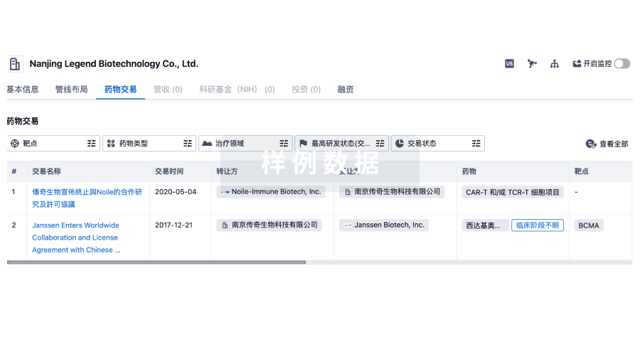
药物交易
使用我们的药物交易数据加速您的研究。
登录
或
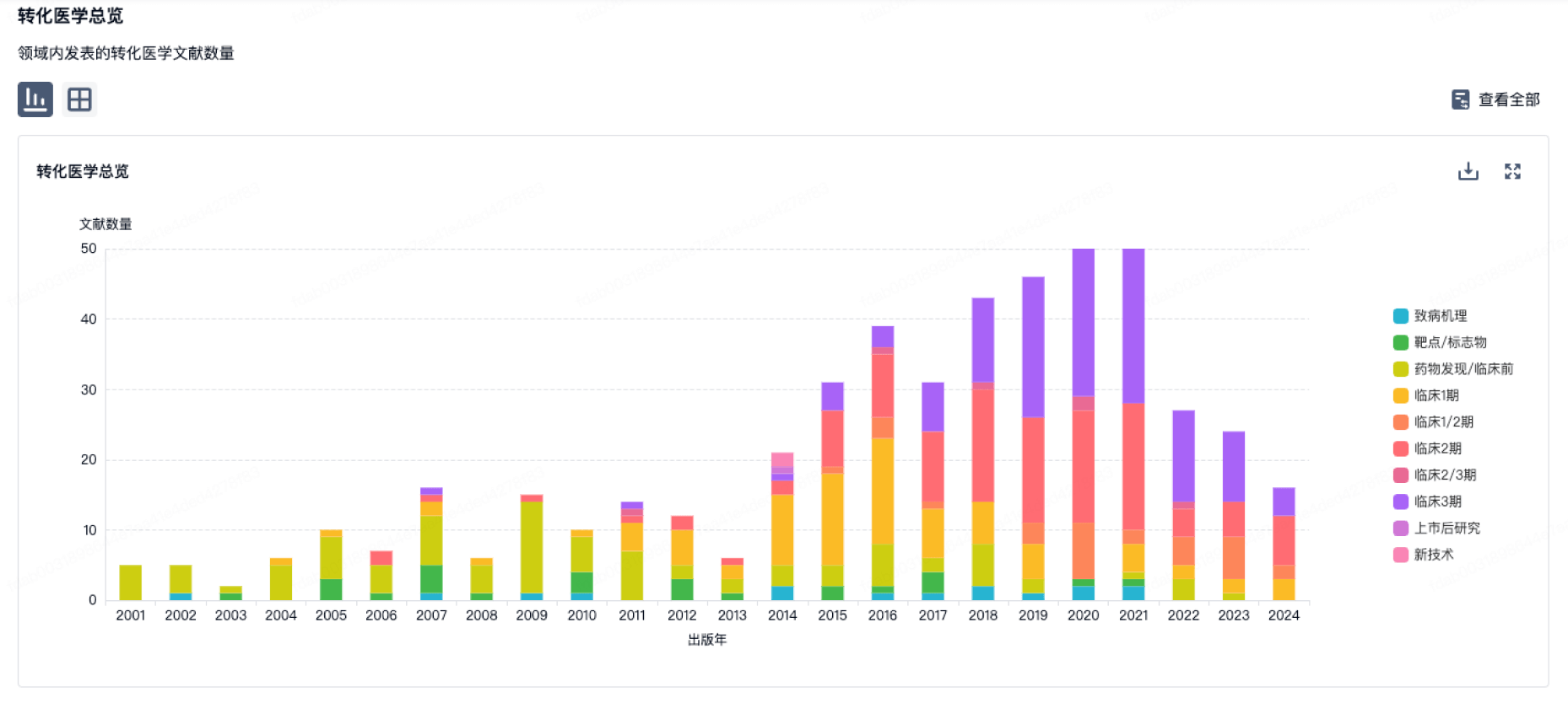
转化医学
使用我们的转化医学数据加速您的研究。
登录
或
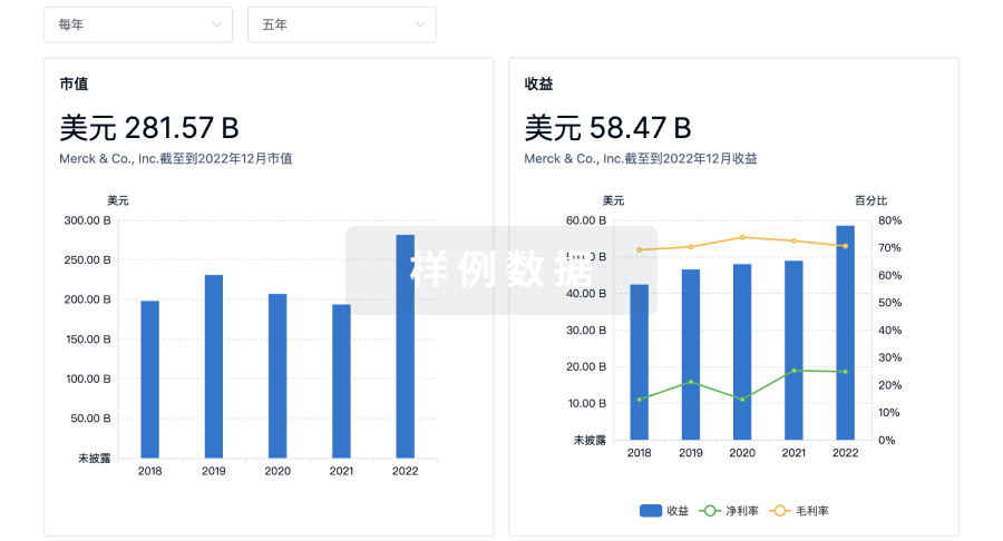
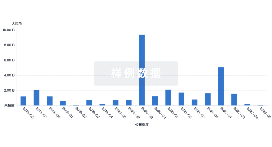
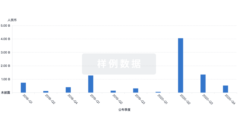
营收
使用 Synapse 探索超过 36 万个组织的财务状况。
登录
或
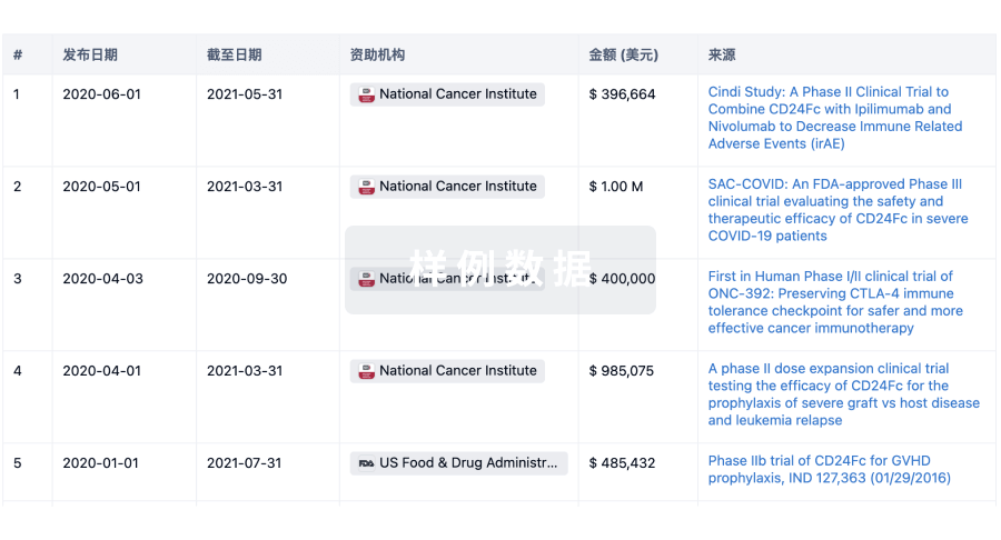
科研基金(NIH)
访问超过 200 万项资助和基金信息,以提升您的研究之旅。
登录
或
投资
深入了解从初创企业到成熟企业的最新公司投资动态。
登录
或
融资
发掘融资趋势以验证和推进您的投资机会。
登录
或
芽仔
全新生物医药AI Agent 覆盖科研全链路,让突破性发现快人一步
立即开始免费试用!
智慧芽新药情报库是智慧芽专为生命科学人士构建的基于AI的创新药情报平台,助您全方位提升您的研发与决策效率。
立即开始数据试用!
智慧芽新药库数据也通过智慧芽数据服务平台,以API或者数据包形式对外开放,助您更加充分利用智慧芽新药情报信息。
生物序列数据库
生物药研发创新
免费使用
化学结构数据库
小分子化药研发创新
免费使用