预约演示
更新于:2026-06-01

Kanazawa Medical University
更新于:2026-06-01
概览
关联
JPRN-UMIN000057250
Efficacy of active learning-based health education for preventing oral frailty - Verification of the effectiveness of health education for preventing oral frailty
JPRN-UMIN000057127
Prediction of tacrolimus blood concentration by CYP3A5 gene polymorphism - Prediction of tacrolimus blood concentration by CYP3A5 gene polymorphism
JPRN-UMIN000057021
Effects of oral or injectable semaglutide on Diabetes Therapy-Related Quality of Life scores in obese patients with type 2 diabetes: A single-center observational study. - An observational study of the effects of oral or injectable semaglutide on DTR-QOL scores.
100 项与 Kanazawa Medical University 相关的临床结果
登录后查看更多信息
登录后查看更多信息
2026-06-01HUMAN PATHOLOGY
Thymic cyst: A clinicopathological, immunohistochemical, and molecular study of 36 cases focusing on squamous- and ciliated-type epithelium
Article
作者: Kumanogoh, Atsushi ; Yamada, Sohsuke ; Nakatani, Yoichiro ; Ishida, Hiroto ; Horie, Masafumi ; Maeda, Daichi ; Morii, Eiichi ; Takata, So ; Ito, Yukinobu ; Goto, Akiteru ; Kudo-Asabe, Yukitsugu ; Yachida, Shinichi ; Shintani, Yasushi ; Miyabe, Ken
BACKGROUND:
Thymic cysts are benign mediastinal lesions lined by various epithelial types, but the distribution of each type is unknown. In this study, we aimed to evaluate the clinicopathological and immunohistochemical features of thymic cysts.
DESIGN:
We retrieved 36 resected primary thymic cysts. The lesions were initially evaluated morphologically, focusing on the type of epithelium. Immunohistochemical staining was performed for cytokeratin 7 (CK7), CK13, CK20, high molecular weight cytokeratin (HMWCK: CK34βE12), p63 and β5t. In addition, we investigated 15 occult cystic lesions (OCLs) associated with thymic epithelial tumors.
RESULTS:
Primary thymic cysts were classified as squamous/cuboidal (SC-type, N = 18), ciliated columnar (CC-type, N = 15), mucinous (N = 2), and mixed (N = 1) types. All cysts were CK7-positive. Linear-luminal pattern CK7 staining was observed more frequently in SC-type cysts (100%) than in CC-type cysts (40%) (P < 0.0001). CK20 was negative in all the cases except for the two mucinous cysts. Full-layer positivity for HMWCK was observed more frequently in SC-type cysts (78%) than in CC-type cysts (6.7%) (P < 0.0001). SC-type cysts were more frequently multilocular than CC-type (10/18, 55.6% vs 1/15, 6.7%; P = 0.0083). CK13 positivity was observed in 70.6% of SC-type cysts and 21.4% of CC-type cysts (P = 0.0113). Radiological findings showed that SC-type cysts were larger than CC-type cysts (P = 0.015), and the computed tomography (CT) values of SC-type cysts were markedly lower than those of CC-type cysts (P < 0.0001). The majority of OCLs were classified as SC-type (13/15).
CONCLUSIONS:
Based on morphological features, thymic cysts mainly comprise two types, SC-type and CC-type, which differ in their sizes, locularity, immunophenotypes, and CT features.
2026-06-01Radiology case reports
Preoperative transcatheter arterial embolization enables safe resection of a giant hypervascular pancreatic acinar cell carcinoma: A case report
Article
作者: Minami, Tetsuya ; Yamada, Sohsuke ; Nobata, Koji ; Ohta, Kiyotaka ; Miura, Seiko ; Mochizuki, Takafumi ; Shibata, Satoshi ; Kadoya, Yoshisuke ; Kondo, Tamaki ; Ueda, Nobuhiko ; Nishino, Yuka
Pancreatic acinar cell carcinoma (ACC) is a rare malignant neoplasm that accounts for 0.4%-0.7% of all pancreatic tumors. It often presents as a large, bulky mass owing to its expansive growth pattern. We report a case of a large pancreatic ACC that achieved remarkable long-term recurrence-free survival after successful surgical resection supported by preoperative interventional radiology (IR). A 64-year-old male presented to our hospital with weight loss and abdominal distension. A firm mass was palpable in the left upper abdomen. CT revealed a giant, heterogeneously enhancing tumor measuring 16.8 cm. Because of the anticipated massive intraoperative hemorrhage associated with tumor size and hypervascularity, Transcatheter Arterial Embolization (TAE) was performed preoperatively. The bilateral inferior diaphragmatic artery, posterior gastric, and splenic arterial branches supplying the tumor were embolized using metal coils and embolic materials. A safe radical resection was successfully performed (distal pancreatectomy, splenectomy, partial gastrectomy, partial colon resection, and left adrenalectomy). The pathological diagnosis confirmed pancreatic ACC (stage IIB). The patient has maintained recurrence-free survival for more than 5 years postoperatively. This case highlights that aggressive surgical resection achieves long-term survival in large pancreatic ACCs. Preoperative IR-TAE effectively controlled bleeding risk, underscoring the crucial role of this technique in safely managing high-risk, large, hypervascular pancreatic tumors. Physicians must consider ACC as a differential diagnosis for large pancreatic masses that may mimic other cystic solid lesions, such as Intraductal Papillary Mucinous Neoplasm (IPMN). Close multidisciplinary collaboration, particularly between interventional radiology and surgery, is essential in managing these challenging cases.
2026-05-01Kidney International Reports
Cystatin C Confirms the Canagliflozin eGFR Slope Benefit in CANPIONE
Article
作者: Hatanaka, Takashi ; Sasaki, Motofumi ; Nakatou, Tatsuaki ; Miyamoto, Satoshi ; Fujimoto, Kei ; Miyatake, Nobuyuki ; Shikata, Kenichi ; Heerspink, Hiddo J L ; Toyoda, Masao ; Kitada, Munehiro ; Takahashi, Yasushi ; Nunoue, Tomokazu ; Wada, Takashi ; Hida, Kazuyuki ; Akai, Hiroaki ; Kamei, Shinji ; Nakamura, Tohru ; Ando, Shinichiro ; Murao, Satoshi ; Sugano, Hisashi ; Kuramoto, Hiromi ; Yoshida, Michihiro ; Sakamoto, Kota ; Kawanami, Daiji ; Nakamura, Akihiko ; de Zeeuw, Dick ; Suzuki, Daisuke
Introduction:
In the canagliflozin in type 2 diabetic patients with microalbuminuria in Japanese population (CANPIONE) study with a novel preintervention slope design, canagliflozin attenuated the decline in creatinine-based estimated glomerular filtration rate (eGFR-creatinine) slope in patients with type 2 diabetes mellitus and microalbuminuria. However, because serum creatinine is affected by muscle mass, the reliability of the eGFR-creatinine slope may be limited. Therefore, this prespecified exploratory analysis aimed to evaluate the effect of canagliflozin on the cystatin C-based eGFR (eGFR-cystatin C) slope, which is less affected by muscle mass, to validate the robustness of the previous findings.
Methods:
The chronic eGFR-cystatin C and eGFR-creatinine slopes were assessed using a 2-slope linear spline mixed-effects model. Pearson correlation coefficients were used to evaluate associations between changes in eGFR and body composition-related parameters.
Results:
eGFR trajectories were directionally consistent across both filtration markers. The chronic eGFR-cystatin C slopes in the canagliflozin and control groups were -0.4 (95% confidence interval [CI], -2.6 to 1.8) and -4.9 (-7.1 to -2.6) ml/min per 1.73 m2/year, respectively, corresponding to a between-group difference of 4.4 (1.3-7.6) ml/min per 1.73 m2/year. Similar trends were observed in the eGFR-creatinine slopes derived from the same time points, yielding a comparable treatment effect on the chronic slope (between-group difference: 3.5 [0.2-6.8] ml/min per 1.73 m2/year). Canagliflozin reduced body weight, body mass index (BMI), and waist circumference, whereas little correlation was observed between these changes and changes in eGFR-cystatin C.
Conclusion:
Incorporating the eGFR-cystatin C slope analysis supports the robustness of the previous creatinine-based slope findings, suggesting that the eGFR-creatinine slope analysis retains reasonable clinical reliability in patients treated with sodium-glucose cotransporter 2 inhibitors (SGLT2is).
2025-10-19
临床2期临床结果临床3期
100 项与 Kanazawa Medical University 相关的药物交易
登录后查看更多信息
100 项与 Kanazawa Medical University 相关的转化医学
登录后查看更多信息
组织架构
使用我们的机构树数据加速您的研究。
登录
或
管线布局
2026年07月21日管线快照
无数据报导
登录后保持更新
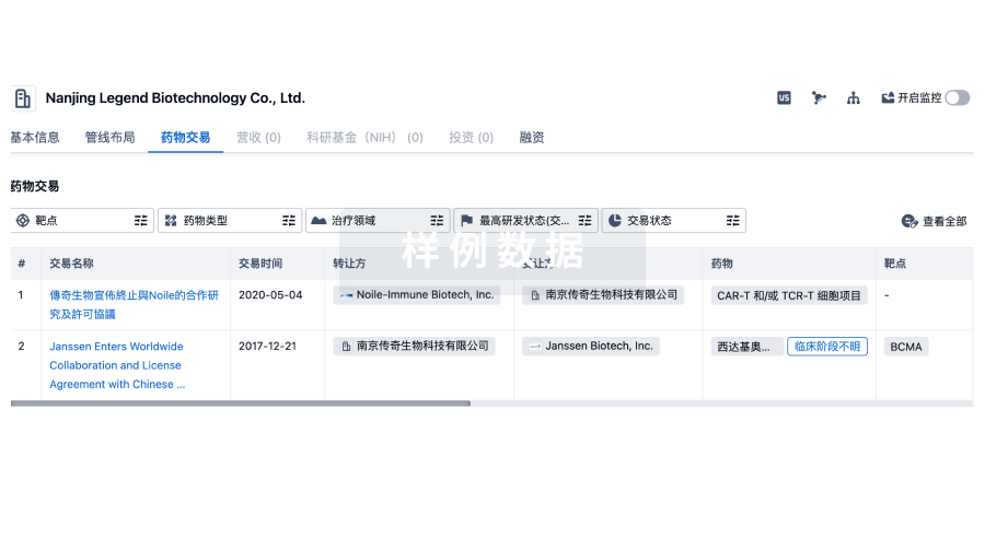
药物交易
使用我们的药物交易数据加速您的研究。
登录
或
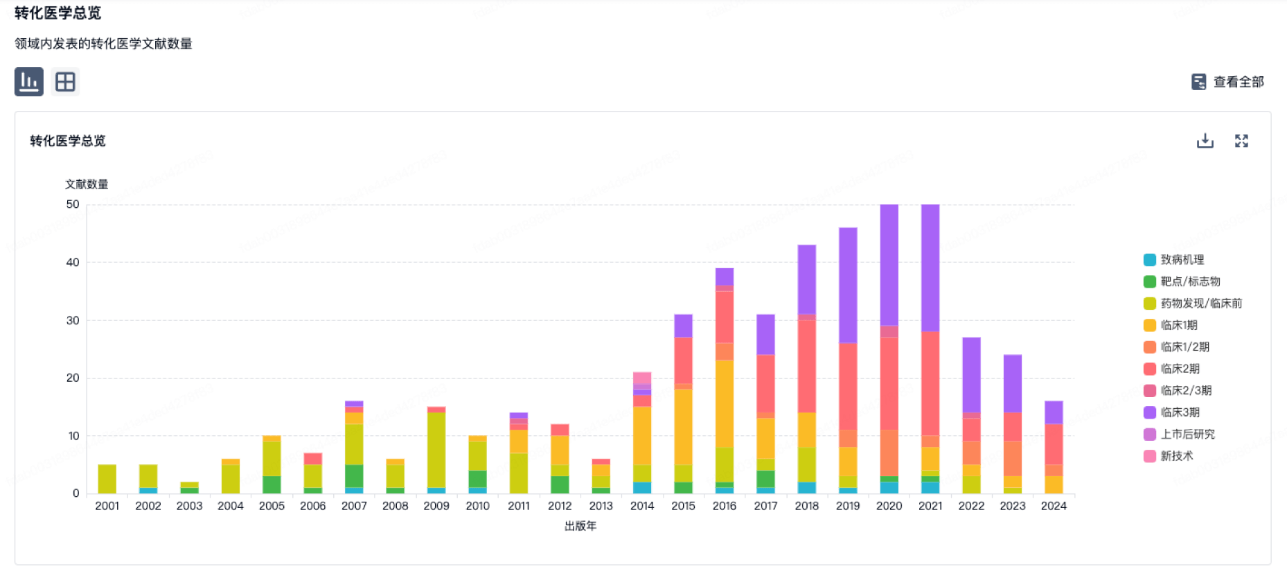
转化医学
使用我们的转化医学数据加速您的研究。
登录
或
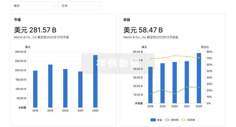
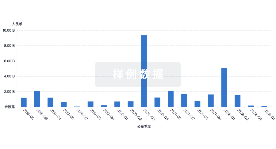
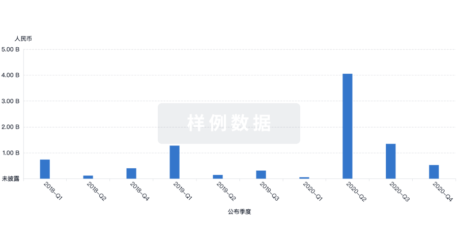
营收
使用 Synapse 探索超过 36 万个组织的财务状况。
登录
或
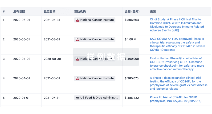
科研基金(NIH)
访问超过 200 万项资助和基金信息,以提升您的研究之旅。
登录
或
投资
深入了解从初创企业到成熟企业的最新公司投资动态。
登录
或
融资
发掘融资趋势以验证和推进您的投资机会。
登录
或
芽仔
全新生物医药AI Agent 覆盖科研全链路,让突破性发现快人一步
立即开始免费试用!
智慧芽新药情报库是智慧芽专为生命科学人士构建的基于AI的创新药情报平台,助您全方位提升您的研发与决策效率。
立即开始数据试用!
智慧芽新药库数据也通过智慧芽数据服务平台,以API或者数据包形式对外开放,助您更加充分利用智慧芽新药情报信息。
生物序列数据库
生物药研发创新
免费使用
化学结构数据库
小分子化药研发创新
免费使用