The ADVISE Study: Advanced Visualization In Corneal Surgery Evaluation Intra-operative Optical Coherence Tomography in Posterior Lamellar Keratoplasty
Rationale: Intra-operative optical coherence tomography (iOCT) is a new technology that incorporates advanced imaging techniques in the ophthalmic operating theatre. This allows surgeons to visualize tissues in a way previously impossible We conceptualized an iOCT-guided surgical protocol for the treatment of endothelial cell dysfunction, that refrains from the current practice of over-pressurizing the eye at the end of surgery.
Objective: The aim of this study is to assess the clinical value of intraoperative OCT (iOCT) for Descemet Membrane Endothelial Keratoplasty (DMEK) by comparing an iOCT-optimized surgical protocol with current practice, where the eye is over-pressurized for a set period of time, in terms of surgical efficiency, clinical outcomes, and adverse events.
Study design: International multicentre non-inferiority randomized clinical trial Study population: Patients scheduled for posterior lamellar corneal surgery for endothelial cell dysfunction above the age of 18 years.
Intervention: Both groups will undergo Descemet Membrane Endothelial Keratoplasty. Patients will be randomized for either the iOCT optimized surgical protocol or current standard surgical protocol using 8 minutes of overpressure to facilitate graft adherence. Both groups will be evaluated with iOCT at the end of surgery.
Main study parameters/endpoints:
The main study parameter is the rate of adverse events (particularly graft dislocations). Secondary parameters/endpoints are surgical time, the recovery of visual acuity and endothelial graft quality at 3 and 6 months follow-up, and a detailed evaluation of the extent/duration of surgical tissue manipulations.
Nature and extent of the burden and risks associated with participation, benefit and group relatedness: The use of iOCT during surgery does not entail additional risk to the patient. Participants to this study will adhere to the standard of care after corneal transplant surgery. In addition, they will receive study specific measurements and questionnaires. The additional measurements and questionnaires will be combined with regular follow up moments.
In Vivo Confocal Microscopy Tumor Atlas Study
This study aims to create an atlas based on the preliminary experience of the first feasibility study in neurosurgery. Hypothesis: That a confocal endomicroscope can be used during neurosurgery to provide in vivo histology that enables documentation of neurological pathology across a range of tumor ypes and grades, suitable for comparison with traditional histopathology from site-matched biopsies.
In Vivo Confocal Endomicroscopy of the Brain
The purpose of the study is to test the feasibility of obtaining interpretable in vivo endomicroscopy images which can be compared with traditional histopathology.
Hypothesis: That a rigid confocal endomicroscope can be used during neurosurgery to provide in vivo histology that enables differentiation of tumour tissue from normal adjacent brain tissue.
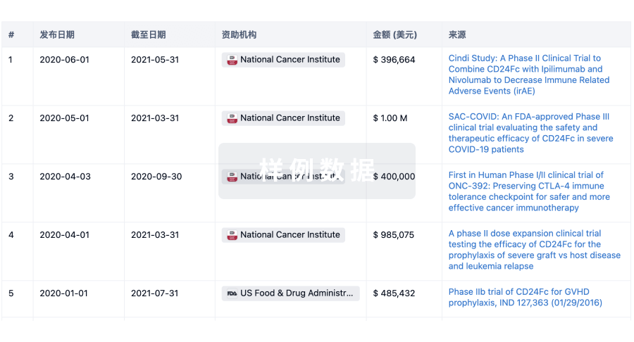
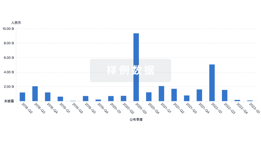
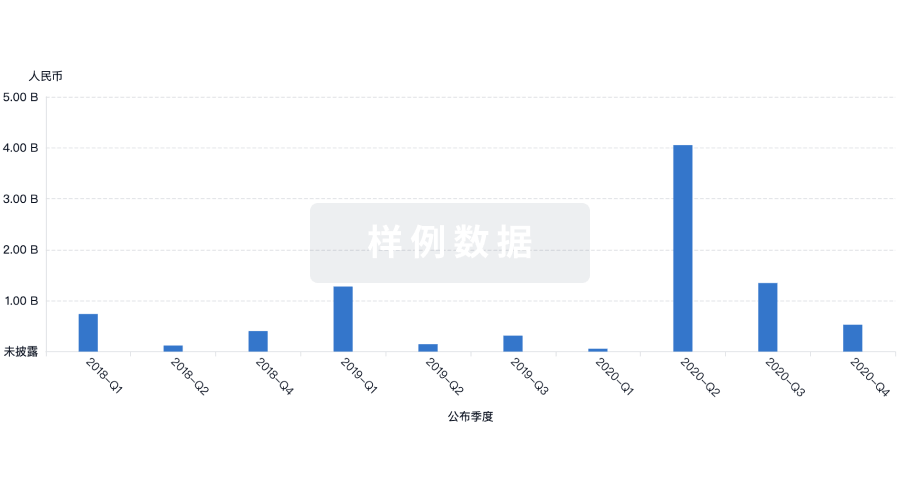
100 项与 Carl Zeiss Surgical GmbH 相关的临床结果
0 项与 Carl Zeiss Surgical GmbH 相关的专利(医药)
100 项与 Carl Zeiss Surgical GmbH 相关的药物交易
100 项与 Carl Zeiss Surgical GmbH 相关的转化医学