预约演示
更新于:2026-05-08
Siemens AG
更新于:2026-05-08
概览
关联
NCT07242690
Advancing PET-CT Imaging With Photon Counting Detector CT

NCT07495332
Siemens Biomarker Multi-modality

NCT07276360
Phase II Randomized Non-Inferiority Trial of Hypofractionated Radiotherapy for Locally Advanced Cervical Cancer in Uganda

100 项与 Siemens AG 相关的临床结果
登录后查看更多信息
登录后查看更多信息
2026-09-01APPLIED ERGONOMICS
Beyond the inbox: Exploring shared cognitive mechanisms underlying susceptibility to and reporting of deception across digital domains
Article
作者: Sarno, Dawn M ; Sabo, Katharine E ; Black, Jeffrey T
As deceptive content proliferates across digital platforms, understanding how users detect and report deception is increasingly important. The present study examined how detection and reporting of deception may be consistent across a variety of digital domains (i.e., emails, news headlines, voicemails) and explored if there are any shared individual differences (e.g., cognitive reflection, digital literacy) underlying user susceptibility. Participants' discrimination abilities, risk tendencies, and reporting behaviors were related across the digital domains, suggesting widespread vulnerability. Cognitive reflection predicted deception detection across digital domains, while digital literacy was only predictive for emails and news headlines. Exploratory analyses revealed that gullibility, social desirability, and age were all related to various aspects of susceptibility across digital domains, highlighting the importance of investigating other broad predictors underlying susceptibility. Our findings indicate similar cognitive mechanisms underlying vulnerability to deception across digital domains, supporting the design of general interventions that target broad skills, like cognitive reflection.
2026-06-01MAGNETIC RESONANCE IMAGING
Improved image quality and reduced acquisition time in brain MRI using deep learning-based reconstruction: A quantitative and subjective assessment compared to standard MPRAGE in 0.55 T MRI
Article
作者: Haag, Nina ; Kroeger, Jan Robert ; Schreck, Julian ; Saeed, Saher ; Moenninghoff, Christoph ; Schoenbeck, Denise ; Woeltjen, Matthias Michael ; Nickel, Dominik ; Reiman, Georg ; Weiland, Elisabeth ; Niehoff, Julius Henning ; Shahzadi, Iram ; Boriesosdick, Jan ; Borggrefe, Jan ; Frohwein, Lynn Johann ; Katz, Maria
PURPOSE:
To assess the impact of deep learning (DL)-based image reconstruction on quantitative and subjective image quality in brain MRI at 0.55 T by comparing DL-reconstructed MPRAGE with standard MPRAGE using comparable acquisition geometry and timing parameters.
METHODS:
In this prospective study, 30 patients underwent two consecutive 3D T1-weighted MPRAGE acquisitions on a 0.55 T MRI system (MAGNETOM Free.Max, Siemens Healthineers): a standard reconstruction and a DL-based reconstruction generated from undersampled k-space data using a variational-network architecture. Identical circular regions of interest (ROIs) were placed in gray matter (caudate nucleus), white matter (centrum semiovale), cerebrospinal fluid (lateral ventricle), and air for noise estimation. Signal-to-noise ratio (SNR) and contrast-to-noise ratio (CNR) were computed. Three blinded radiologists independently evaluated image quality using a 5-point Likert scale based on anatomical detail, gray-white matter differentiation, vascular visibility, artifact burden, and overall diagnostic quality.
RESULTS:
DL-based reconstruction significantly improved objective image quality across all metrics. SNR increased from 40.83 ± 10.11 to 106.30 ± 51.38 in gray matter (p < 0.0001), from 48.95 ± 12.52 to 125.30 ± 58.01 in white matter (p < 0.0001), and from 5.77 ± 1.10 to 7.75 ± 4.76 in CSF (p = 0.021). CNR between gray and white matter increased from 8.12 ± 4.24 to 18.99 ± 11.21 (p < 0.0001). Subjective ratings favored DL reconstruction for anatomical detail (4.05 ± 0.62 vs. 3.68 ± 0.64), vascular visibility (4.37 ± 0.56 vs. 3.63 ± 0.72), and overall image quality (4.07 ± 0.72 vs. 3.57 ± 0.73). Standard reconstruction showed slightly better artifact suppression (3.80 ± 0.61 vs. 3.27 ± 0.78) and gray-white matter contrast (4.00 ± 0.64 vs. 3.87 ± 0.71). Acquisition time was reduced from 6:44 min (standard MPRAGE) to 3:06 min with DL reconstruction, corresponding to a substantial scan time reduction.
CONCLUSION:
DL-based reconstruction markedly enhances quantitative and subjective image quality in low-field brain MRI and enables a markedly shorter acquisition time compared to the standard protocol. This study adds clinical evidence that DL reconstruction can substantially improve 0.55 T MPRAGE imaging, supporting its integration into routine neuroimaging workflows to improve efficiency, patient comfort, and diagnostic confidence.
KEY POINTS:
2026-06-01EUROPEAN JOURNAL OF RADIOLOGY
Non-invasive material characterization after total hip arthroplasty by 4-threshold imaging with photon-counting detector CT
Article
作者: Wulff, Jörg ; Hegmanns, Jan ; Haase, Viktor ; Bäumer, Christian ; Faby, Sebastian ; Schaarschmidt, Benedikt M
2026-05-07
2026-05-06
100 项与 Siemens AG 相关的药物交易
登录后查看更多信息
100 项与 Siemens AG 相关的转化医学
登录后查看更多信息
组织架构
使用我们的机构树数据加速您的研究。
登录
或
管线布局
2026年06月02日管线快照
管线布局中药物为当前组织机构及其子机构作为药物机构进行统计,早期临床1期并入临床1期,临床1/2期并入临床2期,临床2/3期并入临床3期
其他
12
登录后查看更多信息
当前项目
登录后查看更多信息
药物交易
使用我们的药物交易数据加速您的研究。
登录
或
转化医学
使用我们的转化医学数据加速您的研究。
登录
或
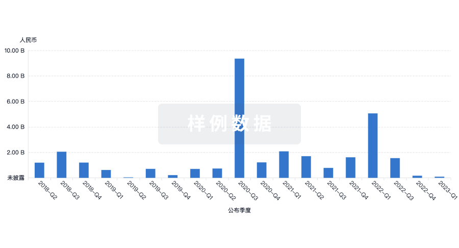
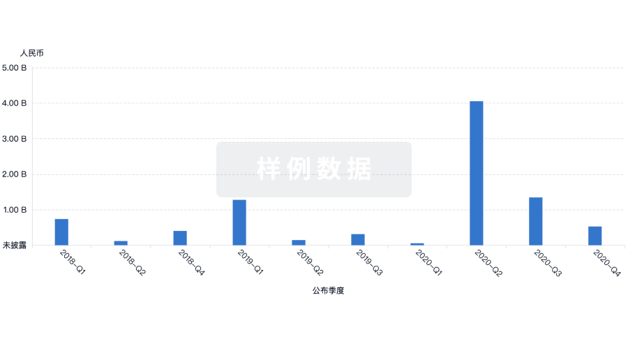
营收
使用 Synapse 探索超过 36 万个组织的财务状况。
登录
或
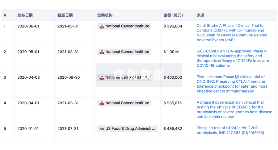
科研基金(NIH)
访问超过 200 万项资助和基金信息,以提升您的研究之旅。
登录
或
投资
深入了解从初创企业到成熟企业的最新公司投资动态。
登录
或
融资
发掘融资趋势以验证和推进您的投资机会。
登录
或
生物医药百科问答
全新生物医药AI Agent 覆盖科研全链路,让突破性发现快人一步
立即开始免费试用!
智慧芽新药情报库是智慧芽专为生命科学人士构建的基于AI的创新药情报平台,助您全方位提升您的研发与决策效率。
立即开始数据试用!
智慧芽新药库数据也通过智慧芽数据服务平台,以API或者数据包形式对外开放,助您更加充分利用智慧芽新药情报信息。
生物序列数据库
生物药研发创新
免费使用
化学结构数据库
小分子化药研发创新
免费使用