预约演示
更新于:2026-02-21

Epistem Ltd.
更新于:2026-02-21
概览
标签
口颌疾病
肿瘤
其他疾病
小分子化药
疾病领域得分
一眼洞穿机构专注的疾病领域
技术平台
公司药物应用最多的技术
靶点
公司最常开发的靶点

暂无数据
关联
靶点 |
作用机制 |
在研机构 |
原研机构 |
在研适应症 |
非在研适应症 |
最高研发阶段 |
首次获批国家/地区 |
首次获批日期 |

NCT02992184
PoC-HCV Genedrive Viral Detection Assay Validation Study

100 项与 Epistem Ltd. 相关的临床结果
登录后查看更多信息
登录后查看更多信息
2022-09-01SINGAPORE MEDICAL JOURNAL4区 · 医学
Change in hair growth-related gene expression profile in human isolated hair follicles induced by 5-alpha reductase inhibitors – dutasteride and finasteride – in the presence of testosterone
4区 · 医学
Article
作者: Mefo, Tim ; Lulic, Zrinka ; Harrison, Elliott ; Ong, Gary ; Hatanaka, Toshiki ; Booth, Cath
INTRODUCTION Dihydrotestosterone (DHT) plays a key role in the pathogenesis of male androgenetic alopecia (AGA) and is converted from the androgen, testosterone, by 5-alpha reductase (5AR). DHT binds to androgen receptors in the hair follicles, causing shortening of the anagen or growing phase of the hair cycle and leading to hair follicle miniaturisation.[1] Over time, large terminal hairs are lost and progressively replaced by thin, short, villus-like hairs, resulting in a characteristic pattern of baldness.[12] Oral treatment with 5AR inhibitors (5ARIs; e.g., dutasteride and finasteride) is used for men with AGA, and is recommended by Japanese guidelines as the first-line treatment for male AGA and female pattern hair loss.[3] Although these agents inhibit testosterone conversion to DHT, their mechanisms of action are different – finasteride specifically inhibits type II 5AR, but dutasteride inhibits both type I and type II 5AR.[45] Consequently, the superiority of dutasteride 0.5 mg to finasteride 1.0 mg, with respect to changes in hair count and width, has been demonstrated in a global phase III clinical trial[6] and documented in review articles.[78] However, it is unclear whether type I 5AR is one of the enzymes driving the hair loss process. Moreover, downstream molecular events following 5AR inhibition are poorly understood. Therefore, in-depth molecular analyses are necessary to clarify the extent of the contribution of type I 5AR to hair growth. This study investigated gene expression changes in growth factors and other related molecules responsible for hair growth using the bulbar portions of hair follicles (BPHF) isolated from plucked human hair and evaluated the involvement of type I 5AR in human hair growth. METHODS This study was approved by the Manchester Consumer Healthcare Research Ethics Committee. In accordance with the Declaration of Helsinki, all volunteer hair donors provided written informed consent. Anagen hair follicles were plucked from frontal, parietal and frontal/temporal areas of five healthy male donors, with varying degrees of AGA (Norwood-Hamilton classification ranging from score 2A–7). Plucked hair follicles were cultured in Dulbecco's Modified Eagle's Medium, containing proprietary growth media, L-glutamine 200 nM (Cat: G7513; Sigma Aldrich, Gillingham, UK) and penicillin-streptomycin (Cat: P0781; Sigma Aldrich), in the presence of DHT or testosterone in 0.1% (v/v) dimethyl sulfoxide (DMSO) or DMSO vehicle only. Hair follicles were incubated at 37°C in a 5% carbon dioxide atmosphere for 24 hours, with and without 5ARIs (dutasteride or finasteride), in the presence of testosterone, as previously described, with modification.[9] Post-culture, anagen hairs were visually assessed for dermal papilla cells (DPCs) and suitable candidates were chosen for lysis and RNA extraction. Total RNA was extracted from DPCs using the Invitrogen RNAqeous total RNA isolation kit (Cat: 10596935; Fisher Scientific, Loughborough, UK). Human fibroblasts derived from skin samples (human abdominal tissue sourced from Tissue Solutions, Glasgow, UK) were used as controls. For quantitative reverse transcriptase polymerase chain reaction (qRT-PCR) analysis, RNA was reverse transcribed into complementary DNA (cDNA) as a template for qRT-PCR. PCR was conducted as follows: hot start at 95°C for 60 seconds (one cycle); denaturation at 96°C for 5 seconds and annealing and extension at 60°C (30 cycles); additional extension at 60°C for 3 minutes and from 60°C to 95°C for 3 seconds (one cycle). The genes assessed in this assay as well as the primers for assessed and housekeeping genes are shown in Supplementary Table 1 [see Appendix]. The focus was on those genes reported to be involved in hair growth: fibroblast growth factor 7 (FGF7), secreted by cells in the dermal papilla, plays a role in stimulating hair matrix cell proliferation and hair growth[10]; insulin-like growth factor 1 (IGF1) promotes maintenance of anagen phase and the stimulation of hair growth[11]; and WNT family member 5A (WNT5a) mediates some of the effects of Sonic hedgehog in hair follicle morphogenesis and is capable of regulating proliferation.[1213] Other related growth factors for hair growth were analysed, including alkaline phosphatase (ALPL), expressed in dermal papilla[14]; fibroblast growth factor 2 (FGF2), expressed in hair matrix cells[10]; vascular endothelial growth factor A (VEGFA), responsible for maintaining vasculature around the hair follicle during the anagen growth phase,[15] and platelet-derived growth factor A (PDGFA), stimulating morphogenesis of new capillaries in anagen.[16] Related surrogate markers were also analysed, including: lymphoid enhancer binding factor 1 (Lef1),[17] involved in hair follicle organogenesis; bone morphogenetic protein 4 (BMP4), found in the hair placode, i.e., in the sites of Lef1/b-catenin expression[18]; Noggin, driving hair follicle morphogenesis and an antagonist for BMP4[1920]; and WNT inhibitory factor 1 (Wif1), an antagonist for WNT family.[2122] The study was conducted sequentially in three steps. In Step 1, we assessed the assay system and confirmed type I and type II 5AR expression in BPHF. As DHT has been reported to change hair growth factor gene expression levels,[23] gene expression levels of FGF7, IGF1, WNT5a and relevant factors were evaluated using human BPHF in the presence of DHT 1.5 nM, 3 nM or 15 nM (or DMSO control). DHT concentrations used were based on a phase II clinical trial of dutasteride in patients with AGA,[24] during which baseline serum DHT concentrations were in the range 371–482 pg/mL (approximately 1.5 nM). Therefore, the starting DHT concentration, in our study, was 1.5 nM, with high concentrations of 3 nM and 15 nM also investigated. In Step 2, we evaluated whether using testosterone in this assay system mimicked the conversion to DHT by 5AR in vivo. Changes in gene expression levels of the hair growth factors assessed in Step 1 (FGF7, IGF1 and WNT5a) plus related factors (VEGFA, Lef1, BMP4, PDGFA, Noggin and Wif1) were investigated in isolated human BPHF following stimulation with testosterone 3 nM, 10 nM or 30 nM (or DMSO control). Testosterone concentrations were based on results of the phase II clinical trial[24] and Step 1. In the phase II study, baseline serum testosterone concentration was around 4.5 ng/mL (approximately 15 nM). In Step 1, DHT 3 nM was suggested as an optimal concentration and considering the tenfold higher potency of DHT than that of testosterone, the submaximal concentration of testosterone was speculated to be 30 nM. In Step 3, we assessed the expected inhibitory effects of dutasteride and finasteride on gene expression of hair growth-related factors in human BPHF following testosterone stimulation (10 nM, as determined in Step 2). Concentrations of dutasteride were 0.3 nM, 0.03 nM and 0.003 nM, and concentrations of finasteride were 1.5 nM, 0.15 nM and 0.015 nM, based on phase II/III trial results.[6] In the previous study, the range of concentrations for dutasteride 0.5 mg and finasteride 1.0 mg in therapeutic plasma steady-state were around 40 ng/mL (76 nM) and 9 ng/mL (24 nM), respectively, which after correcting for protein binding equated to 0.2 ng/mL (0.38 nM) and 0.63 ng/mL (1.69 nM), respectively. Data was analysed by normalising targets to the geomean of two housekeeping genes. Analysis was primarily conducted on the Partek Genomic Suite (Partek, St Louis, MO, USA) using contrasts of vehicle versus treatment samples for intra-patient and global cohort analyses. Statistical significance was calculated by analysis of variance. RESULTS In Step 1, changes were observed in gene expression subsequent to DHT stimulation and optimal DHT concentration was determined. Following DHT simulation, steroid 5AR 1 and 2 (SRD5A1 and SRD5A2) were detected, indicating that both type I and type II 5AR were expressed in BPHF [Figure 1a]. Up to 3 nM, DHT stimulation showed a trend to decrease gene expression levels of FGF7 (p = 0.52), IGF1 (p = 0.85) and WNT5a (p = 0.08) [Figure 1b]. However, 15 nM DHT stimulation did not show consistent changes in the gene expression levels of any assessed factors, except for AR and VEGFA. Therefore, 3 nM was the optimal concentration of DHT in this assay system and was used as the base concentration for Step 2.Figure 1: Charts show changes in gene expression of (a) SRD5A1, SRD5A2 and AR, and (b) other factors related to hair growth induced by DHT. ALPL: alkaline phosphatase, AR: androgen receptor, delta CT: difference of expression between gene of interest and reference sequence, DHT: dihydrotestosterone, DMSO: dimethyl sulfoxide, FGF2: fibroblast growth factor 2, FGF7: fibroblast growth factor 7, IGF1: insulin-like growth factor 1, SRD5A1: steroid 5 alpha-reductase 1, SRD5A2: steroid 5 alpha-reductase 2, VEGFA: vascular endothelial growth factor A, WNT5a: WNT family member 5A.In Step 2, there were changes in gene expression following testosterone stimulation. Testosterone stimulation of BPHF showed a trend to decrease the gene expression levels of three hair growth factors: FGF7 (p = 0.53); IGF1 (p = 0.93); and WNT5a (p = 0.51) [Figure 2a]. Similar changes in the gene expression levels of Lef1 and BMP4 were observed, while Noggin and Wif1 showed opposite changes [Figure 2b]. Although the VEGFA gene expression increased in a concentration-dependent manner alongside that of testosterone, this change was opposite to that observed in Step 1. As observed in Step 1, the maximal testosterone concentration (30 nM) was considered to cause saturation in WNT5a, PDGFA and Noggin; therefore, 10 nM was determined as the optimal concentration to use for Step 3.Figure 2: Charts show changes in gene expression of (a) FGF7, IGF1 and WNT5a, and (b) other factors related to hair growth induced by testosterone. BMP4: bone morphogenetic protein 4, delta CT: difference of expression between two genes, DMSO: dimethyl sulfoxide, FGF7: fibroblast growth factor 7, IGF1: insulin-like growth factor 1, Lef1: lymphoid enhancer binding factor 1, NOG: Noggin, PDGFA: platelet-derived growth factor A, VEGFA: vascular endothelial growth factor A, Wif1: WNT inhibitory factor 1, WNT5a: WNT family member 5A.In Step 3, there was a 5ARI effect on changes in gene expression levels in the presence of testosterone. The expected inhibitory effects of dutasteride and/or finasteride on the observed hair growth factor gene expression changes under testosterone stimulation were investigated, with testosterone at an optimal concentration (10 nM), as determined in Step 2. Gene expression levels of FGF7, IGF1 and WNT5a, with dutasteride or finasteride, under testosterone stimulation showed a consistent trend of re-increasing [Figure 3a]. The fold change (mean ± standard error of the mean) in FGF7 expression, when compared with DMSO control, ranged from 1.00 ± 0.25 to 1.53 ± 10.04 (p = 0.10) for finasteride and from 0.93 ± 0.36 to 1.22 ± 0.10 (p = 0.23) for dutasteride. Similarly, IGF1 expression ranged from 0.56 ± 0.78 to 4.34 ± 2.71 (p = 0.72) with finasteride treatment and from 1.70 ± 0.78 to 2.73 ± 1.24 (p = 0.61) with dutasteride treatment. Meanwhile, WNT5a expression ranged from 1.03 ± 0.99 to 1.14 ± 0.10 (p = 0.26) with finasteride and 1.08 ± 0.25 to 1.21 ± 0.20 (p = 0.65) with dutasteride treatments.Figure 3: Charts show changes in gene expression of (a) FGF7, IGF1 and WNT5a, and (b) other factors related to hair growth under 10 nM testosterone stimulation in the presence of dutasteride or finasteride. BMP4: bone morphogenetic protein 4, delta CT: difference of expression between two genes, DMSO: dimethyl sulfoxide, FGF7: fibroblast growth factor 7, IGF1: insulin-like growth factor 1, Lef1: lymphoid enhancer binding factor 1, NOG: Noggin, PDGFA: platelet-derived growth factor A, VEGFA: vascular endothelial growth factor A, Wif1: WNT inhibitory factor 1, WNT5a: WNT family member 5A.In our study, the expression of Noggin and Wif1 showed a trend of increasing in the presence of testosterone stimulation, whereas WNT5a expression showed a trend of decreasing with testosterone stimulation [Figure 2b]. PDGFA gene expression showed a consistent trend towards decreasing with increasing 5ARI concentrations [Figure 3b]. Changes in VEGFA and Lef1 gene expression were inconsistent [Figure 3b]. Additionally, hair growth factor expression under testosterone stimulation was evaluated in isolated human fibroblasts to confirm whether observed changes in gene expression were induced specifically in BPHF. No genes were shown to decrease under testosterone stimulation in the human fibroblasts (data not shown), indicating that effects on gene expression were specific to BPHF. DISCUSSION By utilising BPHF isolated from plucked human hair in this assay, we investigated why dutasteride was more effective at promoting human hair growth than finasteride, as demonstrated in a clinical trial.[6] We also evaluated the involvement of type I 5AR in human hair growth. Our three-step assay system was considered to detect changes in gene expression of hair growth factors and other related molecules. By measuring changes in the expression levels of targeted genes, new 5ARI candidates and medicines may be evaluated at the gene level. However, the assay system needs further development. In Step 1, stimulation of BPHF with 15 nM DHT resulted in saturation of the trend of decreasing gene expression for three factors – FGF7, IGF1 and WNT5a – possibly due to the over-concentration of DHT and/or DHT-induced down-regulation of 5AR in the cells. Furthermore, changes in gene expression of other related molecules (VEGFA, Lef1 and PDGFA) were shown to be inconsistent in all steps. This could be due to the use of BPHF, with other components, and not purely isolated DPCs. Notably, dutasteride and finasteride showed the trend to cancel suppressive effects of testosterone on hair-related gene expression, presumably in DPCs in Step 3, as the expression levels of targeted genes (FGF7, IGF1 and WNT5a) were increased. Considering the inhibitory potency of dutasteride and finasteride, there was a trend towards differing changes in expression of the investigated genes and factors between the two 5ARIs. The effect of finasteride on FGF7, IGF1 and WNT5a expression plateaued at the middle concentration evaluated (i.e. 0.15 nM), but the effect of dutasteride was linear to the highest concentration tested (or 0.3 nM), although these findings were not statistically significant. This observation suggests that dutasteride may have a stronger inhibitory potency to increase growth factor expression than finasteride, possibly due to the inhibition of type I 5AR by dutasteride or greater inhibition of type II 5AR by dutasteride when compared to finasteride. Our results suggest that type I 5AR may play an important role in hair growth, as well as type II 5AR.[1011] Furthermore, WNT5a expression in the developing hair follicles requires Sonic hedgehog; this suggests that WNT5a may mediate some effects of Sonic hedgehog in hair follicle morphogenesis, a hypothesis supported by the fact that both WNT5a and Sonic hedgehog are capable of regulating proliferation.[12] Noggin is a known inhibitor of BMP4[19]; therefore, results were consistent with BMP4 expression decreasing in a concentration dependent manner. Similarly, results for Wif1, a WNT antagonist,[2225] were consistent with those of WNT5a, showing a trend for reduced expression. Thus, FGF7, IGF1 and WNT5a were confirmed as key factors for hair growth in the study. As with other in vitro/ex vivo investigations, this study had potential limitations. Changes in gene expression were not verified as leading to corresponding changes in protein levels. Although not the focus of this study, effects on protein expression should also be assessed. Furthermore, concentrations of DHT converted from testosterone in the culture medium were not assessed in Steps 1 and 2. Testosterone induced changes in expression of several genes and testosterone conversion to DHT was inhibited by 5AR in BPHF. However, more precise investigations, including the determination of DHT concentration in the medium, may be necessary. Another limitation was the small sample size; larger sample sizes, with over 35 participants, should be considered in the future. Therefore, interpretation of these results for testosterone stimulation, alone and in the presence of dutasteride/finasteride, should be carefully considered. In addition, hair growth ex vivo culture was not assessed directly, given the study design; 24-hour incubation time is too short to reach levels of gene expression changes that would lead to changes in efficacy endpoints (e.g. hair shaft lengths, hair width, hair count). Finally, data analyses and interpretation may be difficult due to our use of BPHF instead of pure DPCs; assays using pure DPCs are expected to show more clear-cut results and provide more in-depth interpretation. Our study indicates that dutasteride and finasteride are potent modulators of the expression of genes encoding hair growth factors and other molecules potentially related to hair growth. Dutasteride may be more potent than finasteride in modulating the expression levels of key hair growth genes (e.g. FGF7, IGF1 and WNT5a). This study provides supporting evidence that type I 5AR may be involved in hair growth in addition to type II 5AR. These findings indicate an in-depth downstream mechanism of action by 5AR and provide further clarification of the importance of type I 5AR function. Acknowledgements We greatly appreciate Professor Manabu Ohyama (Department of Dermatology, Kyorin University School of Medicine, Tokyo, Japan) for his excellent management and scientific advice on the study protocol, as well as reviewing the manuscript, as the scientific advisor of the present study. We are indebted to Masahiro Yamada, formerly of GlaxoSmithKline KK, Medical Affairs, Japan, and Spiro Getsios, formerly of GlaxoSmithKline and now of Aspect Biosystems, Vancouver, BC, Canada, for their contributions to this study and manuscript. We are also grateful to Jonathan Bullman (GlaxoSmithKline R&D Projects Clinical Platforms and Sciences, UK) for his useful scientific advice on the consideration of test compound concentrations. In addition, we appreciate Yoshiaki Kawano (GlaxoSmithKline KK, Medical Affairs, Japan) for his thoughtful and kind review, and Kumiko Kobayashi (GlaxoSmithKline KK, Medical Affairs, Japan), for her vigorous efforts in creating excellent graphs. Medical writing support, in the form of developing/editing drafts based on author input, editorial assistance and submission of the final manuscript, was provided by Katy Tucker, PhD, of Fishawack Indicia Ltd, UK, funded by GlaxoSmithKline. Financial support and sponsorship This study was funded by GlaxoSmithKline. Conflicts of interest Toshiki Hatanaka, Zrinka Lulic and Gary Ong are employees of GlaxoSmithKline. Tim Mefo, Cath Booth and Elliott Harrison are employees of Epistem. Supplementary material
2021-10-01Health physics4区 · 医学
The Natural History of Acute Radiation-induced H-ARS and Concomitant Multi-organ Injury in the Non-human Primate: The MCART Experience
4区 · 医学
Article
作者: Farese, Ann M. ; Hankey, Kim G. ; Booth, Catherine ; MacVittie, Thomas J. ; Cohen, Eric P. ; Cui, Wanchang ; Parker, George A. ; Tudor, Greg L.
Abstract:
The dose response relationship and corresponding values for mid-lethal dose and slope are used to define the dose- and time-dependent parameters of the hematopoietic acute radiation syndrome. The characteristic time course of mortality, morbidity, and secondary endpoints are well defined. The concomitant comorbidities, potential mortality, and other multi-organ injuries that are similarly dose- and time-dependent are less defined. Determination of the natural history or pathophysiology associated with the lethal hematopoietic acute radiation syndrome is a significant gap in knowledge, especially when considered in the context of a nuclear weapon scenario. In this regard, the exposure is likely ill-defined, heterogenous, and nonuniform. These conditions forecast sparing of bone marrow and increased survival from the acute radiation syndrome consequent to threshold doses for the delayed effects of acute radiation exposure due to marrow sparing, medical management, and use of approved medical countermeasures. The intent herein is to provide a composite natural history of the pathophysiology concomitant with the evolution of the potentially lethal hematopoietic acute radiation syndrome derived from studies that focused on total body irradiation and partial body irradiation with bone marrow sparing. The marked differential in estimated LD50/60 from 7.5 Gy to 10.88 Gy for the total body irradiation and partial body irradiation with 5% bone marrow sparing models, respectively, provided a clear distinction between the attendant multiple organ injury and natural history of the two models that included medical management. Total body irradiation was focused on equivalent LD50/60 exposures. The 10 Gy and 11 Gy partial body with 5% bone marrow sparing exposures bracketed the LD50/60 (10.88 Gy). The incidence, progression, and duration of multiple organ injury was described for each exposure protocol within the hematopoietic acute radiation syndrome. The higher threshold doses for the partial body irradiation with bone marrow sparing protocol induced a marked degree of multiple organ injury to include lethal gastrointestinal acute radiation syndrome, prolonged crypt loss and mucosal damage, immune suppression, acute kidney injury, body weight loss, and added clinical comorbidities that defined a complex timeline of organ injury through the acute hematopoietic acute radiation syndrome. The natural history of the acute radiation syndrome presents a 60-d time segment of multi-organ sequelae that is concomitant with the latent period or time to onset of the evolving multi-organ injury of the delayed effects of acute radiation exposure.
2020-02-11Inflammatory bowel diseases2区 · 医学
Differential Expression of Soluble Receptor for Advanced Glycation End-products in Mice Susceptible or Resistant to Chronic Colitis
2区 · 医学
Article
作者: Brass, Andy ; Cruickshank, Sheena M ; Rich, Kevin ; Han, Namshik ; Chakraborty, Ajanta ; McLaughlin, John ; Wilson, James ; Logunova, Larisa ; Bramhall, Michael
Abstract:
Background:
Identifying the factors that contribute to chronicity in inflamed colitic tissue is not trivial. However, in mouse models of colitis, we can investigate at preclinical timepoints. We sought to validate murine Trichuris muris infection as a model for identification of factors that promote development of chronic colitis.
Methods:
We compared preclinical changes in mice with a resolving immune response to T. muris (resistant) vs mice that fail to expel the worms and develop chronic colitis (susceptible). Findings were then validated in healthy controls and patients with suspected or confirmed IBD.
Results:
The receptor for advanced glycation end products (RAGE) was highly dysregulated between resistant and susceptible mice before the onset of any pathological signs. Increased soluble RAGE (sRAGE) in the serum and feces of resistant mice correlated with reduced colitis scores. Mouse model findings were validated in a preliminary clinical study: fecal sRAGE was differentially expressed in patients with active IBD compared with IBD in remission, patients with IBD excluded, or healthy controls.
Conclusions:
Preclinical changes in mouse models can identify early pathways in the development of chronic inflammation that human studies cannot. We identified the decoy receptor sRAGE as a potential mechanism for protection against chronic inflammation in colitis in mice and humans. We propose that the RAGE pathway is clinically relevant in the onset of chronic colitis and that further study of sRAGE in IBD may provide a novel diagnostic and therapeutic target.
2025-11-13
细胞疗法ASCO会议免疫疗法临床结果基因疗法
100 项与 Epistem Ltd. 相关的药物交易
登录后查看更多信息
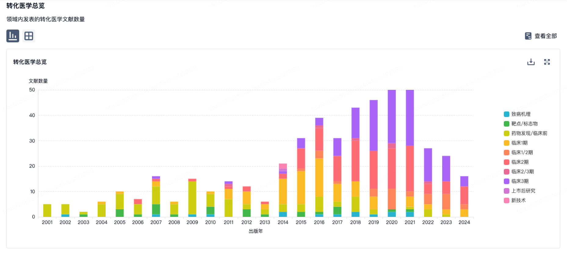
100 项与 Epistem Ltd. 相关的转化医学
登录后查看更多信息
组织架构
使用我们的机构树数据加速您的研究。
登录
或
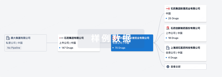
管线布局
2026年07月01日管线快照
管线布局中药物为当前组织机构及其子机构作为药物机构进行统计,早期临床1期并入临床1期,临床1/2期并入临床2期,临床2/3期并入临床3期
临床前
1
登录后查看更多信息
当前项目
登录后查看更多信息
药物交易
使用我们的药物交易数据加速您的研究。
登录
或

转化医学
使用我们的转化医学数据加速您的研究。
登录
或
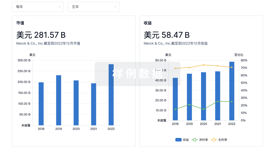
营收
使用 Synapse 探索超过 36 万个组织的财务状况。
登录
或
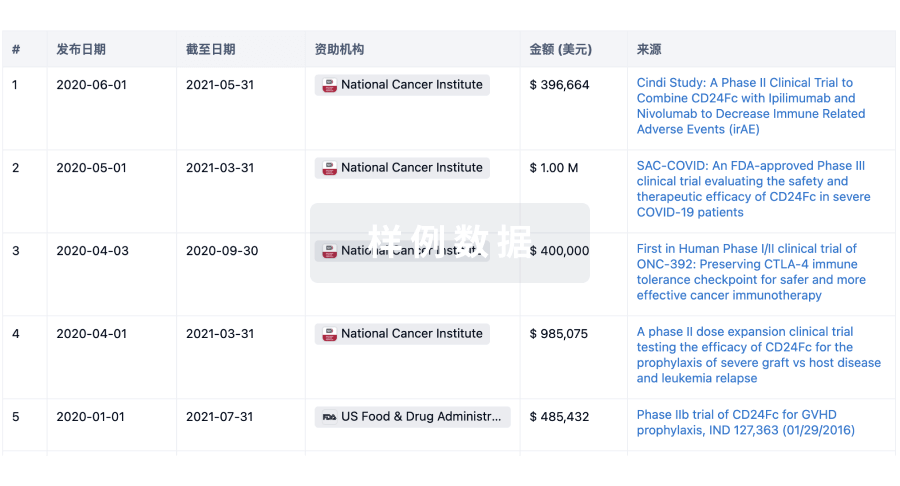
科研基金(NIH)
访问超过 200 万项资助和基金信息,以提升您的研究之旅。
登录
或
投资
深入了解从初创企业到成熟企业的最新公司投资动态。
登录
或
融资
发掘融资趋势以验证和推进您的投资机会。
登录
或
生物医药百科问答
全新生物医药AI Agent 覆盖科研全链路,让突破性发现快人一步
立即开始免费试用!
智慧芽新药情报库是智慧芽专为生命科学人士构建的基于AI的创新药情报平台,助您全方位提升您的研发与决策效率。
立即开始数据试用!
智慧芽新药库数据也通过智慧芽数据服务平台,以API或者数据包形式对外开放,助您更加充分利用智慧芽新药情报信息。
生物序列数据库
生物药研发创新
免费使用
化学结构数据库
小分子化药研发创新
免费使用