预约演示
更新于:2026-03-05

AnaBios Corp.
更新于:2026-03-05
概览
标签
神经系统疾病
心血管疾病
其他疾病
小分子化药
化学药
疾病领域得分
一眼洞穿机构专注的疾病领域

技术平台
公司药物应用最多的技术
靶点
公司最常开发的靶点
关联
靶点 |
作用机制 |
在研机构 |
原研机构 |
在研适应症 |
非在研适应症 |
最高研发阶段 |
首次获批国家/地区 |
首次获批日期 |
靶点 |
作用机制 |
在研机构 |
原研机构 |
在研适应症 |
非在研适应症 |
最高研发阶段 |
首次获批国家/地区 |
首次获批日期 |
100 项与 AnaBios Corp. 相关的临床结果
登录后查看更多信息
登录后查看更多信息
2025-06-03PROCEEDINGS OF THE NATIONAL ACADEMY OF SCIENCES OF THE UNITED STATES OF AMERICA
Modulation of human dorsal root ganglion neuron firing by the Nav1.8 inhibitor suzetrigine
Article
作者: Osorno, Tomás ; Fujita, Akie ; Stewart, Robert G. ; Bean, Bruce P. ; Jo, Sooyeon ; Ferraiuolo, Alyssa ; Carlin, Kevin
Nav1.8 voltage-gated sodium channels are strongly expressed in human primary pain-sensing neurons (nociceptors) and a selective Nav1.8 inhibitor VX-548 (suzetrigine) has shown efficacy for treating acute pain in clinical trials. Nociceptors also express other sodium channels, notably Nav1.7, raising the question of how effectively excitability of the neurons is reduced by inhibition of Nav1.8 channels alone. We used VX-548 to explore this question, recording from dissociated human dorsal root ganglion neurons at 37 °C. Applying VX-548 at 10 nM (about 25 times the IC
50
determined using cloned human Nav1.8 channels at 37 °C) had only small effects on action potential threshold and upstroke velocity but substantially reduced the peak and shoulder. Counterintuitively, VX-548 shortened the refractory period—likely reflecting reduced potassium channel activation by the smaller, narrower action potential—sometimes resulting in faster firing. Generally, repetitive firing during depolarizations was diminished but not eliminated by VX-548. Voltage clamp analysis suggested two reasons that repetitive firing often remains in 10 to 100 nM VX-548. First, many neurons had such large Nav1.8 currents that even 99% inhibition leaves nA-level Nav1.8 current that could help drive repetitive firing. Second, Nav1.7 current dominated during initial spikes and could also contribute to repetitive firing. The ability of human neurons to fire repetitively even with >99% inhibition of Nav1.8 channels may help explain the incomplete analgesia produced by even the largest concentrations of VX-548 in clinical studies.
2025-05-01JOURNAL OF PAIN
Efficient removal of naturally-occurring lipofuscin autofluorescence in human nervous tissue using high-intensity white light
Article
作者: Mannes, Andrew J ; Manalo, Allison P ; Nara, Pranavi ; Ghetti, Andre ; Iadarola, Michael J ; Ramsden, Christopher E ; Talbot, Thomas L ; Ma, Wenting ; King, Diana M ; Maric, Dragan ; Shah, Samay R ; Sapio, Matthew R
Background autofluorescence is enhanced in human tissue relative to small animals and presents a barrier to fully realizing the potential of novel multiplex methods in human studies. In particular, lipofuscin (LF) is an interfering pigment in multiplex fluorescence assays. Lipofuscin (LF) is a highly cross-linked aggregate of oxidized lipids, proteins, sugars, and metal ions that accumulates in lysosomes with age, and is strongly fluorescent across wavelengths that interfere with signals from common fluorophores. This is particularly apparent in dorsal root ganglion (DRG), where the LF deposits occupy up to 80% of the visible neuronal cytoplasm, affecting ∼45% of neurons in a typical section. This report describes a straightforward, scalable, pre-staining, white-light photobleaching method that near-totally reduces LF autofluorescence, and improves signal detection across the color spectrum without negatively impacting the multiplex fluorescence detection assay. It is effective for peripheral and central nervous system structures as well as pathological tissue such as Alzheimer's disease brain, which contains high levels of autofluorescent interference. This demonstrates the broad applicability to improving signal detection in human disease states to enable translational investigations in humans. This low-cost procedure can be rapidly implemented into existing research programs to increase the accessibility of high-plex fluorescent microscopy methodologies to enable direct-in-human research. PERSPECTIVE: White light photobleaching of lipofuscin before multiplex fluorescent in situ hybridization allows for rapid, near-total quenching of autofluorescence in healthy and diseased human nervous system tissue. Given the importance of direct-in-human investigations for validating translational studies and ensuring medical relevance, this simple yet powerful advance enables future anatomical investigations.
2024-10-01Cell Reports Medicine
The μ-opioid receptor differentiates two distinct human nociceptive populations relevant to clinical pain
Article
作者: Iadarola, Michael J. ; Mannes, Andrew J ; King, Diana M. ; Sapio, Matthew R. ; Ghetti, Andre ; Iadarola, Michael J ; Mannes, Andrew J. ; Sapio, Matthew R ; Maric, Dragan ; Staedtler, Ellen S. ; King, Diana M ; Staedtler, Ellen S
The shortfall in new analgesic agents is a major impediment to reducing reliance on opioid medications for control of severe pain. In both animals and man, attenuating nociceptive transmission from primary afferent neurons with a μ-opioid receptor agonist yields highly effective analgesia. Consequently, deeper molecular characterization of human nociceptive afferents expressing OPRM1, the μ-opioid receptor gene, is a key component for advancing analgesic drug discovery and understanding clinical pain control. A co-expression matrix for the μ-opioid receptor and a variety of nociceptive channels as well as δ- and κ-opioid receptors is established by multiplex in situ hybridization. Our results indicate an OPRM1-positive population with strong molecular resemblance to rodent peptidergic C-nociceptors associated with tissue damage pain and an OPRM1-negative population sharing molecular characteristics of murine non-peptidergic C-nociceptors. The empirical identification of two distinct human nociceptive populations that differ profoundly in their presumed responsiveness to opioids provides an actionable translational framework for human pain control.
2026-02-13
·生物谷
2025-11-16
100 项与 AnaBios Corp. 相关的药物交易
登录后查看更多信息
100 项与 AnaBios Corp. 相关的转化医学
登录后查看更多信息
组织架构
使用我们的机构树数据加速您的研究。
登录
或
管线布局
2026年07月02日管线快照
管线布局中药物为当前组织机构及其子机构作为药物机构进行统计,早期临床1期并入临床1期,临床1/2期并入临床2期,临床2/3期并入临床3期
临床前
2
登录后查看更多信息
当前项目
登录后查看更多信息
药物交易
使用我们的药物交易数据加速您的研究。
登录
或
转化医学
使用我们的转化医学数据加速您的研究。
登录
或
营收
使用 Synapse 探索超过 36 万个组织的财务状况。
登录
或
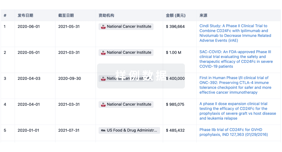
科研基金(NIH)
访问超过 200 万项资助和基金信息,以提升您的研究之旅。
登录
或
投资
深入了解从初创企业到成熟企业的最新公司投资动态。
登录
或
融资
发掘融资趋势以验证和推进您的投资机会。
登录
或
生物医药百科问答
全新生物医药AI Agent 覆盖科研全链路,让突破性发现快人一步
立即开始免费试用!
智慧芽新药情报库是智慧芽专为生命科学人士构建的基于AI的创新药情报平台,助您全方位提升您的研发与决策效率。
立即开始数据试用!
智慧芽新药库数据也通过智慧芽数据服务平台,以API或者数据包形式对外开放,助您更加充分利用智慧芽新药情报信息。
生物序列数据库
生物药研发创新
免费使用
化学结构数据库
小分子化药研发创新
免费使用