预约演示
更新于:2026-05-11
GPC3 x Trop-2
更新于:2026-05-11
关联
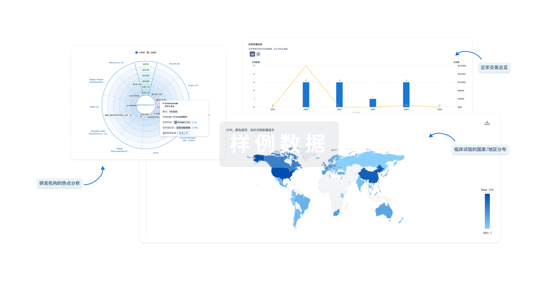
100 项与 GPC3 x Trop-2 相关的临床结果
登录后查看更多信息
100 项与 GPC3 x Trop-2 相关的转化医学
登录后查看更多信息
0 项与 GPC3 x Trop-2 相关的专利(医药)
登录后查看更多信息
3
项与 GPC3 x Trop-2 相关的文献(医药)2026-04-01·PET Clinics
Novel Theranostic Targets for Prostate Cancer Beyond Prostate-Specific Membrane Antigen
Review
作者: Kako, Bashar ; Heidari, Pedram ; Esfahani, Shadi A ; Jadvar, Hossein
Prostate-specific membrane antigen (PSMA)-targeted theranostics have transformed prostate cancer management, but some tumors exhibit low or heterogeneous PSMA expression, limiting eligibility and efficacy for radiopharmaceutical therapy. This review explores emerging biomarkers that may complement or expand beyond PSMA, including gastrin-releasing peptide receptor, fibroblast activation protein, prostate stem cell antigen, CD46, delta-like ligand 3, glypican-3, B7-H3, 6-transmembrane epithelial antigen of the prostate-1, trophoblast cell surface antigen-2, CEACAM5, and vicrostatin. We review their molecular biology, preclinical validation, clinical translation, and ongoing trials. These biomarkers represent diverse biological compartments, broadening the scope of precision radiotheranostics for prostate cancer.
2014-12-01·Archives of pathology & laboratory medicine3区 · 医学
Evolving Practices of Diagnostic Immunohistochemistry
3区 · 医学
Article
作者: Lin, Fan
It has been more than 6 years since the Archives of Pathology & Laboratory Medicine published the special issue on diagnostic immunohistochemistry (IHC) consisting of a series of review articles edited by Jaishree Jagirdar, MD, which was a great, concise, and yet comprehensive review and update on important IHC markers, panels, and diagnostic strategies.1 It has been my great honor and immense privilege to organize this new special issue on IHC, contributed to by pathologists from Geisinger Medical Center (Danville, Pennsylvania) and many expert pathologists from others medical centers. This 2-part special issue features 14 review articles with an attempt to cover IHC automation, standardization of diagnostic IHC, and the role of IHC in diagnosing tumors in major organs and tumors of unknown primary.This series begins with an article emphasizing standardization of diagnostic IHC in the preanalytic, analytic, and postanalytic phases, with a specific focus on (1) newly proposed guidelines on antibody validation from the College of American Pathologists Pathology and Laboratory Quality Center, (2) testing/optimizing a new antibody and troubleshooting, (3) interpreting and reporting IHC assay results, (4) continuing quality improvement programs, and (5) developing and implementing the concept of best practices in IHC. When it comes to the question of how to implement best practices, emphasis is placed on the evidence-based application of IHC markers to practical scenarios, which includes eliminating unnecessary markers, starting with a small IHC panel and continuing with a second panel only when the first panel leads to an inconclusive result. Two questions should be asked before ordering any IHC assay: “Why should I order this marker?” and “Does it change my diagnosis and patient care?” In addition, some external quality control programs and College of American Pathologists checklists for inspecting a clinical IHC laboratory are outlined. The role of digital pathology in the field of diagnostic IHC is briefly discussed as well.The second review article compares and contrasts the recent technological advances in new generation automated IHC platforms from all major companies. Some advantages over the prior automated platforms are stressed, including complete walkaway automation, faster speed, use of less reagents, user friendliness, better integration with laboratory information systems, effective waste control, capability for multiplexing, and more reliable and reproducible results.One of the most frequent and important applications of diagnostic IHC is to assist in working up an undifferentiated neoplasm/tumor of uncertain primary site. The third review article comprehensively reviews some diagnostic strategies and algorithms, updates many recently described diagnostic markers such as GATA binding protein 3 (GATA3), placental S100 (S100P), mammary serine protease inhibitor (maspin), von Hippel-Lindau tumor suppressor gene protein (pVHL), paired box gene (PAX) 8, ETS-related gene (ERG), sal-like protein 4 (SALL4), sex-determining region Y box (SOX) 10, arginase-1, napsin A, special AT-rich sequence-binding protein 2 (SATB2), and cadherin-17, and refines the diagnostic IHC panels frequently used in daily practice.The remaining 11 review articles are devoted to organ-specific diagnostic IHC. In the lung and pleural review article, p40, desmoglein-3, desmocollin-3, and cytokeratin (CK) 5 are described as the most effective panel of IHC markers for squamous cell carcinoma; hepatocyte nuclear factor 4 α (HNF4α) is demonstrated to be a marker for invasive mucinous adenocarcinoma of the lung; and glucose transporter 1 (Glut1), insulin-like growth factor II messenger RNA–binding protein (IMP3), and cluster of differentiation (CD) 146 are shown to be helpful in differentiating reactive conditions from malignant mesothelial proliferations.In breast pathology, GATA3 has been reported in several publications to be expressed in most breast ductal and lobular carcinomas, including more than 50% of ER-negative breast carcinomas and metaplastic carcinomas. Ankyrin repeat domain 30A (NY-BR-1) is another newly described marker useful in identifying a breast primary, although in limited publications. Numerous predictive biomarkers have been evaluated, and additional studies are needed to confirm the initial findings.In genitourinary pathology, PAX8 and PAX2 have proven to be important diagnostic markers for identifying renal cell carcinoma when working on a tumor of unknown primary. ERG is a specific but not sensitive marker for prostatic adenocarcinoma. In contrast, NK3 homeobox 1 (NKX3.1) may become a highly sensitive and specific marker for prostatic adenocarcinoma if additional publications substantiate the initial reports. In limited studies, uroplakin (UP) II has proven to be a more sensitive marker than UPIII for identifying urothelial carcinoma. GATA3 and S100P are useful markers for urothelial carcinoma. A new generation of germ cell tumor markers, including SALL4, octamer-binding transcription factor 4 (OCT4), lin-28 homolog A (LIN28), SOX2, GATA3, and Nanog homeobox (Nanog), has proven to be superior to traditional germ cell tumor markers.In the field of lymphomas, a group of relatively sensitive and specific biomarkers has been identified for various types of lymphomas: (1) human germinal center–associated lymphoma (HGAL)/GCET2, LIM-only transcription factor 2 (LMO2), and stathmin for follicular lymphoma; (2) lymphoid enhancer-binding factor 1 (LEF1), CD200, and membranous CD160 has been present in virtually all neoplastic cells of B-cell chronic lymphocytic leukemia/small lymphocytic lymphoma; (3) SOX11 expression has been found in almost all cases of mantle cell lymphoma, including both cyclin D1–positive and cyclin D1–negative types, as well as cyclin D1–negative blastoid mantle cell lymphoma; (4) immunoglobulin superfamily receptor translocation–associated 1 (IRTA1) is selectively expressed on the surface of neoplastic cells of extranodal and nodal marginal zone lymphomas; (5) c-MYC could detect increased MYC protein expression by both MYC translocation and MYC overexpression, which could be used as a screening test to select the cases for further investigation using the fluorescence in situ hybridization study; and (6) IMP3 cytoplasmic staining has been found in Hodgkin cells of almost all cases, superior to other IHC markers such as CD15, CD30, PAX5, and multiple myeloma oncogene 1 (MUM1).In part II, the following review articles will be presented.In the gastrointestinal, liver, and pancreatobiliary area, 2 new markers, SATB2 and cadherin-17, have been useful in working on gastrointestinal tract tumors. SATB2 appears to be more sensitive than caudal type homeobox 2 (CDX2) and CK20 in identifying colorectal carcinomas; the SATB2 positivity has been reported in CDX2-negative poorly differentiated colorectal carcinomas and medullary carcinomas of the large intestine. SATB2 is frequently positive in colorectal neuroendocrine neoplasms but negative in neuroendocrine neoplasms from other organs. In contrast, cadherin-17 is a positive marker for both lower and upper gastrointestinal adenocarcinomas and neuroendocrine neoplasms. Maspin, S100P, pVHL, and IMP3 have been reported to be a reliable panel of markers to differentiate a carcinoma from reactive ducts in the pancreatobiliary tract, carcinoma being negative for pVHL and positive for S100P, maspin, and IMP3 and reactive ducts positive for pVHL and negative for maspin, S100P, and IMP3. Interestingly, intrahepatic cholangiocarcinoma tends to be positive for pVHL (70% of cases), which can be potentially used in the distinction of a metastatic pancreatic adenocarcinoma from an intrahepatic cholangiocarcinoma.In liver, a panel of 4 markers, including liver fatty acid–binding protein (LFAB), serum amyloid associate protein (SAA), C-reactive protein (CRP), glutamine synthetase (GS), and hepatocyte nuclear factor 1α (HNF1α), has been proposed for further classifying a hepatocellular adenoma (HCA) into 4 major subtypes: HNF1α-mutated HCA, β-catenin–mutated HCA, inflammatory HCA, and unclassified HCA. Within intrahepatic cholangiocarcinomas, 10% of tumors may also be β-catenin mutated. Arginase 1 is a more sensitive and specific marker for identifying a hepatocellular carcinoma than hepatocyte paraffin 1 (HepPar1) and glypican 3. Arginase 1, like HepPar1, is also positive in normal liver and benign hepatic lesions.In gynecologic pathology, the loss of p57 expression in the villous cytotrophoblasts and stromal cells is a characteristic feature of complete moles; hepatocyte nuclear factor 1β (HNF1β) is a useful marker for identifying clear cell carcinoma of the uterus and ovary and is usually negative in papillary serous carcinoma; pVHL and kidney injury molecule 1 (KIM-1) have been reported to be helpful in the distinction between clear cell carcinoma of the uterus and ovary from serous carcinoma; forkhead box L2 (FOXL2) and steroidogenic factor 1 (SF-1) have been reported to be sensitive and specific markers for sex cord stromal tumors. SF-1 is also a useful marker for identifying an adrenal cortical neoplasm.In head and neck pathology, discovered on gastrointestinal stromal tumor protein 1 (DOG1) expression was seen in normal serous acini, mucous acini, and distal intercalated ducts. All acinic cell carcinomas were positive for DOG1, with predominantly apical/luminal membranous staining. Most ductal tumors were negative, but DOG1 immunoreactivity can be seen in mammary analogue secretory carcinomas and pleomorphic low-grade adenocarcinomas. No DOG1 immunoreactivity was seen in salivary duct carcinomas, oncocytomas/oncocytic carcinomas, myoepitheliomas, Warthin tumors, or sebaceous lymphadenoma. GATA3 was positive in 100% of mammary analogue secretory carcinomas and salivary duct carcinomas and a much lower percentage of other salivary gland tumors. Overexpression of p16 is highly correlated with human papillomavirus–positive squamous cell carcinomas of oropharynx and oral cavity; p16 immunostain is considered a very sensitive surrogate biomarker for human papillomavirus infection and has been routinely performed on all squamous cell carcinomas of the oropharynx and oral cavity. Our group has reported that tumor-associated calcium signal transducer 2 (TROP2) is a potential diagnostic marker for papillary thyroid carcinoma; it appears to have a better diagnostic specificity than the traditional markers, such as CK19, Hector Battifora mesothelial 1 (HBME1) and galectin 3. Additional studies from others would need to validate our findings.In the skin review article, androgen receptor and adipophilin expression support the diagnosis of sebaceous carcinoma over squamous cell carcinoma; clonal integration of the newly discovered Merkel cell polyomavirus has been shown in the great majority of Merkel cell carcinomas; claudin-1 and Glut1 are stronger and stain more diffusely in perineurioma than epithelial membrane antigen (EMA); langerin (CD207) has shown diagnostic sensitivity similar to CD1a but with improved specificity for Langerhans cell histiocytosis; friend leukemia virus integration 1 (FLI1) and ERG have been reported as nuclear markers of endothelial differentiation and are highly sensitive for benign and malignant vascular tumors; positive staining for p63, podoplanin (D2-40), and CK15 favors a primary cutaneous adnexal neoplasm over metastatic adenocarcinoma; and Wilms tumor 1 (WT1) cytoplasmic endothelial expression has been reported in vascular tumors but is lacking in lymphatic and venous vascular malformations.In soft-tissue pathology, SATB2 has been reported to be a marker for a sarcoma with osteoblastic differentiation; cancer/testis antigen 1 (NY-ESO-1) has been introduced as a marker for myxoid and round cell liposarcoma; mucin 4 (MUC4) has proven to be a marker for low-grade fibromyxoid sarcoma and sclerosing epithelioid fibrosarcoma; NK2 homeobox 2 (NKX2.2) has been shown to be a more specific marker than CD99 for Ewing sarcoma; mouse double minute 2 homolog (MDM2), cyclin-dependent kinase 4 (CDK4), and p16 are good markers for well-differentiated and dedifferentiated liposarcomas; ERG has been demonstrated to be the most sensitive marker for benign and malignant vascular tumors; subsets of perivascular epithelioid cell tumors and epithelioid hemangioendothelioma are positive for transcription factor E3 (TFE3); succinate dehydrogenase (SDHB) has shown loss of expression in a subset of pediatric gastrointestinal stromal tumors and paragangliomas; signal transducer and activator of transcription 6 (STAT6) has been reported in most solitary fibrous tumors; and loss of integrase interactor 1 (INI-1) is seen in most epithelioid sarcomas and some myoepithelial carcinomas.Because of the limited scope of this 2-part special issue, applications of IHC in other important areas, such as central nervous system tumors and infectious agents, cannot be included. Isocitrate dehydrogenase 1 (IDH1) has been reported to be a useful marker for differentiating glioma from reactive gliosis. The association of viruses with their associated tumors can be demonstrated, such as Merkel cell polyomavirus in Merkel cell carcinoma, Epstein-Barr virus in Burkitt lymphoma, and human herpesvirus 8 in Kaposi sarcoma and body cavity lymphoma.Genetic alterations of tumors are traditionally detected by molecular analysis, such as cytogenetics, fluorescence in situ hybridization, or polymerase chain reaction–based assay. With the advances in IHC technologies and rapidly growing knowledge of molecular pathology in the last few years, IHC has entered a new era and is now able to provide specific genetic information about certain tumors. One of the breakthroughs is the generation of mutation-specific antibodies directly against the mutated proteins, such as epidermal growth factor receptor (EGFR) gene L858R and exon 19 deletion mutation in lung adenocarcinoma; BRAF gene V600E mutation in papillary thyroid carcinoma, melanoma, and other cancers; and IDH1 gene R132H mutation in glioma.2 Specific chromosomal translocations or specific gene rearrangements can be demonstrated through the detection of the related protein products such as B-cell chronic lymphocytic leukemia/lymphoma (Bcl2) in follicular lymphoma, cyclin D1 in mantle cell lymphoma, anaplastic lymphoma kinase (ALK) in lung adenocarcinoma, TFE3 in alveolar soft part sarcoma, and XP11 translocation renal cell carcinoma and c-MYC in Burkitt lymphoma and some postradiation and lymphedema-associated angiosarcomas.2 The deletion or loss of gene function can also be demonstrated by loss of the expression of related protein products, such as INI-1 in epithelioid sarcomas, renal medullary carcinomas, rhabdoid tumors, and atypical teratoid/rhabdoid tumors; E-cadherin in lobular breast carcinomas and some signet ring cell gastric carcinomas; and mismatch repair proteins (MutL homolog 1 [MLH1], MutS protein homolog [MSH]2, MSH6, and postmeiotic segregation increased 2 [PMS2]) in microsatellite instability tumors.2 Gene amplification can be detected through the overexpression of related protein products, such as human epidermal growth factor receptor 2 (Her2) in breast carcinomas and gastroesophageal/gastric carcinomas and MDM2 and CKD4 in well-differentiated liposarcomas/some dedifferentiated liposarcomas.2In summary, there is no doubt that IHC will continue playing an important role in surgical pathology and cytopathology, with additions of more sensitive and specific biomarkers and more well-defined yet smaller IHC panels for each diagnosis and differential diagnosis. Furthermore, more antibodies that provide genetic information or predictive marker information, especially mutation-specific antibodies, are on their way to clinical IHC laboratories.
Frontiers in Immunology
Glypican 3-targeted chimeric antigen receptor T cells secreting TROP2-directed bispecific T cell engagers exhibit potent efficacy against lung squamous cell carcinoma
Article
作者: Lin, Liming ; Ruan, Lianjie ; Lin, Mengqing ; Zhang, Haiqing ; Lin, Dekai ; Weng, Suiyan
Background:
Chimeric antigen receptor T cell (CAR-T) therapy faces multiple challenges in solid tumors, especially the heterogeneity of tumor antigens. Glypican-3 (GPC3) and trophoblast cell-surface antigen 2 (TROP2) are highly expressed antigens in lung squamous cell carcinoma (LUSC) for development of dual-targeted therapy. The absence of GPC3 in any normal tissues of adults makes it an ideal target for CAR-T therapy. However, TROP2 is expressed in the epithelial cells of various normal tissues and thus is not acceptable for direct design of CAR-T therapy due to the high risk of “on-target off-tumor” effects. Here we developed a dual-targeted LUSC therapy featuring a GPC3-targeted CAR-T cell secreting TROP2-directed bispecific T cell engagers (GPC3 CAR-T. TROP2 BiTE), and verified the antitumor activity
in vitro
and
in vivo
, respectively.
Methods:
Immunohistochemistry (IHC) was used to confirm the expression of GPC3 and TROP2 in LUSC and normal tissues. CAR-T cells were produced through lentiviral transduction of CAR genes. Real-time cytotoxicity assay (RTCA) was used to assess the cytotoxic effect of CAR-T cells on LUSC cells. Flow cytometry was utilized to examine the CAR-T cell phenotype, exhaustion and activation. Enzyme-linked immunosorbent assay (ELISA) was performed to detect the release of cytokines. To evaluate the activity of CAR-T cells
in vivo
, tumor-bearing immunodeficient mice were given a single intravenous injection of CAR-T cells, and the tumor burden and CAR-T cell expansion were regularly monitored.
Results:
GPC3 was overexpressed in 70% of LUSC tissues, while negatively expressed in all normal tissues. Positive expression of TROP2 was observed in all LUSC tissues and also in many normal tissues. Compared with GPC3 CAR-T and TROP2 CAR-T, GPC3 CAR-T. TROP2 BiTE exhibited cytotoxicity to both GPC3
+
and TROP2
+
LUSC cells, and thereby showed faster killing and durable antitumor effect against LUSC cells with heterogenous expression of GPC3 and TROP2. In tumor-bearing mice, GPC3 CAR-T. TROP2 BiTE showed strong ability to eliminate tumors.
Conclusions:
This study demonstrated that GPC3 CAR-T. TROP2 BiTE was a potent therapy for LUSC and provided a strategy for overcoming the antigen heterogeneity in solid tumors.
162
项与 GPC3 x Trop-2 相关的新闻(医药)2026-05-08
·泽蓝恒研
Part.01
大会背景:全球肿瘤学界的“风向标”即将开启
2026年美国临床肿瘤学会(ASCO)年会将于5月29日至6月2日在芝加哥盛大举行。本届大会以“转化科学与实践:改善全球癌症结局”为主题,强调基础研究、转化医学与临床实践的高效衔接。
作为全球肿瘤领域规格最高、影响力最大的顶级学术盛会,ASCO年会每年集中展示全球临床肿瘤学的前沿科研成果与创新治疗方案,是全球肿瘤学界的“风向标”
从已披露的摘要标题看,全球肿瘤研发更加聚焦“可转化、可注册、可改变临床实践”的高质量证据,围手术期、一线治疗等前线布局持续升温,PFS(无进展生存期)、OS(总生存期)等硬终点成为创新药价值判断的核心。
Part.02
中国军团的突破:13项研究入选LBA,创历史新高
本届ASCO大会上,中国创新药企的表现呈现鲜明的“质量驱动”特征。据东吴证券统计,12家中国创新药企的13项研究入选LBA(最新突破摘要),创历史新高。
2.1 入选数量与质量双飞跃
年份
中国药企LBA入选数量
入选全体大会研究数
核心特征
2015年
0项
0项
仅1项新药研究入选口头报告
2025年
11项
0项
开始规模化突破2026年13项1项量质齐升,硬终点成锚点
这一数据背后,是中国创新药十年磨一剑的厚积薄发。从2015年仅有1项新药研究入选口头报告,到如今13项研究登顶LBA,中国药企已从“跟跑者”转变为“并跑者”,部分领域甚至实现“领跑”。
2.2 康方生物:唯一入选全体大会的中国研究
本届ASCO最大的亮点之一,是由上海市胸科医院陆舜教授牵头、康方生物主导的HARMONi-6研究,以LBA4的身份重磅亮相全体大会(Plenary Session),成为今年唯一获此殊荣的中国研究,也是ASCO历史上第二个入选全体大会的中国本土创新药。
Plenary Session作为ASCO年会的最高规格发布平台,含金量极高。国金证券研报指出,该环节每年仅遴选5项顶尖LBA研究,代表着当年全球肿瘤治疗领域的最高突破与发展方向
此次国产双抗方案成功跻身该环节,打破了海外药企长期垄断顶级学术核心赛道的格局。
2.3 HARMONi-6研究:头对头挑战“药王”
HARMONi-6是一项大型III期关键临床试验,核心是头对头对照评估依沃西单抗联合化疗,与替雷利珠单抗联合化疗,用于晚期鳞状非小细胞肺癌一线治疗的疗效与安全性。
值得注意的是,替雷利珠单抗目前是国内销量最高的PD-1抑制剂,此次头对头对照研究的结果,对肺癌治疗格局具有重要影响。去年ESMO期间,HARMONi-6研究已公布期中分析结果:依沃西单抗联合化疗组中位无进展生存期(mPFS)达11.14个月,较现有标准治疗方案显著延长4.24个月,PFS风险比0.60,差异具备极高统计学显著性(P<0.0001)。
本届ASCO全体大会上,该研究将公布总生存期(OS)这一核心终点数据,不仅能进一步夯实依沃西单抗的疗效优势,更有望推动晚期肺鳞癌治疗进入双抗联合化疗的新时代。
Part.03
双抗与ADC:中国创新药的两大“王牌”
3.1 IO 2.0时代:双抗数据接棒验证
中国企业正引领全球肿瘤免疫2.0时代。除康方生物外,信达生物IBI363(PD-1/IL-2双抗)、基石药业CS2009(PD-1/VEGF/CTLA-4三抗)等新一代IO品种也将更新数据,为PD-1耐药后的治疗格局提供新的解法。
信达IBI363作为全球首款PD-1/IL-2α-bias双抗,并非简单沿袭旧假设,而是通过自主尝试和生物学研究重新拆解IL-2三受体轴的调控逻辑,为多家跨国药企在IL-2路径上反复受挫的问题提供了新的可行性验证方向,成为下一代IO的有力竞争者。
3.2 ADC赛道:从追赶到并跑
ADC(抗体偶联药物)是中国创新药走向全球舞台最集中的领域之一。本届ASCO,中国ADC管线覆盖多个热门赛道:
科伦博泰的sac-TMT(TROP2 ADC)联合帕博利珠单抗(K药)一线治疗PD-L1阳性NSCLC的III期结果将进行口头报告,这是全球首个在该适应证中取得阳性结果的ADC+免疫联合疗法III期研究,具备里程碑式意义。
百利天恒的iza-bren(BL-B01D1),作为全球首个进入临床III期的EGFR×HER3双抗ADC,在三阴性乳腺癌的III期数据入选LBA,另有HER2 ADC、DLL3 ADC等多条管线集中亮相,充分体现了中国ADC平台从跟随创新向全球竞争力的跃迁。
其他值得关注的研究:
迪哲医药:WU-KONG28研究成果入选LBA口头报告
中国生物制药:贝莫苏拜单抗联合化疗及安罗替尼一线治疗晚期非鳞非小细胞肺癌III期研究入选LBA
百济神州:将公布GPC3/4-1BB双抗数据(入选口头报告)
乐普生物:MRG006A(GPC3 ADC)治疗肝癌的首次人体数据将展示
Part.04
硬终点时代:从“有数据”到“OS说话”
本届ASCO中国药企聚焦PFS、OS等硬终点的高等级证据显著增多。市场和学术界对创新药的评价标准正从机制创新转向能否在随机对照研究中带来明确生存获益。
这意味着数据的“含金量”将直接影响相关公司的价值重估节奏。从数据披露结构看:
PFS优势 → 可推动注册路径和商业化预期
OS获益 → 真正决定能否改写临床指南、成为标准疗法
方正证券指出,康方生物依沃西的OS数据若积极,有望从PFS优势升级为生存获益证据,形成注册路径及商业化预期的重要催化。
Part.05
产业逻辑:BD出海加速与政策红利共振
5.1 BD出海进入“质量驱动”阶段
ASCO数据的密集披露只是催化剂,创新药板块背后有着更深层的产业逻辑支撑。AACR大会刚刚落幕,中国药企在ADC、多抗、TCE等领域的前沿布局已引发全球MNC关注,而ASCO上高等级临床数据的读出,有望促成新一轮重磅License-out交易。
东吴证券强调,数据本身的强度——包括生存获益是否明确、安全性窗口是否可控、结果是否具备海外开发外推性——将是判断后续BD交易和商业价值的关键。
5.2 政策端协同发力
国务院《关于健全药品价格形成机制的若干意见》对创新药首发价格留足空间,商保创新药目录加速落地,支付端从单一医保向多元化转变,为创新药的商业化回报提供了更可预期的制度保障。
5.3 “快与省”的效率飞轮
波士顿咨询(BCG)在2026年3月的深度报告中指出,中国创新药之所以能形成今天的数量级,关键在于“快”与“省”两大支柱:
维度
中国优势
对比美国
药物分子发现成本
仅美国的20%–30%
节省70%以上
临床前开发成本
节省至少一半
效率优势显著
临床入组速度
依托庞大患者基数
显著加速
通过“理解—试错—执行”的闭环,中国将信息、知识与工程能力转化为持续产出的创新成果。
Part.06
市场表现与投资展望
6.1 资本市场提前反应
4月28日,生物医药板块迎普涨,生物医药ETF(512290)大涨超3%,创新药ETF(517110)、疫苗ETF(159643)、医疗ETF(159828)大涨超2%。
港股通创新药ETF(159570)全天成交额超12亿元,此前10日已累计净流入超52亿元,截至4月24日,规模已超309亿元,全市场医药类ETF遥遥领先。
6.2 投资主线
从“数量爆发”到“OS说话”,创新药板块的催化剂密度和深度均在升级。在临床价值兑现与BD出海加速的双重驱动下,市场关注以下几大主线:
双抗赛道:康方生物、信达生物、基石药业等二代IO品种
ADC赛道:科伦博泰、百利天恒、恒瑞医药等
小分子靶向药:迪哲医药、百济神州等
政策受益标的:创新药商业化价值有望充分释放
Part.07
风险提示
尽管前景光明,仍需关注以下风险:
风险类型
具体内容
行业政策风险
集采、医保支付政策变化对行业发展的影响
研发不及预期风险
临床入组进度、疗效及安全性数据的不确定性
审批不及预期风险
资料补充、审批流程变化导致的审批周期延长
宏观环境波动风险
全球经济增速放缓、国际关系、汇率及利率风险
Part.08
结语:中国创新药的“成人礼”
2026年ASCO,中国创新药军团以13项LBA的亮眼成绩,完成了从“拼数量”到“拼硬实力”的华丽蜕变。这不仅是数据量的提升,更是临床证据质量的全面升级——从“有数据”走向“有硬终点”,从“me-too”走向“同类首创”和“同类最优”。
正如波士顿咨询所言,中国医药创新正在经历一轮质量飞跃。本土企业信心显著抬升,越来越多中国新药选择与全球重磅药物开展头对头III期临床试验。当下,价值与竞争力正在被全球市场定价,中国创新正式进入被验证、可兑现的新阶段
ASCO 2026,中国军团已就位。让我们共同期待5月29日-6月2日芝加哥的“中国时刻”。
免责声明:本文内容基于公开信息和券商研报整理,提及个股仅用于行业事件分析,不构成任何投资建议。投资有风险,入市需谨慎。
2026-05-06
摘要:嵌合抗原受体 T 细胞(CAR-T)疗法已在血液系统恶性肿瘤治疗中取得突破性成功,但传统体外制备模式存在制备流程复杂、成本高昂、可及性差、规模化生产受限等核心瓶颈。体内 CAR-T 细胞工程通过在患者体内直接重编程内源性免疫细胞表达 CAR,无需细胞采集、体外扩增与回输,为突破现有局限提供了变革性解决方案。本文系统梳理体内 CAR-T 疗法的核心优势、主流递送系统与工程化设计策略,总结其临床转化进展,分析当前领域面临的关键挑战与优化方向,为该技术的后续研究与临床开发提供参考。
引言
过去十余年,CAR-T 细胞疗法彻底革新了血液肿瘤的治疗格局,多款产品获批用于复发/难治性 B 细胞淋巴瘤、多发性骨髓瘤等疾病,显著延长了患者生存期。目前所有获批的 CAR-T 产品均依赖体外自体细胞制备:从患者外周血分离 T 细胞,体外激活、基因修饰使其表达 CAR,经大规模扩增后回输至患者体内。尽管临床疗效确切,该模式仍存在不可忽视的局限:制备周期长、产品保质期短、对患者 T 细胞质量依赖性高、需清淋巴预处理,且单疗程治疗费用极高,极大限制了疗法的可及性与规模化应用。
体内 CAR-T 细胞工程是指通过靶向递送系统,将 CAR 编码基因直接递送至患者体内的内源性 T 细胞,使其原位重编程为功能性 CAR-T 细胞的技术体系。该技术彻底绕过了体外细胞制备的全流程,从根本上解决了传统 CAR-T 的核心痛点,近年来在载体工程、靶向设计与临床转化方面均取得重大进展,已成为肿瘤免疫治疗领域的前沿研究方向。本文基于最新研究成果,对体内 CAR-T 技术的研究现状与发展前景进行系统综述。
一、体内 CAR-T 疗法相较传统体外 CAR-T 的核心优势
体内 CAR-T 不仅简化了治疗流程,更在生物学特性、安全性、可及性等方面展现出多重优势,核心差异与特点如下:
治疗流程极简,可及性大幅提升:无需细胞采集、体外操作与严格的冷链运输,单剂量静脉/局部给药即可完成治疗,突破了中心化制备的地域限制,且具备规模化生产潜力。行业预估体内 CAR-T 单剂量成本可低至 5000 美元,远低于传统 CAR-T 数十万美元的治疗费用。
保留 T 细胞天然特性,降低耗竭风险:体内原位重编程的 CAR-T 细胞从少量转导 T 细胞渐进式扩增,避免了体外长期扩增导致的细胞分化与耗竭,可维持干细胞样记忆表型,在更低剂量下即可实现强效抗肿瘤应答,且体内存续能力更优。
降低预处理相关毒性,保留免疫完整性:体内 CAR-T 可利用患者内源性 T 细胞实现重编程,无需清淋巴化疗预处理,显著降低了感染、骨髓抑制等并发症风险;完整的免疫系统可促进抗原表位扩散,拓宽抗肿瘤应答,减少靶抗原逃逸导致的治疗失败。
治疗窗口灵活,安全性可控性更强:非病毒载体介导的瞬时 CAR 表达可通过重复给药动态调控表达水平,避免永久整合带来的长期风险;即便出现不良事件,也可快速终止治疗,相较体外 CAR-T 更易管控毒性。
二、体内 CAR-T 的核心递送系统与工程化策略
体内 CAR-T 疗法成功的核心前提是高效、靶向、安全的基因递送,需在复杂的体内环境中精准将 CAR 编码基因递送至 T 细胞,同时最小化脱靶效应。目前主流递送系统分为病毒载体与非病毒载体两大类,其靶向工程化核心策略见图 1,各类载体的核心特征对比见表 1。
图 1 靶向体内 CAR-T 细胞工程的核心策略(慢病毒与非病毒递送系统)
表 1 体内 CAR-T 疗法递送载体平台汇总
2.1 病毒载体递送系统
病毒载体是目前体内 CAR-T 研究中最成熟的递送工具,凭借高效的转导效率与长期转基因表达能力,成为率先进入临床阶段的技术路线,核心类型包括慢病毒、γ- 逆转录病毒、腺相关病毒(AAV)与病毒样颗粒(VLP)。2.1.1 慢病毒(LV)载体
慢病毒是体内 CAR-T 应用最广泛的病毒载体,可高效转导分裂与非分裂细胞,能将大片段 CAR 转基因整合至宿主基因组,实现长期稳定的 CAR 表达。为解决野生型慢病毒广谱嗜性导致的脱靶转染问题,研究者主要通过 3 种工程化策略实现 T 细胞特异性靶向:
包膜蛋白假型化改造:传统 VSV-G 假型化慢病毒通过 LDLR 介导进入细胞,嗜性广、易被血清补体灭活,不适合体内给药。目前主流采用可可病毒(Cocal)包膜、尼帕病毒(NiV)包膜、麻疹病毒包膜替代 VSV-G,其中可可包膜保留了高滴度优势,同时显著提升人血清抗性;尼帕病毒包膜可在细胞膜直接介导膜融合,无需内体逃逸,且抗血清中和能力强,已被证实可在体内高效靶向转导 CD8+ T 细胞。
表面靶向分子修饰:通过基因工程在包膜表面展示抗 CD3/CD4/CD7/CD8 scFv、单克隆抗体或靶向配体,实现 T 细胞特异性识别。例如 Umoja 公司的 VivoVec 平台,在慢病毒表面表达含抗 CD3 scFv、CD58、CD80 的多结构域融合蛋白,同时实现 T 细胞靶向与共刺激激活,在非人灵长类模型中无需清淋巴即可实现高效体内 CAR-T 生成。此外,还可通过点击化学、桥接抗体等化学偶联方式,将靶向抗体共价连接至病毒包膜,实现灵活的靶向重定向。
转录水平靶向调控:通过插入 T 细胞特异性启动子、miRNA 靶位点,使 CAR 仅在 T 细胞中表达,进一步沉默非靶细胞中的转基因表达,降低脱靶风险。2.1.2 其他病毒载体
γ- 逆转录病毒:仅可转导分裂细胞,对静息 T 细胞转导效率低,体内应用受限。目前仅用于植入式支架辅助的局部递送,如 MASTER 系统,通过藻酸盐支架共装载逆转录病毒颗粒与 T 细胞激活抗体,皮下植入后原位生成 CAR-T 细胞,但该方案仍需预加载外源性 T 细胞,并非完全体内重编程。
AAV 载体:无包膜、不整合宿主基因组,通过游离体实现中长期基因表达,免疫原性低、安全性高,但包装容量有限,仅少数研究将其用于体内 CAR-T 递送,目前仍处临床前早期阶段。
VLP 载体:由病毒结构蛋白组装而成,无感染性、不整合基因组,可包裹 mRNA 实现瞬时递送,大幅降低插入突变风险,同时保留了病毒的高效细胞穿透能力。最新研究开发的 T 细胞特异性融合 VLP,可选择性向体内 T 细胞递送 CAR mRNA,实现瞬时 CAR 表达与肿瘤控制,无脱靶转染,是极具潜力的下一代病毒载体。2.2 非病毒载体递送系统
病毒载体存在插入突变风险、免疫原性与规模化生产限制,非病毒载体凭借低免疫原性、合成简便、可规模化、无基因组整合风险等优势,成为体内 CAR-T 领域的核心研发方向,其中脂质纳米颗粒(LNP)与聚合物纳米颗粒是两大主流系统。2.2.1 脂质纳米颗粒(LNP)系统
LNP 是目前最成熟的非病毒核酸递送平台,已在 mRNA 疫苗中实现大规模临床应用,也是体内 CAR-T 非病毒递送的核心工具。标准 LNP 由可电离阳离子脂质、磷脂、胆固醇、PEG 化脂质四部分组成,通过静电作用包裹核酸载荷,进入细胞后通过内体逃逸释放核酸至胞质。
为实现 T 细胞特异性靶向,主流策略是通过马来酰亚胺 - 硫醇化学,将抗 CD3/CD5/CD8 靶向抗体偶联至 LNP 表面,避免肝脏非特异性摄取。例如 Carl June 团队开发的 CD8 靶向 LNP,采用新型可电离脂质 L829 包裹抗 CD19 CAR mRNA,在啮齿类与非人灵长类模型中实现高效体内 CAR-T 生成,完成肿瘤控制与 B 细胞耗竭;Zhou 等开发的 CD3 靶向 LNP,可共递送 CAR 质粒与 IL-6 shRNA,体内生成 CAR-T 的同时降低 CRS 风险。2.2.2 聚合物纳米颗粒系统
以聚 β- 氨基酯(PBAE)、聚乙烯亚胺(PEI)为代表的阳离子聚合物,可通过静电作用与核酸自组装形成纳米颗粒,具备载荷容量大、制备简便、可生物降解等优势。通过在颗粒表面修饰抗 CD3/CD8 抗体,可实现 T 细胞靶向递送,在小鼠模型中成功原位生成 CAR-T 细胞并清除肿瘤。目前该系统仍需优化内体逃逸效率与批次稳定性,临床前研究仍处早期阶段。2.3 递送核酸载荷的选择与优化
非病毒载体递送的核酸载荷直接决定 CAR 的表达时效、效率与安全性,主要分为 3 类:
质粒 DNA:可实现长期转基因表达,稳定性高、生产成本低,但需进入细胞核完成转录,转染效率低,且存在意外基因组整合风险,体内应用逐渐减少。
mRNA:无需入核,在胞质即可直接翻译,转染效率高、无基因组整合风险,是目前 LNP 递送的首选载荷。通过假尿苷修饰、5' 帽结构与 3'poly (A) 尾优化,可显著提升 mRNA 稳定性、降低固有免疫原性、延长翻译时效。
环状 RNA(circRNA):共价闭环结构使其具备极强的核酸酶抗性,稳定性远高于线性 mRNA,可实现更长时间的翻译表达,且免疫原性极低,无需复杂的端基修饰,生产成本更低。目前已有临床试验采用 circRNA 平台开展体内 CAR-T 递送,是下一代核酸载荷的核心研发方向。
三、体内 CAR-T 的临床转化进展
截至 2026 年,全球已有多款体内 CAR-T 产品进入临床试验,分为慢病毒载体介导的永久表达型、mRNA-LNP 介导的瞬时表达型两大类别,核心临床管线汇总见表 2。
表2 全球已进入临床阶段的体内 CAR-T 产品汇总
3.1 慢病毒载体类临床管线
INT2104(Interius 公司):全球首个进入人体临床试验的体内 CAR-T 疗法,2024 年 7 月获澳大利亚 TGA 批准开展 1 期临床。该产品为 CD7 靶向慢病毒载体,编码抗 CD20 CAR,可同时转导 T 细胞与 NK 细胞,临床前数据显示 16 只食蟹猴中 15 只实现循环 B 细胞降低≥75%,目前用于复发 / 难治性 B 细胞恶性肿瘤的治疗。
UB-VV111(Umoja 公司/艾伯维):基于 VivoVec 平台开发的 CD3 靶向慢病毒产品,编码抗 CD19 CAR,2024 年获美国 FDA IND 批准进入 1 期临床,可通过淋巴结内或静脉给药,用于复发/难治性 B 细胞恶性肿瘤。
ESO-T01(EsoBiotec 公司):全球首个进入临床的体内 BCMA-CAR-T 产品,2025 年 1 月在中国启动 1 期临床,用于复发/难治性多发性骨髓瘤。2025 年《柳叶刀》发布的早期数据显示,最低剂量组 4 例患者中 2 例实现严格完全缓解(sCR),2 例部分缓解(PR),所有患者 28 天骨髓 MRD 均为阴性,展现出显著的早期疗效。3.2 mRNA-LNP 类临床管线
CPTX2309(Capstan 公司):CD8 靶向 LNP 包裹抗 CD19 CAR mRNA,2025 年 6 月在澳大利亚完成健康志愿者首例 1 期临床给药,无需清淋巴预处理,临床前数据显示最高 80% 的 CD8+ T 细胞可表达 CAR,用于 B 细胞介导的自身免疫病与 B 细胞恶性肿瘤。
MT-302/MT-303(Myeloid 公司):MT-302 是全球首个进入临床的体内 CAR 疗法,采用非靶向 LNP 递送 TROP2 靶向 CAR mRNA,通过髓系细胞特异性启动子限制 CAR 仅在髓系细胞表达,2023 年进入 1 期临床用于晚期上皮源性肿瘤;MT-303 靶向 GPC3,用于肝细胞癌,2024 年完成首例患者给药。
HN2301(魔录 RNA):CD8 靶向 LNP 包裹抗 CD19 CAR mRNA,用于难治性系统性红斑狼疮。2025 年《新英格兰医学杂志》发布的早期临床数据显示,输注后 6 小时即可在 5%-50% 的循环 CD8+ T 细胞中检测到 CAR 表达,高剂量组患者实现完全 B 细胞耗竭,无 3/4 级 CRS、ICANS 等严重不良事件,证实了 LNP 体内 CAR-T 在自身免疫病中的安全性与可行性。
四、体内 CAR 工程向其他免疫细胞的拓展
除 T 细胞外,体内 CAR 工程已逐步拓展至巨噬细胞、NK 细胞等固有免疫细胞,突破了传统体外工程的技术限制,尤其为实体瘤治疗提供了新方案。
体内 CAR - 巨噬细胞(CAR-M):巨噬细胞可高效浸润实体瘤微环境,通过重编程 M2 型肿瘤相关巨噬细胞为 M1 型,实现抗肿瘤免疫。多项研究通过靶向 LNP、聚合物纳米颗粒,在胰腺癌、肝癌、脑胶质瘤小鼠模型中实现体内原位 CAR-M 生成,显著抑制肿瘤生长、延长生存期,是目前体内 CAR 工程在实体瘤领域最具潜力的方向。
体内 CAR-NK 细胞:NK 细胞无需 HLA 匹配,抗肿瘤谱广、毒性低,是现货型细胞治疗的核心方向。目前体内 CAR-NK 仍处早期研究阶段,慢病毒载体与 LNP 均可实现体内 NK 细胞的 CAR 转导,但转导效率仍低于 T 细胞,需进一步优化靶向与递送效率。
五、现存核心挑战与优化策略
尽管体内 CAR-T 技术已取得重大突破,但其临床转化仍面临多重挑战,病毒与非病毒平台的核心优劣势对比见表 3,当前核心挑战与对应优化策略如下:
表3 病毒与非病毒递送平台的核心差异对比
脱靶转染与特异性不足:全身给药后,载体易在肝脏、脾脏富集,或转染非靶细胞,不仅稀释疗效,还可能导致抗原遮蔽、免疫逃逸、非特异性毒性。优化策略包括:优化载体靶向配体,选择 T 细胞特异性高、内化能力强的靶标;通过转录 / 转录后调控限制 CAR 仅在靶细胞表达;采用植入式支架、瘤内注射等局部递送方式,降低全身暴露。
免疫原性与载体稳定性问题:病毒载体易被预存抗体中和、激活补体系统,导致体内活性丧失;LNP 重复给药易引发抗载体抗体,降低疗效并触发炎症反应。优化策略包括:筛选抗血清中和的包膜蛋白(如尼帕病毒、可可病毒包膜);对包装细胞进行基因编辑去除同种抗原,降低病毒免疫原性;优化 LNP 脂质配方,降低固有免疫原性,提升重复给药可行性。
安全性风险管控:慢病毒载体存在插入突变导致继发恶性肿瘤的潜在风险,需长期随访患者;体内 CAR-T 激活可能引发 CRS、ICANS 等毒性。优化策略包括:采用非整合型载体、瞬时 mRNA 递送降低基因毒性;选择 CD5 等低激活风险的靶向配体,避免 T 细胞过度激活;构建可控开关系统,实现 CAR 表达的动态调控。
实体瘤应用瓶颈:实体瘤微环境存在免疫抑制、基质纤维化、血管异常等问题,体内生成的 CAR-T 难以浸润肿瘤组织,疗效受限。优化策略包括:开发可同时递送 CAR 与免疫调节因子的载体,重塑肿瘤微环境;采用局部注射、植入式支架递送,提升肿瘤部位 CAR-T 生成效率;拓展 CAR-M、CAR-NK 等固有免疫细胞的体内工程,突破实体瘤微环境限制。
六、总结与展望
体内 CAR-T 细胞工程彻底颠覆了传统 CAR-T 的制备模式,从根本上解决了现有疗法可及性差、成本高昂、流程复杂的核心痛点,同时在细胞特性、安全性、适应症拓展方面展现出巨大潜力。目前该领域已实现从临床前研究到早期临床转化的跨越,多款产品在血液肿瘤与自身免疫病中展现出积极的疗效与安全性。
未来,体内 CAR-T 技术的发展将聚焦三大方向:一是开发更精准、更高效、更安全的递送系统,通过载体工程与靶向设计,实现绝对细胞类型特异性的基因递送;二是突破实体瘤治疗瓶颈,拓展 CAR-M、CAR-NK 等新型细胞类型的体内工程,结合免疫调节策略重塑肿瘤微环境;三是建立完善的监管框架与质量标准,推动技术的规范化临床转化。随着技术的不断优化,体内 CAR-T 有望将细胞免疫治疗从个体化定制疗法升级为可及性更高的大众化药物,惠及更广泛的患者群体。
识别微信二维码,添加生物制品圈小编,符合条件者即可加入
生物制品微信群!
请注明:姓名+研究方向!
版
权
声
明
本公众号所有转载文章系出于传递更多信息之目的,且明确注明来源和作者,不希望被转载的媒体或个人可与我们联系(cbplib@163.com),我们将立即进行删除处理。所有文章仅代表作者观不本站。
2026-05-04
·沪鄂咨询
license in
1 一家风投在关注AOC 领域赛道机会,寻找有布局该领域的投资标的;
2 一家美元基金寻找小众领域资产(已组局Newco),优先罕见病领域,临床后期优先,
3 多家欧洲企业(Almirall-刚买华奥泰的皮肤科项目; Sobi; 辉凌 等)在华寻找临床期资产,具体方向私聊
4 国字号大药企业寻找资产:包括眼科 呼吸,消化代谢,抗感染,骨科,皮肤这类的临床III期或者NDA的项目
5 韩国LG寻找资产:靶点方面:HBSL1、泛KRAS靶点;肿瘤免疫方面:双抗或多抗、TCE、CD28等;平台方面:最好有临床数据的ADC link-payload平台;适应症领域:肾细胞癌、非小细胞肺癌、头颈癌、乳腺癌、卵巢癌、AML等;分子形式方面:抗体、ADC、小分子抑制剂都可以;希望能有早期的资产,pcc阶段到pre-ind都可以
6 韩国G2Gbio / VSPT / Abion等 寻找中国合作伙伴 ,具体大家可以官网查询
license out /寻求融资
1 某Biotec 过脑下一代BTK抑制剂,针对免疫疾病 ,已到临床期,寻找合作伙伴
2 某Biotec有一个治疗hemophilia A/B的小核酸药,感兴趣的私聊
3 某Biotec由澳门大学创新创业中心(国家级众创空间)孵化企业,聚焦神经退行性疾病创新药研发,重点布局具有明确致病机制、显著未满足临床需求及国际授权价值的小分子候选药物。公司当前以 C9orf72 基因相关肌萎缩侧索硬化症/额颞叶痴呆症(ALS/FTD)项目为核心推进方向,围绕 C9orf72 基因 G4C2六核苷酸重复扩增形成的G-四链体结构,开发具备差异化机制和全球合作潜力的创新药资产。
4 一家美国公司双抗ADC资产介绍:拥有2+2双抗ADC平台和亲水连接子, 双抗不需要重新筛抗体,大部分的VH就能即插即用,不需要Common Light Chain,也可以做成四抗(如需添加PDL1 arm)。最前端的项目今年提交IND;其中和Trop2搭配的双抗ADC(包括Trop2-Nectin4)有多个项目都在临床前,有体外和动物CDX体内数据;用的自己的亲水Exatecan连接子;此前已和知名美国公司过上亿美金交易;目前考虑开放中国资本/合作方进入,对合作方式非常flexiable,可以融资/Newco 也可以BD License;
5 一家Biotec 三抗资产简介:拥有多条创新型生物三抗管线,拥有多功能药物设计开发平台-1基于CD3相关的双抗及三抗组合(CD19 CD22 HER2 VEGFR BAFF等)围绕自免及血液实体肿瘤; 2 基于CD3/41BB 组合的实体瘤(ROR1 HER2等)感兴趣联系(编者认为CD3/41BB/TAA 是一个很好赛道,国内代表新锐Biotech艾科联AACR亮相:CD3/41BB/CDH17数据优异,开启下一代TCE ,他的CD3/41BB/DLL3已经进入临床)可以融资/BD License;
6 某Biotec专注于重组蛋白药物开发已有20多年历史,聚焦眼科、肿瘤赛道;产品管线;形成以眼科、肿瘤为主丰富的产品储备;1款肿瘤核心产品完成关键临床研究,用于联合大剂量甲氨蝶呤(HD-MTX)进行肿瘤治疗,实现快速解毒增效,15分钟MTX血浆清除率超过95%,首发针对骨肉瘤,计划2026年申报上市;1款眼科核心产品处于关键临床,计划2027年申报上市;1款肿瘤生物新药计划2026年申报IND,并进入II期临床
7 一家Biotec 资产简介:拥有多条创新型生物大分子管线,其中一条眼科管线2026 Q2将NDA;长效EPO 项目针对肾性贫血, 1期已完成,对外合作;2 多款融合蛋白涉及 IL7(只国内对外合作) IL15 PD-1/CD80等 3 多款双抗及双抗ADC CD3/MSLN; CD3/EGFR等;GPC3/PDL1 B7H3/PDL1 ;
8 某药企多款KRAS G12D的 ADC处于PCC阶段,国内权益考虑合作;另有多款pan RAS在开发阶段,感兴趣联系---DAC方向可以考虑
9 一家Biotec ADC资产介绍:极具差异化的Trop2ADC管线,目前处于动物实验药效验证阶段,马上开展正式药学或IND申报相关研究,明年底着手临床相关工作。公司建立了葡萄糖醛酸酶连接子的ADC技术平台,适应症特异血液稳定性好,肿瘤微环境释放,非特异性内吞低,半衰期长,DAR4定点偶联技术,双毒素Trop2 ADC。
10 某药企一款全新FIC分子,三靶点全人重组融合蛋白,临床1期已完成,临床 2 期计划是 IgAN、SLE 或白塞,海外权益在谈,国内权益考虑授权,中美欧专利在审查,
11 某药企一款高亲和力高选择性长效MASP-2单抗 针对肾病,IND已获批,寻找BD合作(此前该公司已做过大out 交易)
12 一家Biotec ADC资产简介;一款新一代双靶点抗体,专门设计用于同时结合 HER2 细胞外域上的不同表位,目前在向ADC方向开发;另外还有几款新靶点分子(如SIRPa、BTN3A、IL31R双抗等进入PCC/IND,感兴趣联系(此前该公司已做过多个国内外out 交易)
为了更好的服务大家,编者特建立 “穿山甲”俱乐部; 感兴趣私聊(仅限于各企业BD总监及以上)
寓意解读: 穿山甲以其强大的挖掘能力著称,能够穿越岩石和土壤,这种不屈不挠的精神象征着坚韧不拔的毅力和决心。 在面对困难和挑战时,人们可以从穿山甲身上汲取力量,学会坚持和努力,不断突破自我限制。 应用场景: 在个人成长、团队建设或企业发展中,可以引用穿山甲的形象来激励成员保持坚韧不拔的精神,勇于面对各种困难,最终取得成功。大家知道 BD的旅行都不是一帆风顺的,会遇到各种各样的困难,我们要向穿山甲学习,坚韧不拔,才会迎来自己的辉煌;
分析
对领域进行一次全面的分析。
登录
或
芽仔
全新生物医药AI Agent 覆盖科研全链路,让突破性发现快人一步
立即开始免费试用!
智慧芽新药情报库是智慧芽专为生命科学人士构建的基于AI的创新药情报平台,助您全方位提升您的研发与决策效率。
立即开始数据试用!
智慧芽新药库数据也通过智慧芽数据服务平台,以API或者数据包形式对外开放,助您更加充分利用智慧芽新药情报信息。
生物序列数据库
生物药研发创新
免费使用
化学结构数据库
小分子化药研发创新
免费使用