预约演示
更新于:2025-07-16
HER2 x Muc18
更新于:2025-07-16
关联
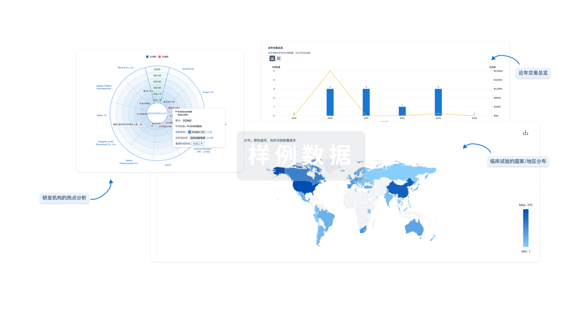
100 项与 HER2 x Muc18 相关的临床结果
登录后查看更多信息
100 项与 HER2 x Muc18 相关的转化医学
登录后查看更多信息
0 项与 HER2 x Muc18 相关的专利(医药)
登录后查看更多信息
13
项与 HER2 x Muc18 相关的文献(医药)2022-12-01·BMC cancer2区 · 医学
Characterization of surface markers on extracellular vesicles isolated from lymphatic exudate from patients with breast cancer
2区 · 医学
ArticleOA
作者: Pétursson, Hafsteinn Ingi ; Johansson, Junko ; Olofsson Bagge, Roger ; Ekström, Karin ; Crescitelli, Rossella ; Lässer, Cecilia
Abstract:
Background:
Breast cancer is the most common cancer, and the leading cause of cancer-related deaths, among females world-wide. Recent research suggests that extracellular vesicles (EVs) play a major role in the development of breast cancer metastasis. Axillary lymph node dissection (ALND) is a procedure in patients with known lymph node metastases, and after surgery large amounts of serous fluid are produced from the axilla. The overall aim was to isolate and characterize EVs from axillary serous fluid, and more specifically to determine if potential breast cancer biomarkers could be identified.
Methods:
Lymphatic drain fluid was collected from 7 patients with breast cancer the day after ALND. EVs were isolated using size exclusion chromatography, quantified and detected by nanoparticle tracking analysis, electron microscopy, nano flow cytometry and western blot. The expression of 37 EV surface proteins was evaluated by flow cytometry using the MACSPlex Exosome kit.
Results:
Lymphatic drainage exudate retrieved after surgery from all 7 patients contained EVs. The isolated EVs were positive for the typical EV markers CD9, CD63, CD81 and Flotillin-1 while albumin was absent, indicating low contamination from blood proteins. In total, 24 different EV surface proteins were detected. Eleven of those proteins were detected in all patients, including the common EV markers CD9, CD63 and CD81, cancer-related markers CD24, CD29, CD44 and CD146, platelet markers CD41b, CD42a and CD62p as well as HLA-DR/DP/DQ. Furthermore, CD29 and CD146 were enriched in Her2+ patients compared to patients with Her2- tumors.
Conclusions:
Lymphatic drainage exudate retrieved from breast cancer patients after surgery contains EVs that can be isolated using SEC isolation. The EVs have several cancer-related markers including CD24, CD29, CD44 and CD146, proteins of potential interest as biomarkers as well as to increase the understanding of the mechanisms of cancer biology.
2018-07-01·Annals of anatomy = Anatomischer Anzeiger : official organ of the Anatomische Gesellschaft3区 · 医学
Stromal cells/telocytes and endothelial progenitors in the perivascular niches of the trigeminal ganglion
3区 · 医学
Article
作者: Creţoiu, S M ; Rusu, M C ; Vrapciu, A D ; Creţoiu, D ; Mănoiu, V S
Stromal cells/telocytes (SCs/TCs) were recently described in the human adult trigeminal ganglion (TG). As some markers are equally expressed in SCs/TCs and endothelial cells, we hypothesized that a subset of the TG SCs/TCs is in fact represented by endothelial progenitor cells of a myelomonocytic origin. This study aimed to evaluate whether the interstitial cells of the human adult TG correlate with the myelomonocytic lineage. We used primary antibodies for c-erbB2/HER-2, CD31, nestin, CD10, CD117/c-kit, von Willebrand factor (vWF), CD34, Stro-1, CD146, α-smooth muscle actin (α-SMA), CD68, VEGFR-2 and cytokeratin 7 (CK7). The TG pial mesothelium and subpial vascular microstroma expressed c-erbB2/HER-2, CK7 and VEGFR-2. SCs/TCs neighbouring the neuronoglial units (NGUs) also expressed HER-2, which suggests a pial origin. These cells were also positive for CD10, CD31, CD34, CD68 and nestin. Endothelial cells expressed CD10, CD31, CD34, CD146, nestin and vWF. We also found vasculogenic networks with spindle-shaped and stellate endothelial progenitors expressing CD10, CD31, CD34, CD68, CD146 and VEGFR-2. Isolated mesenchymal stromal cells expressed Stro-1, CD146, CK7, c-kit and nestin. Pericytes expressed α-SMA and CD146. Using transmission electron microscopy (TEM), we found endothelial-specific Weibel-Palade bodies in spindle-shaped stromal progenitors. Our study supports the hypothesis that an intrinsic vasculogenic niche potentially involved in microvascular maintenance and repair might be present in the human adult trigeminal ganglion and that it might be supplied by either the pial mesothelium or the bone marrow niche.
2014-01-01·BioMed research international3区 · 生物学
Expression of Mesenchymal Stem Cells-Related Genes and Plasticity of Aspirated Follicular Cells Obtained from Infertile Women
3区 · 生物学
ArticleOA
作者: Novakovic, Srdjan ; Virant-Klun, Irma ; Stimpfel, Martin ; Cerkovnik, Petra ; Dzafic, Edo
After removal of oocytes forin vitrofertilization, follicular aspirates which are rich in somatic follicular cells are discarded in daily medical practice. However, there is some evidence that less differentiated cells with stem cell characteristics are present among aspirated follicular cells (AFCs). The aim of this study was to culture AFCsin vitroand to analyze their gene expression profile. Using the RT2Profiler PCR array, we investigated the expression profile of 84 genes related to stemness, mesenchymal stem cells (MCSs), and cell differentiation in AFCs enriched by hypoosmotic protocol from follicular aspirates of infertile women involved in assisted reproduction programme in comparison with bone marrow-derived mesenchymal stem cells (BM-MSCs) and fibroblasts. Altogether the expression of 57 genes was detected in AFCs: 16 genes (OCT4,CD49f,CD106,CD146,CD45,CD54,IL10,IL1B,TNF,VEGF,VWF,HDAC1,MITF,RUNX2,PPARG, andPCAF) were upregulated and 20 genes (FGF2,CASP3,CD105,CD13,CD340,CD73,CD90,KDR,PDGFRB,BDNF,COL1A1,IL6,MMP2,NES,NUDT6,BMP6,SMURF2,BMP4,GDF5, andJAG1) were downregulated in AFCs when compared with BM-MSCs. The genes which were upregulated in AFCs were mostly related to MSCs and connected with ovarian function, and differed from those in fibroblasts. The cultured AFCs with predominating granulosa cells were successfullyin vitrodifferentiated into adipogenic-, osteogenic-, and pancreatic-like cells. The upregulation of some MSC-specific genes andin vitrodifferentiation into other types of cells indicated a subpopulation of AFCs with specific stemness, which was not similar to those of BM-MSCs or fibroblasts.
3
项与 HER2 x Muc18 相关的新闻(医药)2025-06-09
Cytiva智荟专线400-810-9118,2号键#智荟锦囊循环肿瘤细胞CTCs简介1869年,Ashworth,T.R等【1】首次在一例转移性肿瘤患者血液中观察到循环肿瘤细胞(Circulating Tumor Cells, CTCs)。CTCs即是指从原发肿瘤或转移灶脱落,进入血液循环系统的肿瘤细胞【2】,如图1。图1:循环肿瘤细胞CTCs进入血液循环原发肿瘤释放CTCs进入循环中,大部分CTCs死亡,但是少部分CTCs存活并在远端形成转移灶【9】。每天每g原位瘤可能会释放多达百万的肿瘤细胞进入血液循环【3】,但大部分因血流剪切力、失巢凋亡、免疫系统识别【4】而被清除,只有极少数(0.01%)【4】【5】存活下来,通过血液或淋巴到达远端器官形成新的转移灶,是肿瘤转移(Metastasis)和复发的重要生物学基础之一。因此,CTCs是肿瘤精准医疗的重要靶点【6】,作为液体活检(其它如cfDNA、cfRNA、microRNA、外泌体、肿瘤代谢物)重要组成之一,其完整细胞的特有属性,允许通过表型、基因型(如单细胞测序)、细胞功能以及异种移植模型,用于肿瘤早期诊断、疾病进展、复发检测、转移癌监控、肿瘤亚型改变、耐药性研究以及预后评估【7】,促进个体化治疗实施。此外,CTCs携带了肿瘤的基因组、转录组和代谢组等多组学信息,可用于研究肿瘤微环境、转移微环境PMN、免疫逃逸机制,还可用于培养类器官【8】进行体外药物筛选和检测。CTCs的分离方法CTCs的分离方法,主要分为非标记法和标记法,如图2。图2:CTCs分离、培养和分析方法【10】非标记法主要依赖CTCs自身的物理性质,如大小、密度、粘附、介电性质和变形性【11】【12】。被广泛使用的Cytiva Ficoll-Paque 和Percoll分离液即基于密度分离。大小和变形性等则常用于微流控技术【13】。标记法主要为阳性富集(如上皮细胞粘附分子EpCAM【14】、HER2、束丝蛋白3【15】、前列腺特异性抗原PSA【16】等或Cocktail)和阴性富集(如CD45抗体去除白细胞、CD61去除血小板和巨核细胞【17】、CD14等或Cocktail)。基于抗EpCAM免疫磁珠技术的CellSearch CTC Test方法,是目前唯一被美国FDA批准作为肿瘤预后评估工具用于转移性乳腺癌(2004年)【18】、结直肠癌(2007年)【19】和前列腺癌(2008年)【20】。中国CFDA在2012年批准了CellSearch系统用于转移性乳腺癌的预后评估。其它本土获批产品包括CytoSorter循环肿瘤细胞检测系统等,更多检测方法也在兴起和验证中。CTCs的分离也可以联合非标记和标记法【21】,并受到肿瘤类型的影响【17】。但是,CTCs分离仍面临挑战:肿瘤细胞本身即具有异质性。并且,CTCs因上皮-间质转化(Epithelial-Mesenchymal Transition, EMT)失去极性和黏附性,获得更强的迁移和侵袭能力,与基质互作并抵抗治疗,同时表现出上皮和间质表型【22】,也造成细胞表面Marker的动态变化。此外,CTCs还会和血液中其它细胞如白细胞形成CTC微血栓促进自身生存和转移。CTCs在血液中的密度很低,~1-10 CTCs/mL血液【10】,分离富集挑战大。一般,转移性癌症中每10 mL血液中有0-100个单体CTCs以及0-5个CTCs Cluster(CTCs簇),并与癌症类型、血液收集位置以及治疗阶段有关【23】。CTCs半衰期通常只有 1.0‑2.4 h【24】。使用密度梯度离心法分离CTCs早在1959年,Seal SH等【25】即使用硅油配制密度梯度离心介质,分离血液中的游离肿瘤细胞。目前,商品化的梯度介质,如Cytiva的Ficoll-Paque进行CTCs的分离,在临床研究中被广泛使用【21】【26】【27】【28】。作为非标记的分离方法,利于富集表面Marker表达异质性CTCs以及未知CTCs亚型,操作简单、低成本、快速,处理通量大。使用密度梯度介质分离CTCs,文献报道中主要包括4种方法:常规标准操作、抗体预先结合杂质细胞、抗体预先结合CTCs目的细胞,以及使用Percoll不连续密度梯度进行分离。使用Ficoll-Paque进行CTCs分离的常规操作根据斯托克斯定律,如图3,颗粒的沉降速率v,和颗粒直径(d)、样品密度(ρp)和溶液密度(ρl)之差成正比;与溶液的粘稠度(η)成反比。当不同细胞经过同一密度离心介质,不同颗粒直径和密度的细胞,沉降速率不同,从而被分开。外周血中,红细胞、粒细胞密度大【38】如图6,穿过Ficoll-Paque密度梯度介质(1.077 g/mL)沉于底层;外周血单个核细胞PBMCs和CTCs密度小,一起停留在白膜层(buffy coat)中,最上面是血浆。后续可以使用阳性富集【29】或阴性富集(如anti-CD45磁珠去除白细胞)【30】【39】进一步回收CTCs。图3:颗粒沉降速率公式【31】Ficoll-Paque密度梯度介质标准操作及细胞分布,请参考文章:☞ Ficoll-Paque应用解析:PBMCs分离中病人样本的特殊性抗体结合杂质细胞后,再使用密度梯度介质分离CTCs在密度梯度分离之前,进行杂质细胞预标记,促进其沉降。例如采用anti-CD45(白细胞共同抗原)【8】【32】【33】、anti-66b(粒细胞)与样品进行孵育,使白细胞与红细胞形成免疫玫瑰花状结构,密度和大小增加,在后续密度梯度离心过程中,将白细胞群拉到管底,则在白膜层可以回收更纯的CTCs,如图4,少量CTCs可能会渗漏到血浆层,也可以一起回收【6】【32】。相对于阳性筛选,基于去除白细胞的阴性筛选更有优势,避免了使用抗体激活CTCs以及某些CTCs因为不表达相应的抗原而被遗漏【32】。图4:使用Ficoll-Paque 密度梯度介质,分离CTCs将血液和白细胞抗体如RosetteSep抗体孵育,使用DPBS+2% FBS稀释血样,然后小心地铺到Ficoll-Paque 密度梯度介质上面,离心(关闭刹车)后在白膜层收获CTCs【32】。抗体结合目的CTCs后,再使用密度梯度介质分离CTCs在密度梯度分离之前,进行CTCs目的细胞预标记,促进其沉降。Huang Q等【34】使用同时偶联anti‑EpCAM抗体和anti‑CD146抗体(后者作为肿瘤标志物以及上皮间质转化EMT诱导剂,利于捕获EpCAM无/低表达间质CTCs)的SiO2微珠(表面包被可降解明胶),先和血样孵育以结合CTCs,之后将样品铺在Cytiva Percoll密度梯度介质(配制成1.15 g/mL)的表面,离心,CTCs结合在微珠表面而沉降于管底,之后酶解表层明胶释放CTCs,结果显示CTCs收率>80%,纯度大于85%,如图5。也可以使用Ficoll-Paque密度梯度介质,采用类似方法离心沉降CTCs目的细胞,收集管底组分并裂红,回收CTCs。图5:基于微珠和密度梯度介质进行CTCs分离(部分图片)A:通过可降解明胶包被的微珠(偶联了anti-EpCAM和anti-CD146抗体),高效吸附CTCs。 B:经过Percoll密度梯度离心,将结合CTCs的二氧化硅微珠沉淀到管底,从而和血细胞分离。C:通过基质金属蛋白酶MMP-9酶解明胶,释放结合在微珠表面的CTCs。除了CTCs单细胞外,CTC Cluster【35】以及CTCs与其它血液成分组成的细胞团,往往具有更高的转移潜能。由于细胞团的密度更大,容易在离心后沉降,也可以采用类似方法进行分离,或使用Cluster-Chip【36】。 另外,具有干性的CTCs,即循环癌干细胞(Circulating Cancer Stem Cells,CCSCs)同样值得关注。使用Percoll不连续密度梯度分离CTCs此外,也可以将Percoll密度梯度介质,配制成多个不连续密度梯度,以便将杂质细胞拦截在不同的密度梯度中,CTCs则集中在夹层中,利于回收【37】。Cytiva有两种产品可供选择:Percoll于1978年发布,为聚乙烯吡咯烷酮(PVP)涂层二氧化硅胶体介质,适用于科研;Percoll PLUS于2006年推出,是共价偶联烷硅涂层二氧化硅胶体介质,具有低水平的内毒素,提供法规支持文件,适用于临床研究。具体的密度梯度配制,可以参考Boyum, A等【38】绘制出的外周血各个细胞组分的密度分布,如图6。图6:人外周血中细胞密度分布图Percoll密度梯度配制方法,请参考文章:☞ 智荟锦囊之Percoll分离液配制指南细胞分离液相关产品文章内相关中文数据文件和操作说明,如Ficoll-Paque PLUS、不同浓度的Ficoll-Paque PREMIUM、Percoll以及Percoll PLUS产品等欢迎扫描下方二维码即刻下载如果大家对产品和技术感兴趣,欢迎拨打智荟专线:智荟专线:400-810-9118(转1号线询价购买或2号线技术支持)相关阅读:☞Ficoll-Paque应用解析:PBMCs分离中病人样本的特殊性☞Ficoll-Paque应用解析:间充质干细胞的分离参考文献1. Ashworth, T.R. A case of cancer in which cells similar to those in the tumors were seen in the blood after death. Aust. Med. J. 1869, 14, 146–149.2. Prasanna BK, Balakrishnan A and Kumar P: Circulating tumor cell clusters and circulating tumor cell derived explant models as a tool for treatment response. Biotechniques 69: 362 363, 2020.3. Chang YS, di Tomaso E, McDonald DM, Jones R, Jain RK, Munn LL. Mosaic blood vessels in tumors: frequency of cancer cells in contact with flowing blood. Proc Natl Acad Sci U S A. 2000 Dec 19;97(26):14608-13. doi: 10.1073/pnas.97.26.14608. PMID: 11121063; PMCID: PMC18966.4. Tayoun T, Faugeroux V, Oulhen M, Aberlenc A, Pawlikowska P and Farace F: CTC derived models: A window into the seeding capacity of circulating tumor cells (CTCs). Cells 8: 1145, 20195. Leone K, Poggiana C and Zamarchi R: The interplay between circulating tumor cells and the immune system: From immune escape to cancer immunotherapy. Diagnostics (Basel) 8: 59, 2018.6. Mahmoud Labib 1, Shana O Kelley. Circulating tumor cell profiling for precision oncology. Mol Oncol. 2021 Feb 1;15(6):1622–1646.7. Smerage JB, Barlow WE, Hortobagyi GN, Winer EP, Leyland-Jones B, Srkalovic G, Tejwani S, Schott AF, O'Rourke MA, Lew DL, Doyle GV, Gralow JR, Livingston RB, Hayes DF. Circulating tumor cells and response to chemotherapy in metastatic breast cancer: SWOG S0500. J Clin Oncol. 2014 Nov 1;32(31):3483-9. doi: 10.1200/JCO.2014.56.2561. Epub 2014 Jun 2. PMID: 24888818; PMCID: PMC4209100.8. Gao D, Vela I, Sboner A, Iaquinta PJ, Karthaus WR, Gopalan A, Dowling C, Wanjala JN, Undvall EA, Arora VK, Wongvipat J, Kossai M, Ramazanoglu S, Barboza LP, Di W, Cao Z, Zhang QF, Sirota I, Ran L, MacDonald TY, Beltran H, Mosquera JM, Touijer KA, Scardino PT, Laudone VP, Curtis KR, Rathkopf DE, Morris MJ, Danila DC, Slovin SF, Solomon SB, Eastham JA, Chi P, Carver B, Rubin MA, Scher HI, Clevers H, Sawyers CL, Chen Y. Organoid cultures derived from patients with advanced prostate cancer. Cell. 2014 Sep 25;159(1):176-187. doi: 10.1016/j.cell.2014.08.016. Epub 2014 Sep 4. PMID: 25201530; PMCID: PMC4237931.9. Ruchi Agashe, Razelle Kurzrock. Circulating Tumor Cells: From the Laboratory to the Cancer Clinic. Cancers (Basel). 2020 Aug 21;12(9):2361.10. Xiuxiu Hu 1, Xiaojuan Zang 2, Yanguan Lv 3. Detection of circulating tumor cells: Advances and critical concerns (Review). Oncol Lett. 2021 May;21(5):422.11. Chudziak J, Burt DJ, Mohan S, et al. Clinical evaluation of a novel microfluidic device for epitope-independent enrichment of circulating tumour cells in patients with small cell lung cancer. Analyst 2016;141:669-78.12. Mehdi Rahmanian 1, Omid Sartipzadeh Hematabad 2, Esfandyar Askari, et al. A micropillar array-based microfluidic chip for label-free separation of circulating tumor cells: The best micropillar geometry? J Adv Res. 2023 May:47:105-121.13. Tyler A Allen. The Role of Circulating Tumor Cells as a Liquid Biopsy for Cancer: Advances, Biology, Technical Challenges, and Clinical Relevance. Cancers (Basel). 2024 Mar 31;16(7):1377.14. Ferreira MM, Ramani VC, Jeffrey SS. Circulating tumor cell technologies. Mol Oncol 2016;10:374-94.15. Yokobori T, Iinuma H, Shimamura T, Imoto S, Sugimachi K, Ishii H, Iwatsuki M, Ota D, Ohkuma M, Iwaya T, Nishida N, Kogo R, Sudo T, Tanaka F, Shibata K, Toh H, Sato T, Barnard GF, Fukagawa T, Yamamoto S, Nakanishi H, Sasaki S, Miyano S, Watanabe T, Kuwano H, Mimori K, Pantel K, Mori M. Plastin3 is a novel marker for circulating tumor cells undergoing the epithelial-mesenchymal transition and is associated with colorectal cancer prognosis. Cancer Res. 2013 Apr 1;73(7):2059-69. doi: 10.1158/0008-5472.CAN-12-0326. Epub 2013 Feb 1. PMID: 23378342.16. Krebs MG, Metcalf RL, Carter L, Brady G, Blackhall FH, Dive C. Molecular analysis of circulating tumour cells-biology and biomarkers. Nat Rev Clin Oncol. 2014 Mar;11(3):129-44. doi: 10.1038/nrclinonc.2013.253. Epub 2014 Jan 21. PMID: 24445517.17. 中华医学会检验医学分会分子诊断学组. 循环肿瘤细胞临床应用与实验室检测专家共识.中华检验医学杂志 2021 年11 月第 44 卷第 11 期.18. Hayes DF, Cristofanilli M, Budd GT et al. Circulating tumor cells at each follow-up time point during therapy of metastatic breast cancer patients predict progression-free and overall survival. Clin. Cancer Res. 12(14 Pt 1), 4218–4224 (2006).19. Cohen SJ, Punt CJ, Iannotti N et al. Relationship of circulating tumor cells to tumor response, progression-free survival, and overall survival in patients with metastatic colorectal cancer. J. Clin. Oncol. 26(19), 3213–3221 (2008).20. de Bono JS, Scher HI, Montgomery RB et al. Circulating tumor cells predict survival benefit from treatment in metastatic castration-resistant prostate cancer. Clin. Cancer Res. 14(19), 63026309 (2008).21. Yali Wang 1, Yucheng Zhang, Zhenwu Du, et al. Detection of micrometastases in lung cancer with magnetic nanoparticles and quantum dots. Int J Nanomedicine. 2012:7:2315-24.22. De T, Goyal S, Balachander G et al. A novel ex vivo system using 3D polymer scaffold to culture circulating tumor cells from breast cancer patients exhibits dynamic E-M phenotypes. J.Clin. Med. 8(9), 1473 (2019).23. Au SH, Edd J, Haber DA, Maheswaran S, Stott SL and Toner M: Clusters of circulating tumor cells: A biophysical and techno¬logical perspective. Curr Opin Biomed Eng 3: 13 19, 201724. Meng S, Tripathy D, Frenkel EP, Shete S, Naftalis EZ, Huth JF, Beitsch PD, Leitch M, Hoover S, Euhus D, et al: Circulating tumor cells in patients with breast cancer dormancy. Clin Cancer Res 10: 8152 8162, 2004.25. SEAL SH. Silicone flotation: a simple quantitative method for the isolation of free-floating cancer cells from the blood. Cancer. 1959 May-Jun;12(3):590-5. doi: 10.1002/1097-0142(195905/06)12:3<590::aid-cncr2820120318>3.0.co;2-n. PMID: 13652106.26. Zahra Eslami-S, Luis Enrique Cortés-Hernández, Frédéric Thomas, et al. Functional analysis of circulating tumour cells: the KEY to understand the biology of the metastatic cascade. Br J Cancer. 2022 Sep;127(5):800-810.27. Wan Shi Low, Wan Abu Bakar Wan Abas. Benchtop Technologies for Circulating Tumor Cells Separation Based on Biophysical Properties. Biomed Res Int. 2015:2015:239362.28. Yap K, Cohen EN, Reuben JM and Khoury JD: Circulating tumor cells: State of the art update on technologies and clinical applications. Curr Hematol Malig Rep 14: 353 357, 2019.29. Fizazi K, Morat L, Chauveinc L, Prapotnich D, De Crevoisier R, Escudier B, et al. High detection rate of circulating tumor cells in blood of patients with prostate cancer using telomerase activity. Ann Oncol. 2007;18(3):518–521. doi: 10.1093/annonc/mdl419.30. Morgan TM, Lange PH, Vessella RL. Detection and characterization of circulating and disseminated prostate cancer cells. Front Biosci. 2007 May 1;12:3000-9. doi: 10.2741/2290. PMID: 17485277.31. Methodology and applications Cell separation media. CY14836-24Jan21-HB.32. Alexandra Soler, Laure Cayrefourcq, Martine Mazel, et al. EpCAM-Independent Enrichment and Detection of Viable Circulating Tumor Cells Using the EPISPOT Assay. Methods Mol Biol. 2017:1634:263-276.33. Drucker A, Teh EM, Kostyleva R, Rayson D, Douglas S and Pinto DM: Comparative performance of different methods for circulating tumor cell enrichment in metastatic breast cancer patients. PLoS One 15: e0237308, 2020.34. Huang Q, Wang FB, Yuan CH, He Z, Rao L, Cai B, Chen B, Jiang S, Li Z, Chen J, et al: Gelatin nanoparticle coated silicon beads for density selective capture and release of heteroge¬neous circulating tumor cells with high purity. Theranostics 8: 1624 1635, 2018.35. Danova M, Torchio M, Mazzini G. Isolation of rare circulating tumor cells in cancer patients: technical aspects and clinical implications. Expert Rev Mol Diagn 2011;11:473-85.36. Sarioglu AF, Aceto N, Kojic N, et al. A microfluidic device for label-free, physical capture of circulating tumor cell clusters. Nat Methods. 2015 Jul;12(7):685-91. doi: 10.1038/nmeth.3404. Epub 2015 May 18. PMID: 25984697; PMCID: PMC4490017.37. Wenjun Le, Shuyuan Hu, Pengbo Zhang, et al. Capture of live circulating tumor cells via unique surface charges from cancer patients by a novel nanotechnology. Interdiscip. Med. 2024, 2, e20240045.38. Bøyum A, Løvhaug D, Tresland L, Nordlie EM. Separation of leucocytes: improved cell purity by fine adjustments of gradient medium density and osmolality. Scand J Immunol. 1991 Dec;34(6):697-712. doi: 10.1111/j.1365-3083.1991.tb01594.x. PMID: 1749920.39. Gakhar G, Navarro VN, Jurish M, Lee GY, Tagawa ST, Akhtar NH, Seandel M, Geng Y, Liu H, Bander NH, Giannakakou P, Christos PJ, King MR, Nanus DM. Circulating tumor cells from prostate cancer patients interact with E-selectin under physiologic blood flow. PLoS One. 2013 Dec 27;8(12):e85143. doi: 10.1371/journal.pone.0085143. PMID: 24386459; PMCID: PMC3874033. 本期关键词:#FicollPaque
疫苗临床研究免疫疗法
2025-04-28
·小药说药
-01-引言缺氧诱导因子(HIFs)是高度保守并受氧气丰度调节的转录因子,对各种生物适应有限的氧气供应至关重要。在缺氧发生时,HIFs可以直接促进靶基因的转录,例如红细胞生成素(EPO)、血管内皮生长因子(VEGFs)或对细胞外腺苷的产生和信号传导至关重要的基因产物。HIF激活的作用包括红细胞生成刺激、缺氧期间的细胞代谢优化以及缺血和炎症期间的适应性反应。相比之下,HIF抑制可以用于治疗各种癌症、视网膜新生血管和肺动脉高压。在过去的十年里,人们已经开发出多种有效调节HIFs功能的药物,并在临床试验中进行了测试。这些药物在治疗癌症、心脏的缺血再灌注损伤、急性肾损伤、急性呼吸窘迫综合征(ARDS)、炎症性肠病、病原体感染、出血性休克和肾贫血等疾病方面展现出良好的应用前景。同时, HIF激活剂或抑制剂的广泛应用也面临着具体的挑战。-02-一、HIFs的分子结构和功能HIF途径的氧依赖性调节是细胞和系统适应低氧水平的核心。HIFs的紧密调节是由几种关键酶介导的,包括因子抑制HIF(FIH)、HIF PHDs和von Hippel–Lindau蛋白(pVHL)。HIFs的活性转录形式由一个α单元(HIFα)和一个β单元(HIF1β)组成。HIFα对调节HIFs的转录活性至关重要。目前,已鉴定出HIFα的三种亚型:HIF1α、HIF2α和HIF3α。所有三种异构体在N末端都含有一个碱性螺旋-环-螺旋和两个Per-Arnt-Sim结构域(bHLH-PAS),在C末端都含有氧依赖性降解结构域,而只有HIF1α和HIF2α含有反式激活结构域。在这三种异构体中,HIF1α被鉴定为与EPO启动子区结合的转录因子。HIF2α和HIF3α后来被发现是基于它们与HIF1α的序列同源性。人HIF1α和HIF2α的氨基酸同源性为53.4%,人HIF1α和HIF3α的氨基酸序列同源性为57.1%。有趣的是,三种HIFα亚型根据组织和细胞的特异性具有不同的功能作用。HIF1α在所有细胞类型中都表达,而HIF2α最初被认为仅在内皮细胞中表达,然而,最近的研究表明HIF2α具有在血管内皮细胞外的功能。HIF1α和HIF2α协同增强共享靶基因如EPO或VEGF的表达。然而,HIF1α和HIF2α可能具有相反的生物学功能。例如,HIF1α特异性调节诱导型一氧化氮合酶(iNOS)基因,而HIF2α控制精氨酸酶1的表达,从而对巨噬细胞极化或癌症转移的调节产生相反的作用。而HIF3α缺乏反式激活结构域,因此,它被认为是一种抑制元件。与HIF1α和HIF2α相比,HIF3α及其各种异构体和剪接变体的功能作用还有待进一步阐明。所有三个HIFα亚基都需要与其异二聚体伴侣HIF1β相互作用才能具有转录活性。HIF1β在N末端含有一个bHLH PAS结构域,用于DNA与HIFα的结合和二聚化。与对氧依赖性降解敏感的HIFα不同,HIF1β在所有哺乳动物细胞中结构性表达,与氧的可用性无关。-03- 二、HIFs的缺氧调节HIF1α或HIF2α的氧依赖性失活是一个由几个关键酶和分子通过羟基化和泛素化控制的多步骤过程。一旦HIFα与HIF1β二聚化,其随后转移到细胞核中以调节基因转录。HIFα和HIF1β上的bHLH PAS结构域对二聚化和DNA结合至关重要。同时,辅因子p300和CBP的募集对反式激活至关重要。进入细胞核后,HIFα–HIF1β–p300–CBP复合物与位于HIF靶基因启动子区的缺氧反应元件(HRE)结合,并激活转录。HIF的调节首先通过FIH——一种氧依赖性天冬酰胺羟化酶——通过在C-末端反式激活结构域(C-TAD)结构域内羟基化Asn803来抑制HIF系统,阻止共激活物、CBP和组蛋白乙酰转移酶p300的结合。HIF途径的中央调控与含有HIF脯氨酰羟化酶结构域的蛋白(HIF-PHDs)的酶活性有关。HIF-PHD有三种亚型:PHD1(也称为EGLN2)、PHD2(也称为EGLN1)和PHD3(也称为EGLN3)。PHD1和PHD2促进Pro402和Pro564在人HIF1α上的羟基化,而PHD3仅羟基化Pro564。羟基化过程高度依赖于氧和α-酮戊二酸作为底物和铁作为催化剂的可用性。羟基化后,HIFα与pVHL结合,并被延伸蛋白B、延伸蛋白C、cullin 2(CUL2)、环盒蛋白1(RBX1)和E3泛素连接酶形成的复合物泛素化,导致随后的蛋白酶体降解。-04-三、HIFs激动剂 目前,临床上开发的激动剂主要通过抑制HIF-PHDs来阻止脯氨酸羟基化和随后的蛋白酶体降解来激活HIF。有五种HIF PHD小分子抑制剂已进入III期临床,包括roxadustat(阿斯利康)、vadaustat(阿科比亚)、daprodustat(葛兰素史克)、molidustat(拜耳)和desidustat(Zydus)。自从发现靶向HIF PHDs的小分子HIF激活剂以来,临床焦点一直集中在肾性贫血上,这主要是由于HIF激动对EPO的大量调节。一些化合物显示出与红细胞生成刺激剂(ESA)相当的治疗效果。日本已经批准了五种HIF PHD抑制剂,此外,roxadustat还在其他国家或地区获得批准,包括欧盟、中国、韩国和智利。2023年2月2日,daprodustat在美国获得了FDA的批准,用于治疗接受透析的成年患者中与慢性肾脏疾病(CKD)相关的贫血。其他HIF PHD抑制剂,如vadadustat已在35个国家获得批准,包括日本、澳大利亚和许多欧洲国家。HIF PHD抑制剂的其他临床适应症,如ARDS,仍处于早期阶段。除了肾性贫血,ARDS是另一个临床适应症,正在进行使用HIF激活剂治疗的临床安全性和有效性试验。ARDS期间的HIF是一种内源性保护途径,可在药理学上进一步用于早期ARDS治疗或预防。在ARDS期间,HIF1α在肺泡上皮细胞中具有优化其碳水化合物代谢和细胞外腺苷信号的功能。HIF2α也有助于ARDS期间的肺部保护,例如,通过维持内皮细胞的屏障完整性。HIF-PHD抑制剂在大型III期临床试验中显示出良好的短期安全性记录,并且与严重的副作用无关。长期使用(3个月以上)HIF PHD抑制剂可能会产生不必要的副作用,包括慢性HIF激活引起的靶向效应和与HIF PHD抑制剂对单个羟化酶的特异性相关的脱靶效应。最重要的一点,由于HIF激活与癌症进展相关,尤其是肾透明细胞癌(ccRCC),长期HIF激活可能导致癌症的发展或进展。HIF PHD抑制剂的另一个潜在靶向副作用涉及过度的红细胞生成和血栓栓塞并发症,重大心血管不良事件(MACE)风险的增加也是与长期使用HIF-PHD抑制剂相关的一个潜在问题。-05-四、HIF抑制剂HIF在许多疾病中可能是有害的,例如某些癌症和肺动脉高压。因此,已经开发出抑制HIFs的药物,正在进行临床测试。早期的药物干预侧重于抑制HIF表达或促进HIF降解。最近的小分子抑制剂,如PT2977,抑制HIF2α和HIF1β转录活性异二聚体的形成。癌症HIFs通常在大多数人类癌症中观察到,特别是在实体瘤中。肿瘤内缺氧和炎症是HIF的主要机制,以及常见于VHL疾病相关癌症(如ccRCC)的基因突变。许多癌症激活途径可以导致HIF激活,包括TP53、PTEN、EGFR和HER2的体细胞突变或表观遗传学改变。癌细胞利用HIF途径通过各种机制驱动癌症进展,如促进血管形成、代谢重编程、免疫逃避、细胞存活以及治疗耐药性。HIF1α在实体瘤中高水平表达,且与预后不良有关。在癌症中HIF1α的治疗靶向已经取得了相当大的努力和进展。例如,PX-478已在癌症患者的I期剂量递增研究中进行了测试(NCT00522652),并显示出有效的HIF1α抑制和合理的安全性。ASO EZN-2968在难治性实体瘤患者中研究HIF1A RNAi的早期努力由于赞助者的兴趣丧失而提前终止。然而,EZN-2968/RO7070179最近在晚期HCC患者中进行了研究;总体而言,EZN-2968/RO7070179在癌症患者中具有良好的耐受性。肺动脉高压肺动脉高压是指平均肺动脉压高于20 mmHg,其特征是由内皮细胞和肺动脉平滑肌细胞的异常增殖、细胞外基质沉积和炎症细胞的募集引起的肺血管阻力增加。在肺动脉高压期间,HIF1α通过诱导细胞周期蛋白依赖性激酶抑制剂1B减少内皮集落形成细胞的增殖,并通过驱动miR-9-1、miR-9-3和miR-322的表达促进肺动脉平滑肌细胞增殖。HIF1α还诱导肺动脉平滑肌细胞中CD146的表达,从而导致血管重塑。小分子HIF2α抑制剂,如belzutifan,可减轻VhlR200W小鼠的肺动脉高压,这些小鼠在Vhl中具有功能缺失突变。然而,HIF抑制剂尚未进入临床试验,以评估其治疗人类肺动脉高压的疗效。视网膜新生血管视网膜新生血管是许多视网膜疾病的发病机制,包括糖尿病视网膜病变和视网膜静脉阻塞。在眼部新生血管形成过程中,新生成的血管缺乏紧密连接,导致血浆渗漏、玻璃体出血和牵引性视网膜脱离,导致严重的视网膜功能障碍和视力丧失。临床前研究以及对缺血性视网膜疾病患者的研究表明,HIF1α和HIF2α在视网膜新生血管形成过程中发挥重要作用。HIF抑制剂地高辛抑制视网膜中的HIF1α和HIF2α,并且在视网膜病小鼠模型中使用减少了视网膜新生血管。此外,在氧诱导的缺血性视网膜病变中,HIF抑制剂阿哌拉韦的治疗减少了小鼠的视网膜新生血管并改善了视力。此外,眼内注射HIF抑制剂32-134D改善了糖尿病眼病小鼠模型中的视网膜新生血管形成。总之,这些研究证明了HIF1α和HIF2α的药理学抑制在各种类型视网膜病变的视网膜新生血管形成中的潜在有益作用,未来有必要进行这方面的临床研究。-06-结语从20世纪90年代初首次发现HIF到最近FDA批准HIF激活剂和HIF抑制剂用于治疗人类疾病,这是一段非凡的旅程。这一成功源于对HIF调节途径的广泛研究,对HIF和PHD功能作用机制的深入理解。未来还需要很多努力来确定如何将这些药物最佳地用于其他疾病条件或其他潜在的药理学策略,以实现更特异的靶向。参考资料:1.Targeting hypoxia-inducible factors: therapeutic opportunities and challenges. Nat Rev Drug Discov.2023 Dec 20.公众号内回复“ADC”或扫描下方图片中的二维码免费下载《抗体偶联药物:从基础到临床》的PDF格式电子书!公众号已建立“小药说药专业交流群”微信行业交流群以及读者交流群,扫描下方小编二维码加入,入行业群请主动告知姓名、工作单位和职务。
临床结果基因疗法
2024-09-02
关注并星标CPHI制药在线
近日,普众发现医药科技(上海)有限公司1类新药「注射用AMT-676 」在国内获得临床试验默示许可,用于治疗晚期实体瘤。据公开资料,AMT-676是普众发现研发的一款CDH17靶向抗体偶联药物(ADC),其针对晚期实体瘤的1期临床研究已在澳大利亚启动。
普众发现是一家专注于ADC药物开发的临床阶段公司,拥有MabArray、T-Moiety两大技术平台,其中MabArray技术平台用于发现新颖的细胞表面抗肿瘤靶点以构建First-in-Class靶标,其可以实现传统方法无法完成高分辨率抗原筛选,并识别高难度抗原如多次跨膜,特殊构象,糖蛋白等,一次性覆盖人类细胞表面膜蛋白(潜在药靶)~50%,推动细胞表面抗原识别抵达新境界。T-Moiety是一种高亲水性自裂解连接子修饰技术,用于开发ADC的新型Linker。T-Moiety连接子具有更优的“疏水屏蔽效应”与稳定性,可以大幅度提高ADC治疗效果,以及延长药物作用时间,且能够克服多重肿瘤耐药性,不增加毒副作用。
基于上述技术平台,普众发现已开发出多款创新ADC,如靶向FRα的AMT-151、靶向MUC18的AMT-253、靶向HER3的AMT-562、靶向CDH6的AMT-707/CUSP06、靶向CD44v9的AMT-116、靶向TF的AMT-754。
值得一提的是,普众发现对外达成两次授权:2022年6月,将AMT-707在大中华区以外的全球开发和商业化权利独家授权给昂阔医药;2024年8月,将AMT-754在大中华区以外的全球开发和商业化权利独家授权给Adcendo。
关于CDH17
CDH17,即钙黏蛋白17,最早于1994年由Dietmar等通过分子克隆技术从鼠肝细胞cDNA文库中分析得到,由于在小鼠仅表达于肝脏和小肠,故命名为肝肠钙黏蛋白。
人CDH17基因位于染色体8q22. 1,其结构与钙黏蛋白家族成员具有同源性,但又有其独特性:CDH17的胞外部分含有7个钙黏蛋白重复区域,而经典钙黏蛋白和桥粒钙黏蛋白为5个;CDH17氨基末端的细胞黏附识别区,决定细胞黏附特性的HAV序列由AAL序列代替;CDH17细胞质尾仅含有20个氨基酸残基,经典钙黏蛋白有150-160个。
CDH17作为多肽转运器和细胞粘附分子来维持上皮组织的完整性,但与经典钙粘蛋白的粘附机制不同,CDH17在不与其他细胞质成分相互作用的情况下仍能保持其粘合功能,而经典钙粘蛋白需要与α和β-钙粘蛋白复合。
CDH17主要表达于胚胎、成人肠上皮细胞和部分胰腺导管上皮细胞,在健康人群肝细胞、食管上皮细胞及胃黏膜中几乎不表达。在肿瘤组织中,CDH17在胃、胰腺、肝、食道、胆管癌和结直肠癌中过表达。研究发现,CDH17在不同肿瘤组织中的表达程度不同,而且其表达水平与患者预后和风险评估密切相关。
目前,CDH17在肿瘤中的作用机制还未完全阐明。研究发现:在在胃癌中,CDH17通过激活NF-κB 信号通路,诱导胃癌的发生和淋巴结转移;在结直肠癌组织中,CDH17处于低表达状态,使得肿瘤发生及进展时肿瘤细胞间的黏附减弱,导致结直肠癌细胞浸润和转移;在肝癌中,CDH17 mRNA过表达与肝癌的发生及侵袭密切相关,应用RNA干扰技术使CDH17基因沉默,发现Wnt/β-catenin信号通路失活,肿瘤生长受到抑制。
CDH17靶向药进展
CDH17被认为是消化道肿瘤药物研发的潜力靶点,目前全球药企针对该靶点已开发出多款新药,如ARB-101、ARB-102、ARB-202、ARB-001、BI-905711、CHM-2101、CHM-2301、TORL-3-600。其中ARB-101和ARB-102是Abele开发的CDH17靶向单抗。
ARB202是Abele开发的一款CDH17/CD3靶向双抗,其对CDH17和CD3独特的差异结合亲和力使其具有高特异性和细胞毒性,同时避免了T细胞的“脱靶”过度激活。
临床前数据显示:ARB202能有效增强T细胞与表达CDH17的胃肠道肿瘤细胞系之间的相互作用,从而导致T细胞的活化并释放IL-2,有效的产生肿瘤细胞杀伤,通过体内注射低剂量ARB202 抗体可以有效地抑制肿瘤的生长。
1a期研究的初步数据显示:ARB 202在血液循环中的耐受性Cmax高达150ng/ml,这表明在这些浓度下没有临床显著的脱靶T细胞活化。
BI905711是勃林格殷格翰开发的双特异性、四价治疗性抗体,可同时靶向促凋亡肿瘤坏死因子相关凋亡诱导配体受体2(TRAILR2)和CDH17。TRAILR2与CDH17交联诱导CDH17依赖性TRAILR2聚集,在共表达TRAILR2和CDH17的肿瘤细胞中诱导激活选择性凋亡。
临床前研究显示:BI 905711在体外可诱导CDH17阳性肿瘤细胞凋亡,抑制pt来源的结直肠癌(CRC)异种移植物中的肿瘤生长,并且未观察到肝毒性。
CHM-2101是Chimeric Therapeutics开发一款第三代 CDH17靶向CAR-T疗法,具有CD28和4-1BB共刺激结构域。临床前研究显示:CHM-2101能够根除七种癌症模型中已形成的肿瘤,并且对正常组织没有毒性。
CHM-2301是Chimeric Therapeutics开发的一款CDH17靶向CAR-NK疗法,目前处于临床前研究阶段。
TORL-3-600是TORL BioTherapeutic开发的一款新型CDH17靶向ADC。临床前研究表明,TORL-3-600在CDH17阳性人结直肠癌模型中能诱导显著消退和肿瘤生长抑制,而且这些模型的反应在停止治疗后可持续长达九周。此外,本研究中测试的每种剂量在小鼠中均具有良好的耐受性,未观察到剂量限制性毒性。
总结
作为消化道肿瘤药物研发的潜力靶点,相较Claudin 18.2、HER2等,CDH17靶点进展较慢,在研药物大多处于临床早期阶段。不过,在研CDH17靶向药类型比较丰富,涉及单抗、双抗、CAR-T、CAR-NK、ADC等。目前,布局CDH17靶点的企业还比较少,期待在药企的不懈坚持下,CDH17靶点可以早日取得重大突破。
END
【智药研习社直播预告】
来源:CPHI制药在线
声明:本文仅代表作者观点,并不代表制药在线立场。本网站内容仅出于传递更多信息之目的。如需转载,请务必注明文章来源和作者。
投稿邮箱:Kelly.Xiao@imsinoexpo.com
▼更多制药资讯,请关注CPHI制药在线▼
点击阅读原文,进入智药研习社~
抗体药物偶联物临床1期
分析
对领域进行一次全面的分析。
登录
或
芽仔
全新生物医药AI Agent 覆盖科研全链路,让突破性发现快人一步
立即开始免费试用!
智慧芽新药情报库是智慧芽专为生命科学人士构建的基于AI的创新药情报平台,助您全方位提升您的研发与决策效率。
立即开始数据试用!
智慧芽新药库数据也通过智慧芽数据服务平台,以API或者数据包形式对外开放,助您更加充分利用智慧芽新药情报信息。
生物序列数据库
生物药研发创新
免费使用
化学结构数据库
小分子化药研发创新
免费使用