预约演示
更新于:2025-05-07
DSG3 x DSG1
更新于:2025-05-07
关联
靶点 |
作用机制 |
在研机构 |
原研机构 |
在研适应症 |
非在研适应症 |
最高研发阶段 |
首次获批国家/地区 |
首次获批日期 |


靶点 |
作用机制 |
在研机构 |
原研机构 |
在研适应症 |
非在研适应症 |
最高研发阶段 |
首次获批国家/地区 |
首次获批日期 |
100 项与 DSG3 x DSG1 相关的临床结果
登录后查看更多信息
100 项与 DSG3 x DSG1 相关的转化医学
登录后查看更多信息
登录后查看更多信息
2025-04-03Expert Review of Clinical Immunology
Diagnostic methods and strategies for autoimmune bullous diseases
Review
作者: Li, Xiaoguang ; Nakama, Takekuni ; Ishii, Norito ; Tsuruta, Daisuke ; Teye, Kwesi ; Tsuchisaka, Atsunari ; Mine, Mako ; Hashimoto, Takashi ; Qian, Hua ; Tateishi, Chiharu ; Koga, Hiroshi ; Hirako, Yoshiaki
2025-04-01Journal of Biological Chemistry
Epitomic profiling and functional characteristics of pemphigus vulgaris autoantibody binding to keratinocyte M3 muscarinic acetylcholine receptor
Article
作者: Chernyavsky, Alex ; Glabe, Charles ; Reyes-Ruiz, Jorge Mauricio ; Grando, Sergei A
2025-04-01JAAD International
Antidesmoglein 1 and 3 serum IgG and positivity by direct immunofluorescence microscopy is associated with relapse in pemphigus in a prospective bicontinental study
Article
作者: Lazaridou, Elisabeth ; Handa, Sanjeev ; Patsatsi, Aikaterini ; van Beek, Nina ; De, Dipankar ; Shahid, Martin ; Radotra, Bishan Dass ; Sachdeva, Naresh ; Mahajan, Rahul ; Vassileva, Snejina ; Drenovska, Kossara ; Shilpa, Shipla ; Mehta, Hitaishi ; Giannakou, Anastasia ; Lesichkova, Spaska ; Kumar, Sheetanshu ; Kishore, Kamal ; Kyriakou, Aikaterini ; Fleva, Alexandra ; Naumova, Elissaveta ; Schmidt, Enno
2023-12-30
引进/卖出上市批准临床2期医药出海
2023-12-24
抗体药物偶联物引进/卖出高管变更
2023-12-21
临床2期临床结果临床3期抗体药物偶联物申请上市
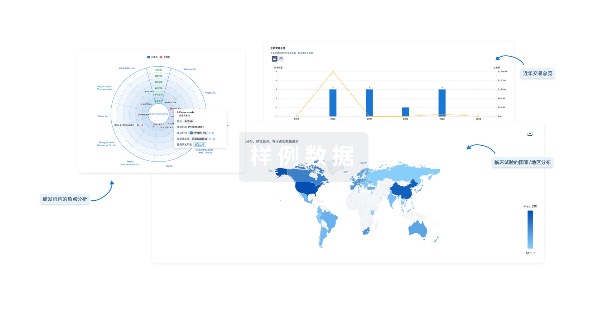
分析
对领域进行一次全面的分析。
登录
或
芽仔
全新生物医药AI Agent 覆盖科研全链路,让突破性发现快人一步
立即开始免费试用!
智慧芽新药情报库是智慧芽专为生命科学人士构建的基于AI的创新药情报平台,助您全方位提升您的研发与决策效率。
立即开始数据试用!
智慧芽新药库数据也通过智慧芽数据服务平台,以API或者数据包形式对外开放,助您更加充分利用智慧芽新药情报信息。
生物序列数据库
生物药研发创新
免费使用
化学结构数据库
小分子化药研发创新
免费使用