预约演示
更新于:2026-05-22
Insulin (Rezolute)
精蛋白牛胰岛素(Rezolute)
更新于:2026-05-22
概要
基本信息
原研机构 |
最高研发阶段临床1期 |
首次获批日期- |
最高研发阶段(中国)- |
特殊审评- |


登录后查看时间轴
关联
100 项与 精蛋白牛胰岛素(Rezolute) 相关的临床结果
登录后查看更多信息
100 项与 精蛋白牛胰岛素(Rezolute) 相关的转化医学
登录后查看更多信息
100 项与 精蛋白牛胰岛素(Rezolute) 相关的专利(医药)
登录后查看更多信息
6
项与 精蛋白牛胰岛素(Rezolute) 相关的文献(医药)2024-05-01·Journal of diabetes
Continuous subcutaneous insulin infusion versus multiple daily injection therapy in pregnant women with type 1 diabetes
Article
作者: Jinhua Yan ; Sihui Luo ; Jianping Weng ; Tian Wei ; Xueying Zheng ; Jing Wang ; Yixin Gong ; Yujie Liu ; Daizhi Yang
Abstract:
Introduction:
The study aimed to compare glycemic control and pregnancy outcomes in women with type 1 diabetes mellitus (T1DM) using multiple daily injection therapy (MDI) and continuous subcutaneous insulin infusion (CSII) and to compare outcomes of women treated with long‐acting insulin or neutral protamine Hagedorn (NPH).
Methods:
This multicenter prospective cohort study involved women with pregestational T1DM treated with MDI and CSII. Primary outcome was glycated hemoglobin (HbA1c) before and during pregnancy. Secondary outcomes included maternal and neonatal outcomes and quality of life.
Results:
Of the 121 studied women, the average age was 28.48 years, and the average body mass index was 21.29 kg/m2 at conception and 26.32 kg/m2 at delivery. Of the studied women, 78.51% had planned pregnancy. Women treated with MDI and CSII had comparable HbA1c before pregnancy or in the first and second trimesters. In the third trimester, women on CSII therapy had significantly lower HbA1c (6.07 ± 0.62 vs 6.20 ± 0.88%, p = .017), higher HbA1c on‐target rate (71.43% vs 64.62%, p = .030), and greater decline of HbA1c from preconception to the third trimester (−0.65 vs −0.30%, p = .047). Fewer daily insulin requirements were observed in those used CSII compared with MDI‐treated women (0.60 ± 0.22 vs 0.73 ± 0.25 U/kg/day, p = .004). Newborns born of mothers treated with the CSII method were more likely to have neonatal jaundice (adjusted odds ratio [OR] 2.76, 95% confidence interval [CI] 1.16–6.57) and neonatal intensive care unit (adjusted OR 3.73, 95%CI 1.24–11.16), and women on CSII had lower scores in patient‐reported quality of life (p = .045). In the MDI group, those receiving long‐acting insulin had nonsignificant lower HbA1c and higher HbA1c on‐target rate in the second and third trimesters, compared with those treated with NPH.
Conclusions:
Insulin pump users may achieve better glycemic control than multiple daily insulin injections, which did not substantially improve pregnancy outcome.image
2012-04-01·Diabetes research and clinical practice3区 · 医学
Insulin analogues and severe hypoglycaemia in type 1 diabetes
3区 · 医学
Article
作者: L. Tarnow ; H. Perrild ; H. Beck-Nielsen ; H.-H. Parving ; K. Nørgaard ; P.L. Kristensen ; B. Thorsteinsson ; L.S. Hansen ; U. Pedersen-Bjergaard ; M.J. Jespersen ; J.S. Christiansen
INTRODUCTION:
The effect of insulin analogues on glycaemic control is well-documented, whereas the effect on avoidance of severe hypoglycaemia remains tentative. We studied the frequency of severe hypoglycaemia in unselected patients with type 1 diabetes treated with insulin analogues, human insulin, or mixed regimens.
METHODS:
A questionnaire was posted from six Danish diabetes clinics to 6112 unselected patients with type 1 diabetes and filled in by 3861 patients (63.2%). Primary endpoint was number of episodes of severe hypoglycaemia in the preceding year. Mild hypoglycaemia was also reported.
RESULTS:
The frequency of severe hypoglycaemic episodes per patient-year in patients receiving long-acting insulin analogues was 1.47±0.18 versus 1.09±0.10 in patients on long-acting human insulin (p=0.01). The frequency of severe hypoglycaemic episodes per patient-year was 1.09±0.11 in patients on short-acting insulin analogues versus 1.26±0.13 in patients on short-acting human insulin (p=0.15), which was statistically significant in an adjusted analysis.
CONCLUSIONS:
Severe hypoglycaemia is more frequent in patients with type 1 diabetes treated with long-acting insulin analogues. Confounding by indication may be involved. Clinical intervention trials using insulin analogues in patients prone to severe hypoglycaemia are highly needed.
2009-09-01·Diabetologia1区 · 医学
The influence of glucose-lowering therapies on cancer risk in type 2 diabetes
1区 · 医学
Article
作者: Poole, C. D. ; Currie, C. J. ; Gale, E. A. M.
AIMS/HYPOTHESIS:
The risk of developing a range of solid tumours is increased in type 2 diabetes, and may be influenced by glucose-lowering therapies. We examined the risk of development of solid tumours in relation to treatment with oral agents, human insulin and insulin analogues.
METHODS:
This was a retrospective cohort study of people treated in UK general practices. Those included in the analysis developed diabetes >40 years of age, and started treatment with oral agents or insulin after 2000. A total of 62,809 patients were divided into four groups according to whether they received monotherapy with metformin or sulfonylurea, combined therapy (metformin plus sulfonylurea), or insulin. Insulin users were grouped according to treatment with insulin glargine, long-acting human insulin, biphasic analogue and human biphasic insulin. The outcome measures were progression to any solid tumour, or cancer of the breast, colon, pancreas or prostate. Confounding factors were accounted for using Cox proportional hazards models.
RESULTS:
Metformin monotherapy carried the lowest risk of cancer. In comparison, the adjusted HR was 1.08 (95% CI 0.96-1.21) for metformin plus sulfonylurea, 1.36 (95% CI 1.19-1.54) for sulfonylurea monotherapy, and 1.42 (95% CI 1.27-1.60) for insulin-based regimens. Adding metformin to insulin reduced progression to cancer (HR 0.54, 95% CI 0.43-0.66). The risk for those on basal human insulin alone vs insulin glargine alone was 1.24 (95% CI 0.90-1.70). Compared with metformin, insulin therapy increased the risk of colorectal (HR 1.69, 95% CI 1.23-2.33) or pancreatic cancer (HR 4.63, 95% CI 2.64-8.10), but did not influence the risk of breast or prostate cancer. Sulfonylureas were associated with a similar pattern of risk as insulin.
CONCLUSIONS/INTERPRETATION:
Those on insulin or insulin secretagogues were more likely to develop solid cancers than those on metformin, and combination with metformin abolished most of this excess risk. Metformin use was associated with lower risk of cancer of the colon or pancreas, but did not affect the risk of breast or prostate cancer. Use of insulin analogues was not associated with increased cancer risk as compared with human insulin.
18
项与 精蛋白牛胰岛素(Rezolute) 相关的新闻(医药)2026-05-21
·知乎专栏
本周概览头条推荐: 2 篇深度解读: 56 篇简要提及: 109 篇跨界启发: 0 篇已过滤: 246 篇 评分分布评分区间文章数量0-141-242-373-434-5145-6296-7407-8408-9249-103总计: 168 篇评分文章 各领域文章分布领域深度解读简要提及总计临床与转化神经科学43337分子与细胞神经科学121628系统与环路神经科学13821认知神经科学81220计算与理论神经科学6915感觉与运动神经科学41115发育神经科学279未知领域099社会与情感神经科学437方法学314合计: 深度解读 56 篇,简要提及 109 篇,共 165 篇 头条推荐(9.0+分)Neuroinflammation and neurodegeneration trigger a specific splice form of ribosomal protein S24.中文标题: 神经炎症与神经退行性变触发核糖体蛋白S24的一种特异性剪接形式作者: Magadi SS, Jonson M, Lucena PB et al.期刊: Brain | 发表日期: 06 May 2026研究领域: 分子与细胞神经科学 / [‘分子与细胞神经科学’, ‘神经退行性疾病与神经炎症’]关键词: 可变剪接, 核糖体, 转录组学, 原位杂交, 小胶质细胞, 阿尔茨海默病推荐等级: 头条推荐评分: 9.5⁄10亮点: 首次揭示神经炎症与神经退行性变通过特异性剪接开关,产生一种此前未知的核糖体蛋白S24亚型,将蛋白质稳态调控与先天免疫直接联系起来。核心优势: 在体内疾病模型中鉴定出疾病特异性的RPS24剪接事件,并证明其作为翻译调控新节点的功能,连接了RNA加工与神经病理学。研究局限: 该剪接形式在人类临床样本中的普遍性、以及其作为治疗靶点的可成药性尚待验证。目标读者: 神经退行性疾病、RNA生物学和新兴核糖体病领域的转化与基础研究者。推荐理由: 本研究揭示了一个极具转化潜力的分子开关:核糖体蛋白RPS24在神经炎症和神经退行性病变中产生一种特定的剪接异构体。利用高精度的原位杂交和单细胞转录组学技术,作者精准定位该异构体仅在阿尔茨海默病患者脑内的小胶质细胞和退行性神经元中富集,而在正常组织或系统性炎症中几乎不表达。这一发现突破了传统神经炎症标志物缺乏特异性的瓶颈,不仅为核糖体异质性在疾病中的功能提供了新的细胞类型特异性证据,更可能成为神经退行性疾病早期诊断和病情监测的生物标志物。其将表观转录组调控与神经病理学直接关联的创新思路,为理解局部翻译调控在神经变性中的作用开辟了新方向。Rapid directed evolution guided by protein language models and epistatic interactions中文标题: 蛋白质语言模型与上位性互作引导的快速定向进化作者: Vincent Q. Tran, Matthew Nemeth, Liam J. Bartie et al.单位: Arc Institute, Palo Alto, Ca, Usa期刊: Science + Europe PMC | 发表日期: 07 May 2026资深研究者: Brian L. Hie (h指数=25); Silvana Konermann (h指数=25)研究地区: United States研究领域: None / [‘域外高影响’]关键词: 计算方法, 深度学习, 蛋白质工程, 定向进化, 上位效应建模推荐等级: 头条推荐评分: 9.2⁄10亮点: 融合蛋白质语言模型与上位性建模,仅需单轮实验即可实现多突变体的快速定向进化,颠覆传统多轮突变的低效范式。核心优势: 将零样本计算预测与高通量多突变组装无缝整合,在三种蛋白上验证了高达10倍的功能提升,展示了端到端的普适性工程化能力。研究局限: 尽管实验验证扎实,论文未深入探讨模型在多亚基复合物或变构蛋白等极端复杂体系中的适用边界及潜在脱靶效应。目标读者: 系统与合成生物学家、计算生物学家、蛋白质工程师,以及对高通量平台驱动进化感兴趣的神经技术开发者。推荐理由: 这篇论文虽然属于合成生物学和蛋白质工程领域,但其开发的计算框架对神经科学具有深远的启示。MULTI-evolve 通过整合蛋白质语言模型与上位效应建模,高效探索高维序列空间,这一思路可直接迁移到神经科学中的核心难题——例如,系统性理解离子通道、神经递质受体或光遗传学工具的突变-功能景观。目前,神经科学家在优化光激活通道的动力学特性或受体的配体敏感性时,同样面临组合突变的搜索瓶颈,而本文的方法有望突破传统逐步叠加突变的低效性,大幅减少实验轮次。对于计算与系统神经科学社区,该项研究提供了一种将大数据驱动预测与生物物理约束相结合的强大范式,值得深入关注。 精选文献(按领域分类)临床与转化神经科学Biochemical and brain heterogeneity characterizes psychiatric and non-psychiatric illness中文标题: 生化和脑异质性表征精神与非精神疾病作者: Maria A. Di Biase, William R. Reay, Andrew Zalesky期刊: Nature Communications | 发表日期: 08 May 2026资深研究者: Maria A. Di Biase (h指数=28); William R. Reay (h指数=18)研究领域: 临床与转化神经科学 / [‘临床与转化神经科学’, ‘计算与系统神经科学’]关键词: 磁共振成像, 人类, 全脑, 机器学习, 多变量模式分析, 跨诊断推荐等级: 深度解读评分: 7.8⁄10亮点: 通过整合多模态脑影像与血液生化数据,系统揭示精神疾病与非精神疾病共享及特异的生物学异质性,为跨诊断精准医学提供新框架。核心优势: 大规模、跨诊断的多模态实证设计,将血液生化与脑结构异质性直接关联,分析严谨且结果具有理论深度。研究局限: 主要基于横断面关联,因果推断能力有限;发现的异质性模式对临床个体化决策的即时转化路径尚不清晰。目标读者: 生物精神病学研究者、计算神经科学家、以及从事跨诊断精准医学与生物标志物开发的学者。推荐理由: 这篇研究以严密的计算框架挑战了精神疾病神经影像学中‘病例-对照’二分法的固有局限。通过对多中心大样本结构MRI数据的系统分析,论文揭示了脑结构异质性并非精神疾病所独有,而是同样广泛存在于类风湿关节炎等非精神疾病中,颠覆了将其作为精神病理学核心生物标志物的传统认知。其核心价值在于提出了一种量化个体偏离规范模型程度的新方法,将研究焦点从寻找群体平均差异推向刻画个体水平的跨诊断生物异质性。对于寻求精神疾病精准诊疗的计算神经科学家和临床转化研究者而言,这项工作提供了一个可跨病种、跨模态推广的分析范式,并为理解脑-体共病的复杂机制开辟了新的量化途径。Transcranial magnetic stimulation with intracranial recording in humans and primates: a review.中文标题: 人类与灵长类动物的经颅磁刺激与颅内记录:综述作者: Tsang EW, Tatz JR, Hassan U et al.期刊: Brain | 发表日期: 05 May 2026研究领域: 临床与转化神经科学 / [‘临床与转化神经科学’, ‘系统与环路神经科学’]关键词: 经颅磁刺激 (TMS), 颅内电生理记录, 人类与非人灵长类, 多模态整合, 前额叶/运动皮层, 方法学综述推荐等级: 深度解读评分: 7.6⁄10亮点: 首次系统性地比较侵入性与非侵入性神经调控技术结合范式在人类与非人灵长类动物中的跨物种应用全景,为破解TMS作用机制提供了独特的方法学评判框架。核心优势: 极具方法学洞察力地整合了跨物种颅内电生理记录与TMS的稀缺数据,批判性地揭示了动物模型与人类研究的转化鸿沟与技术瓶颈。研究局限: 作为综述,缺乏原创实验数据支撑,且对最新计算模型与闭环刺激范式的覆盖深度尚有提升空间。目标读者: 从事系统神经科学、神经调控临床转化及非侵入性脑刺激机制研究的科研人员与临床医生。推荐理由: 这篇综述系统性地梳理了经颅磁刺激(TMS)与颅内电生理记录相结合的研究范式,横跨人类与非人灵长类动物模型。对于神经科学领域读者,其核心价值在于精准地总结了一项关键的因果验证技术:如何利用颅内记录直接测量TMS诱发的时空精确的神经响应,从而超越纯粹的行为或间接影像学推论。文章涵盖了从诱发电位、振荡活动调制到皮质连接性评估等多个维度,并特别强调了将人类研究中的复杂高阶认知效应与动物模型中的细胞机制进行桥接的重要性。这为从事系统神经科学、脑图谱绘制以及开发闭环神经调控策略的研究者提供了不可或缺的方法论参考,清晰地展示了该交叉领域的技术现状、主要争议与未来潜力。Electrophysiological Brain Connectivity and Subjective States Evoked by Electrical Stimulation of the Human Mid-Thalamus.中文标题: 人类中丘脑电刺激诱发的电生理脑连接与主观状态作者: Pantis S, Togo M, Lyu D et al.期刊: Journal of Neuroscience | 发表日期: 05 May 2026研究领域: 临床与转化神经科学 / [‘临床与转化神经科学’, ‘系统与环路神经科学’]关键词: 颅内电生理学, 人类, 丘脑, 电刺激, 功能连接, 意识推荐等级: 深度解读评分: 7.5⁄10亮点: 首次揭示人类丘脑中央区电刺激所诱发的电生理连接动态与主观状态的直接关联,为闭环神经调控提供了功能连接层面的生物标志物。核心优势: 将颅内电刺激、高时空分辨率电生理记录与即时的主观报告进行桥接,方法学设计优雅且罕见,直接揭示了因果性脑功能连接。研究局限: 受临床样本限制,核团定位精准度和结论普适性可能受限,且主观状态的定量化评估维度较为单一。目标读者: 系统与认知神经科学家、闭环深部脑刺激(DBS)研究者、计算精神病学家以及脑机接口开发者。推荐理由: 这篇论文以罕见的人类丘脑中央区颅内电刺激与同步电生理记录数据,揭示了电生理脑连接组与主观状态体验之间的直接关联。研究成果在方法学上极具稀缺性:其结合了局部场电位、皮层-皮层下功能连接分析与刺激诱发的主观报告,搭建了宏观神经活动模式到现象学体验的因果桥梁。对于关注意识、觉醒及脑网络动态的计算与系统神经科学家而言,该工作提供了丘脑作为’意识开关’的关键实证证据,并提出了可用于大规模脑网络建模的特定丘脑-皮层连接通道。其核心价值在于超越了传统的纯相关性观察,迈向机制性理解的第一步,为难治性神经精神疾病的神经调控靶点选择提供了电生理生物标志物。Beyond human-initiated screening: the imperative for fully autonomous brain health surveillance.中文标题: 超越人工启动筛查:完全自主脑健康监测的必然性作者: Rezaii N, Estiri H期刊: Brain | 发表日期: 08 May 2026研究领域: 临床与转化神经科学 / [‘临床与转化神经科学’, ‘系统与计算神经科学’]关键词: 人工智能/机器学习, 人类, 全脑, 神经影像技术, 数字健康推荐等级: 深度解读评分: 7.1⁄10亮点: 本文提出从人类主导筛查向全自主脑健康监测范式转换的迫切性和概念框架,为神经退行性疾病的早筛研究指引新方向。核心优势: 敏锐地捕捉到现有筛查范式的局限性,并系统性地论证了基于被动数据流、AI驱动的全自主监测系统作为下一代解决方案的必然性,具有前瞻性的领域整合与批判。研究局限: 作为综述/观点文章,缺乏实证数据验证所提出框架的可行性与临床效能,对潜在的技术瓶颈(如隐私、误报)和落地路径的讨论深度不足。目标读者: 从事神经退行性疾病、数字健康与临床人工智能研究的计算神经科学家、系统生物学家和临床研究者。推荐理由: 这篇观点文章深刻而及时地论证了神经科学和医疗保健范式转变的必要性:从依赖人为启动的筛查转向全自主的脑健康持续监控。作者巧妙地运用了当前技术在自动驾驶和可穿戴设备中的成功案例作为类比,勾勒出了一个“神经环路等级自主性”框架,为理解并逐步实现无感、连续的脑健康评估提供了清晰的路线图。对于神经科学家和计算生物学家而言,其核心价值在于挑战了我们对生物信号采样方式的根本假设——间歇性、低维度的测量方式天生无法捕捉大脑动态系统的真实状态或进行有效的超早期风险分层。文章不仅凝练地总结了当前的技术瓶颈,更重要的是提出了一种以个体化动态基线为核心的、结合多元生理信号与环境传感器融合的全新研究策略,这可能是开发下一代神经疾病早期检测系统的关键突破点。本文对于正在探索大规模脑数据应用和个性化神经医学的研究者具有极强的启发性。A next-generation HDAC6 inhibitor for amyotrophic lateral sclerosis and frontotemporal dementia.中文标题: 用于肌萎缩侧索硬化症和额颞叶痴呆的新一代HDAC6抑制剂作者: James RE, Bekier M, Lee PJ et al.期刊: Brain | 发表日期: 05 May 2026研究领域: 临床与转化神经科学推荐等级: 简要提及评分: 6.9⁄10 | 摘要: 新一代HDAC6抑制剂为渐冻症和额颞叶痴呆提供了兼具脑渗透性与高选择性的新型治疗分子,其临床前验证展现了显著的神经保护潜力。优势: 开发并验证了具有高度选择性和优良脑渗透性的下一代HDAC6抑制剂,解决了该靶点药物开发中的核心药理学瓶颈。 局限: 研究限于临床前模型,缺乏人体概念验证数据,且对神经保护机制的深层分子网络解析相对不足。 受众: 神经退行性疾病研究者、药物化学家以及神经药理学与转化医学领域的计算与系统生物学家。Long-read sequencing identifies FGF14 repeat expansions in Parkinson’s disease.中文标题: 长读长测序识别帕金森病中FGF14重复扩增作者: Akçimen F, Daida K, Lange LM et al.期刊: Brain | 发表日期: 05 May 2026研究领域: 临床与转化神经科学推荐等级: 简要提及评分: 6.9⁄10 | 摘要: 利用长读长测序技术,首次在帕金森病中发现FGF14基因重复扩增,架起了共济失调与帕金森病遗传架构的桥梁。优势: 采用长读长测序这一方法学手段,解决了传统短读长测序难以准确解析重复扩增区域的技术难题,从而发现了一个新的风险基因座。 局限: 发现的FGF14重复扩增在帕金森病中的整体频率预计较低,其对疾病的遗传解释度和临床转化价值仍有待更大规模、多民族队列的验证。 受众: 神经遗传学研究者、运动障碍疾病专家、以及从事复杂疾病基因组学和生物信息学分析的科学家。Multi-focal ultrasound neuromodulation to the dorsal anterior cingulate cortex disrupts behavioural and neural pain processing中文标题: 多焦点超声神经调控作用于背侧前扣带皮层可干扰疼痛的行为与神经加工过程作者: Sophie Clarke, Samuel Mugglestone, Sam Hughes期刊: Nature Communications + Europe PMC | 发表日期: 09 May 2026资深研究者: Sophie Clarke (h指数=24)研究领域: 临床与转化神经科学推荐等级: 简要提及评分: 6.8⁄10 | 摘要: 首次通过TUS-fMRI与MRS联用,揭示多靶点超声神经调控对疼痛神经编码的延迟性重塑机制。优势: 结合多模态影像(fMRI、MRS)与行为学指标,在严格控制条件下阐明了dACC超声刺激对疼痛感知的动态、多维神经调控效应。 局限: 未见显著的主治疗效应,其核心结论依赖事后时间窗口分析和健康被试,限制了向临床镇痛的直接推论强度。 受众: 非侵入性神经调控、疼痛神经影像及计算神经科学研究者。The cascade to pathogenicity in autoantibody-mediated CNS diseases.中文标题: 自身抗体介导的中枢神经系统疾病的致病级联机制作者: Montini F, Wing E, Herman M et al.期刊: Brain | 发表日期: 04 May 2026研究领域: 临床与转化神经科学推荐等级: 简要提及评分: 6.7⁄10 | 摘要: 提出了一个从可逆效应到不可逆损伤的统一级联框架,为理解自身抗体介导的CNS疾病发病机制提供了整合性视角。优势: 系统性地整合了从分子层面到临床表型的致病级联反应,并批判性地指出了领域内尚未解决的关键问题。 局限: 作为观点/综述类文章,未提供新的实验数据,其提出的理论框架有待独立实验验证。 受众: 神经免疫学家、临床神经科医生以及对自身免疫性脑炎和脱髓鞘疾病致病机制感兴趣的系统生物学家。MYC-driven gliosis impairs neuron-glia communication in amyotrophic lateral sclerosis.中文标题: ALS中MYC驱动的胶质增生损害神经元-胶质细胞通讯作者: Fioretti PV, Barbieri A, Migazzi A et al.期刊: Brain | 发表日期: 05 May 2026研究领域: 临床与转化神经科学推荐等级: 简要提及评分: 6.6⁄10 | 摘要: 揭示了MYC在ALS中驱动反应性胶质增生并损害神经元-胶质细胞通讯的非细胞自主性致病新机制。优势: 在ALS背景下将经典癌基因MYC与胶质细胞功能障碍直接联系起来,为神经退行性疾病机制提供了跨领域的新视角。 局限: 仅凭摘要无法评估其体内模型、人类样本验证及多模态证据的完整性;MYC在胶质细胞中的具体下游效应网络可能并未完全阐明。 受众: 专注于神经退行性变、神经胶质细胞生物学以及神经-免疫交互作用的研究人员。Successful treatment of neonatal COQ2 deficiency with 4-hydroxybenzoic acid.中文标题: 4-羟基苯甲酸成功治疗新生儿COQ2缺乏症作者: Münch J, Strahleck T, Seibt A et al.期刊: Brain | 发表日期: 05 May 2026研究领域: 临床与转化神经科学推荐等级: 简要提及评分: 6.5⁄10 | 摘要: 罕见病治疗:用廉价膳食补充剂逆转新生儿神经退行性病变,提供COQ2缺乏症的精准生化治疗新范式。优势: 提供了从基因诊断、生化机制到口服小分子有效治疗的完整转化医学证据链,在致命性新生儿线粒体病中实现了功能性治愈。 局限: 作为单中心病例报告,方法学创新有限,且疗效和普适性需更大规模队列验证以排除自发缓解或基因型特异性。 受众: 儿童神经科医生与神经代谢病专家;线粒体生物学与辅酶Q10合成通路研究者;罕见病药物研发与转化医学科学家。Brainstem pathology in anti-IgLON5 disease: new insights into early events and tau progression.中文标题: 抗IgLON5疾病中的脑干病理学:对早期事件和tau蛋白进展的新见解作者: Reinecke R, Nitsch S, Faber F et al.期刊: Brain | 发表日期: 05 May 2026研究领域: 临床与转化神经科学推荐等级: 简要提及评分: 6.4⁄10 | 摘要: 通过精细的脑干神经病理映射,揭示了抗IgLON5病中tau蛋白进展的早期事件和区域特异性模式,为靶向tau病理提供了新窗口。优势: 对罕见病脑干不同核团tau病理的早期时空演进进行了高分辨率、系统性的描绘,填补了该领域病理生理链条的关键空白。 局限: 作为纯病理描述性研究,样本量天然受限且缺乏与活体生物标志物或细胞/动物模型的机制性功能验证,因果性推论强度不足。 受众: 神经病理学家、tauopathy研究者及自身免疫性神经病学专家。Apathy, effort-based decisions and brain integrity in Alzheimer’s and Parkinson’s diseases.中文标题: 阿尔茨海默病与帕金森病中的淡漠、基于努力的决策与大脑完整性作者: Morris LA, Suzuki H, Lee S et al.期刊: Brain | 发表日期: 05 May 2026研究领域: 临床与转化神经科学推荐等级: 简要提及评分: 6.3⁄10 | 摘要: 通过连接冷漠症、努力决策与多模态大脑结构完整性的新颖框架,为阿尔茨海默病和帕金森病的认知-动机障碍提供了可量化的神经行为机制解释。优势: 跨疾病(AD vs. PD)和多模态成像(灰质、白质)的综合分析,将行为经济学范式与结构神经科学紧密结合。 局限: 缺乏纵向数据或干预性(如药物、神经调控)调节,无法确证脑结构变化与冷漠行为的因果方向性。 受众: 研究认知神经科学、神经精神症状及神经退行性疾病的计算精神病学与行为神经学领域研究者。Complex I protein NDUFB9 is a metabolic vulnerability in triple negative breast cancer brain metastases中文标题: 复合体I蛋白NDUFB9是三阴性乳腺癌脑转移中的代谢脆弱点作者: Mingxi Lin, Zhexu Wen, Jian Zhang期刊: Nature Communications | 发表日期: 09 May 2026资深研究者: Mingxi Lin (h指数=9); Jian Zhang (h指数=109)研究领域: 临床与转化神经科学推荐等级: 简要提及评分: 6.2⁄10 | 摘要: 首次揭示线粒体复合体I蛋白NDUFB9是三阴性乳腺癌脑转移的特异性代谢脆弱点,为靶向脑转移瘤提供了新思路。优势: 清晰的转化医学逻辑与明确的临床痛点结合,鉴定了具有治疗潜力的新靶点。 局限: 发现层面为主,缺乏深入的神经科学或系统生物学机制解析,方法学创新性有限。 受众: 肿瘤代谢、脑转移瘤和转化医学研究者。Genotype-structure-phenotype correlations define divergent natural history in early-onset spastic paraplegia type 4.中文标题: 基因型-结构-表型相关性定义了早发性4型痉挛性截瘫的分化自然史作者: Alecu JE, Schierbaum LM, Tam A et al.期刊: Brain | 发表日期: 05 May 2026研究领域: 临床与转化神经科学推荐等级: 简要提及评分: 6.2⁄10 | 摘要: 通过整合基因型-结构-表型相关性,重新定义早发型SPG4型痉挛性截瘫的自然史异质性优势: 将基因变异类型、神经影像结构特征与长期临床结局进行深度关联,提供了疾病分层的多模态证据 局限: 单中心回顾性设计可能引入选择偏倚,缺乏多中心前瞻性验证以确认所提出分层的普适性 受众: 神经遗传学、运动障碍疾病及神经退行性疾病自然史研究领域的临床和科研人员Targeting the HMGB1-TLR4 Axis Alleviates Neuropathic Pain-Associated Cognitive Deficits.中文标题: 靶向HMGB1-TLR4轴可缓解神经病理性疼痛相关的认知缺陷作者: Li J, Liu Y, Liao Z et al.期刊: Journal of Neuroscience | 发表日期: 08 May 2026研究领域: 临床与转化神经科学推荐等级: 简要提及评分: 6.2⁄10 | 摘要: 揭示了HMGB1-TLR4轴在神经病理性疼痛所致认知缺陷中的关键作用,为同时靶向疼痛与认知共病提供了新靶点。优势: 功能与机制验证层次清晰(膜/核/分泌型HMGB1区分、钙信号、小胶质细胞激活),在神经病理性疼痛领域将‘疼痛-认知’共病机制推进到具体的分子通路层面。 局限: 分子机制缺乏结构生物学或单细胞分辨率层面的创新性(TLR4激活下游通路未做深度解析),研究范式未超出经典的‘疼痛模型+分子干扰+行为学’框架。 受众: 慢性疼痛与神经免疫交叉领域研究人员,特别是关注神经炎症与认知损伤共病机制的学者。Distinct cutaneous α-synuclein signatures in body-first and brain-first Parkinson’s disease subtypes.中文标题: 躯体优先与脑优先帕金森病亚型的皮肤α-突触核蛋白特征差异作者: Liu M, Wen T, Luo H et al.期刊: Brain | 发表日期: 05 May 2026资深研究者: Cheng L (h指数=75)研究领域: 临床与转化神经科学推荐等级: 简要提及评分: 6.1⁄10 | 摘要: 首次在皮肤活检中建立α-synuclein病理与帕金森病‘脑优先’及‘体优先’亚型的对应关系,为外周生物标志物分型提供概念验证。优势: 将临床分型假设(脑优先/体优先)与易于获取的外周组织病理学直接关联,验证了病人分层概念的生物学基础。 局限: 缺乏正交技术(如RT-QuIC, PMCA)验证皮肤α-synuclein聚集体的生化特性;队列规模与纵向追踪数据可能有限。 受众: 帕金森病临床研究者与生物标志物开发专家。Alzheimer’s Disease blood biomarkers measured through remote capillary sampling correlate with cognition in older adults中文标题: 通过远程毛细血管采样测量的阿尔茨海默病血液生物标志物与老年人认知功能相关作者: Anne Corbett, Millie Sander-Long, Clive Ballard期刊: Nature Communications + Europe PMC | 发表日期: 06 May 2026资深研究者: Anne Corbett (h指数=50); Clive Ballard (h指数=140)研究领域: 临床与转化神经科学推荐等级: 简要提及评分: 5.9⁄10 | 摘要: 远程毛细血管采样p-tau217检测的临床可行性和认知相关性验证,为阿尔茨海默病规模化早期筛查提供新途径。优势: 将已确立的p-tau217生物标志物与可扩展的远程采样技术结合,并提供了认知功能、诊断性能及患者接受度的多维现实世界证据。 局限: 技术原创性不足,核心检测方法(p-tau217)和采样技术均非首创,研究本质是对已有工具的临床应用验证,缺乏生物学新发现或颠覆性技术突破。 受众: 临床神经科医生、老年医学专家、卫生健康政策制定者及诊断技术转化研究人员。King’s stages of amyotrophic lateral sclerosis: an 18F-FDG-PET study of brain connectivity.中文标题: 肌萎缩侧索硬化症的King分期:一项18F-FDG-PET脑连接研究作者: Di Pede F, Cabras S, Manera U et al.期刊: Brain | 发表日期: 08 May 2026研究领域: 临床与转化神经科学推荐等级: 简要提及评分: 5.9⁄10 | 摘要: 结合King临床分期,用FDG-PET脑网络成像探索肌萎缩侧索硬化症(ALS)的跨阶段代谢连接特征,试图找到分子影像标记物。优势: 将经典临床分期与全脑代谢连接图谱进行系统关联,为ALS的神经退行性扩展提供了更客观的影像学证据链条。 局限: 本质上是一种横断面关联分析,缺乏新算法或颠覆性生物学发现,更像是对已知框架的验证而非突破性发现。 受众: ALS临床研究人员、神经影像学专家以及关注神经退行性疾病分期的计算神经科学家。Longevity Factor Klotho and Resistance to Cognitive Deficits in Individuals with Parkinson’s Disease and in an α-Synuclein Mouse Model.中文标题: 长寿因子Klotho与帕金森病患者及α-突触核蛋白小鼠模型中认知缺陷抵抗力的研究作者: Luthra NS, Bonham LW, Moreno AJ et al.期刊: Journal of Neuroscience | 发表日期: 06 May 2026研究领域: 临床与转化神经科学推荐等级: 简要提及评分: 5.9⁄10 | 摘要: 首次在人类帕金森病和α-突触核蛋白小鼠模型中关联长寿因子Klotho与认知韧性,为认知衰退提供了跨物种生物标志物证据。优势: 跨物种(人类队列+转基因小鼠模型)的双向验证设计,增强了Klotho作为认知保护因子这一发现的转化可信度。 局限: 缺乏Klotho发挥保护作用的深层分子机制探索,因果性证据薄弱,可能仅为相关性发现。 受众: 神经退行性疾病与认知老化研究者,以及从事生物标志物开发和系统生物学的计算神经科学家。Enhanced adenoviral reactivity in Guillain-Barré syndrome after SARS-CoV-2 infection and vaccination.中文标题: SARS-CoV-2感染和疫苗接种后吉兰-巴雷综合征中增强的腺病毒反应性作者: Bellanti R, Iserte AC, Johnson CB et al.期刊: Brain | 发表日期: 05 May 2026研究领域: 临床与转化神经科学推荐等级: 简要提及评分: 5.6⁄10 | 摘要: 揭示新冠感染与疫苗接种后格林-巴利综合征中腺病毒免疫反应的增强现象,为感染后与疫苗后神经免疫并发症的鉴别提供新线索优势: 将临床观察与血清学免疫反应检测相结合,直接对比了新冠感染后与疫苗接种后GBS患者的腺病毒反应性差异 局限: 样本量可能有限,且对腺病毒交叉反应的分子机制缺乏深入的实验验证,结论主要基于关联性而非因果性 受众: 神经免疫学者、临床神经科医师及关注新冠相关神经系统并发症的流行病学研究者Ferroptosis-armed dendritic cell vaccines for glioma immunotherapy中文标题: 铁死亡武装树突状细胞疫苗用于胶质瘤免疫治疗作者: Mariia Saviuk, Victoria D. Turubanova, Dmitri V. Krysko期刊: Nature Communications | 发表日期: 07 May 2026资深研究者: Dmitri V. Krysko (h指数=63)研究领域: 临床与转化神经科学推荐等级: 简要提及评分: 5.6⁄10 | 摘要: 利用诱导铁死亡的肿瘤细胞开发树突状细胞疫苗,为胶质瘤免疫治疗提供了新策略。优势: 将铁死亡与DC疫苗概念结合,提出了增强胶质瘤免疫原性的一种潜在新途径。 局限: 标题与摘要未体现任何神经科学机制层面的探索(如神经免疫互作、血脑屏障、神经元-胶质瘤微环境),作为治疗策略,其领域贡献度对神经科学读者极为有限。 受众: 肿瘤免疫治疗研究者,而非核心神经科学或系统生物学读者。Alzheimer’s disease subtyping approaches and the role of co-pathologies.中文标题: 阿尔茨海默病亚型分类方法及共病理的作用作者: Gawor K, Statz K, Schaeverbeke J et al.期刊: Brain | 发表日期: 04 May 2026研究领域: 临床与转化神经科学推荐等级: 简要提及评分: 5.5⁄10 | 摘要: 系统梳理并批判性整合AD亚型分类方法,强调共病理作为关键混淆变量亟需纳入未来分型框架。优势: 对AD异质性中常被忽视的共病理角色进行了及时且关键的系统性整合与批判。 局限: 作为综述,未提供新的实验数据或方法,且其提出的新框架尚未经过量化或实证检验。 受众: 临床神经科学家、痴呆症研究者和生物标志物开发人员。Large-scale genetic characterization of Parkinson’s disease in the African and African admixed populations.中文标题: 非洲及非洲混血人群帕金森病的大规模遗传特征研究作者: Akçimen F, Paquette K, Wild Crea P et al.期刊: Brain | 发表日期: 05 May 2026研究领域: 临床与转化神经科学推荐等级: 简要提及评分: 5.4⁄10 | 摘要: 首次大规模绘制非洲及非洲混血人群帕金森病遗传图谱,填补了该领域重大的入种多样性空白。优势: 对严重缺乏代表性的非洲裔人群进行了大规模、系统的遗传表征,为精准医学提供了关键的数据基础。 局限: 偏向于描述性的遗传关联发现,缺乏功能性验证实验或深入的机制解析,未能开辟新的生物学范式。 受众: 神经遗传学家、人群遗传学家、致力于减少健康差异的转化神经科学研究人员。Electroencephalography and Electromyography as a Non-Invasive Biomarker of Neural Regeneration: A Review of Central and Peripheral Nervous System Injury and Regeneration中文标题: 脑电图与肌电图作为神经再生的非侵入性生物标志物:中枢与外周神经系统损伤及再生的综述作者: Maryam Kheyrollah, Reza Khanbabaie, Chris Ullrich et al.期刊: arXiv | 发表日期: 03 May 2026研究领域: 临床与转化神经科学推荐等级: 简要提及评分: 5.2⁄10 | 摘要: 从双系统视角(CNS与PNS)重新定位EEG和EMG,从诊断工具升维为实时功能再生生物标志物。优势: 提供了一个清晰的双系统平行分析框架,有效整合了中枢和外周损伤后电生理标志物的变化。 局限: 作为综述,缺乏基于顶级期刊最新文献的系统性批判和原创性范式提炼,观点整合深度不足。 受众: 临床神经康复医生、计算神经科学家及寻求非侵入性电生理生物标志物的转化研究人员The presymptomatic and early manifestations of semantic dementia.中文标题: 语义性痴呆的症状前及早期表现作者: Whiteside DJ, Rouse MA, Jones PS et al.期刊: Brain | 发表日期: 05 May 2026研究领域: 临床与转化神经科学推荐等级: 简要提及评分: 5.0/10 | 摘要: 系统描绘了语义性痴呆从症状前到临床早期的认知与神经影像渐变轨迹,为极早期识别提供了数据基础。优势: 基于无症状遗传携带者的前瞻性设计,揭示了临床症状出现前就已经存在的细微认知和影像改变。 局限: 样本量受限且主要集中于特定基因突变携带者,研究结果的外推性存疑;属于常规纵向观察,缺乏机制性或干预性洞见。 受众: 痴呆症与神经退行性疾病专家、认知神经学家。Extracellular vesicles as liquid biopsy tools for personalized therapeutic goals in glioblastoma.中文标题: 细胞外囊泡作为胶质母细胞瘤个性化治疗目标的液体活检工具作者: Menaceur Vandenbroucke C, Unnikrishnan K, Ita MI et al.期刊: Brain | 发表日期: 05 May 2026资深研究者: Unnikrishnan K (h指数=15)研究领域: 临床与转化神经科学推荐等级: 简要提及评分: 4.7⁄10 | 摘要: 一篇针对胶质母细胞瘤液体活检的及时综述,整合了细胞外囊泡的工具价值与临床转化挑战。优势: 时效性强,系统梳理了液体活检工具在个性化治疗中的现状与瓶颈。 局限: 作为综述,缺乏原创数据和方法学创新,批判性深度不足,未提出突破性的新分析框架。 受众: 神经肿瘤学临床研究者及液体活检转化医学专家。Autologous adipose-derived mesenchymal stromal cells for chronic traumatic brain injury.中文标题: 自体脂肪来源间充质基质细胞治疗慢性创伤性脑损伤作者: Cox CS Jr, Ashley JR, Juranek J et al.期刊: Brain | 发表日期: 07 May 2026研究领域: 临床与转化神经科学推荐等级: 简要提及评分: 4.5⁄10 | 摘要: 首次展示自体脂肪间充质基质细胞治疗慢性创伤性脑损伤的长期安全性与初步疗效的II期随机对照试验优势: 针对慢性TBI这一巨大且缺乏有效疗法的患者群体,提供了人体内长期(48周)的随机、双盲、假性对照的临床数据 局限: 非顶刊级科学突破;仅为细胞疗法的改良应用与临床转化,且基于摘要信息,方法学常规(标准间充质基质细胞制备与输注),证据强度受限于临床II期试验规模和单中心设计未知 受众: 神经创伤临床研究者、康复医学专家以及对干细胞疗法临床转化感兴趣的转化神经科学家Compound heterozygous mutations in the SSPOP gene lead to epilepsy and developmental disorders.中文标题: SSPOP基因复合杂合突变导致癫痫和发育障碍作者: Cai A, Zhang F, Li J et al.期刊: Brain | 发表日期: 05 May 2026研究领域: 临床与转化神经科学推荐等级: 简要提及评分: 3.5⁄10 | 摘要: 首次将SSPOP基因的复合杂合突变与人类癫痫及发育障碍联系起来,扩展了相关基因疾病谱。优势: 报道了全新的致病基因SSPOP及其与癫痫/发育障碍的临床关联。 局限: 缺乏深入的分子机制和功能验证实验,仅停留在临床遗传学关联层面,证据链不完整。 受众: 临床遗传学家、儿科神经病学家及致力于癫痫基因发现的神经遗传学研究者。A chicken-or-egg dilemma resolved: autoantibodies initiate neurodegeneration.中文标题: 先有鸡还是先有蛋的困境已解:自身抗体启动神经退行性变作者: Di Liberto G, Irani SR期刊: Brain | 发表日期: 05 May 2026研究领域: 临床与转化神经科学推荐等级: 简要提及评分: 2.6⁄10 | 摘要: 一篇拨开自身免疫性神经退行性疾病迷雾,提出自身抗体作为神经变性触发因素这一新框架的及时观点。优势: 极具批判性地整合了当前领域的矛盾证据,提出了一个清晰、可验证的“自身抗体先行”的假说框架,对指导未来转化研究有很强的针对性。 局限: 作为一篇观点文章,不包含任何新的实验数据,其核心假说有待严格的纵向队列研究和干预性实验来证实。 受众: 神经免疫学家、神经退行性疾病研究者以及对自身免疫和神经变性交叉方向感兴趣的临床转化科学家。Clinical prognostic indicators in multiple system atrophy.中文标题: 多系统萎缩的临床预后指标作者: Goh YY, Chelban V, Vijiaratnam N et al.期刊: Brain | 发表日期: 07 May 2026研究领域: 临床与转化神经科学推荐等级: 简要提及评分: 2.5⁄10 | 摘要: 为多系统萎缩症提供基于大型队列的精细化预后预测指标,直接辅助临床分层与试验设计。优势: 基于大型临床队列的长期随访数据,提供了稳健的预后统计证据。 局限: 纯观察性、回顾性的临床统计分析,缺乏任何机制性、计算模型或生物标志物层面的验证,无方法学创新。 受众: 运动障碍专科临床医生与临床试验设计者Gradually, then suddenly: the precarious position of UK preclinical neuroscience.中文标题: 逐渐地,然后突然地:英国临床前神经科学岌岌可危的处境作者: Walton ME期刊: Brain | 发表日期: 05 May 2026研究领域: 临床与转化神经科学推荐等级: 简要提及评分: 2.3⁄10 | 摘要: 一篇及时且尖锐的观点文章,警示英国临床前神经科学因资金和制度问题而面临的存亡危机。优势: 对系统性危机进行了清晰有力的批判性诊断,并提出了具体但艰难的政策建议。 局限: 作为观点文章,缺乏原创数据或解决方案的经验验证,仅限于作者视角的定性论述。 受众: 神经科学政策制定者、科研管理人员、基金评审者和关心学科生态的研究人员。Forget processing speed: cognitive changes in multiple sclerosis call for new measurement tools.中文标题: 忘掉处理速度:多发性硬化的认知变化需要新的测量工具作者: Leavitt VM期刊: Brain | 发表日期: 05 May 2026研究领域: 临床与转化神经科学推荐等级: 简要提及评分: 2.2⁄10 | 摘要: 审视多发性硬化症认知评估的范式局限,呼吁开发超越信息处理速度的测量工具。优势: 精准识别了现有核心测量指标的不足,提出了具有临床和科研启发性的批判性质询。 局限: 为观点类文章,缺乏原创实验数据、系统性元分析或可操作的新工具原型,观点本身尚未构成新范式。 受众: 多发性硬化症临床医生、神经心理学评估方法学研究者及认知康复领域学者。Of time, space, and closed-loop deep brain stimulation.中文标题: 时间、空间与闭环深部脑刺激作者: Hart MG期刊: Brain | 发表日期: 05 May 2026研究领域: 临床与转化神经科学推荐等级: 简要提及评分: 2.0/10 | 摘要: 标题含“时间、空间与闭环”,但这是Opinion文章,无新数据,严格按顶刊标准仅具基础科普引导价值优势: 概念框架可能清晰,对非专家理解闭环DBS的时空维度有通俗解释潜力 局限: 作为Opinion/评论,缺乏原创数据、方法学创新、系统性证据与严谨论证,对领域前沿推进贡献极微弱 受众: 初步接触闭环DBS概念的临床医生、神经科学低年级学生或跨学科入门者Paediatric functional seizures: when care models meet clinical reality.中文标题: 儿童功能性癫痫发作:当护理模式遇上临床现实作者: Parikh A, Albert DVF, Yel I et al.期刊: Brain | 发表日期: 07 May 2026研究领域: 临床与转化神经科学推荐等级: 简要提及评分: 1.9⁄10 | 摘要: 从临床现实出发,审视儿科功能性癫痫发作现有诊疗模式的局限性与改进方向。优势: 选题具有明确的临床针对性和现实意义,可能整合了多学科视角。 局限: 非原创研究,缺乏数据驱动的新发现;作为观点/综述,其洞见深度和对未来研究的指导性未知。 受众: 儿科神经科医生、癫痫专科医生、精神科医生及相关领域的医疗政策制定者。Neuropsychiatric symptoms are part of the diagnostic criteria for sporadic Creutzfeldt-Jakob disease.中文标题: 神经精神症状是散发性克雅氏病诊断标准的一部分作者: Appleby BS, Schonberger LB, Maddox RA期刊: Brain | 发表日期: 05 May 2026研究领域: 临床与转化神经科学推荐等级: 简要提及评分: 1.9⁄10 | 摘要: 将神经精神症状正式纳入散发型克雅氏病诊断标准,可能对临床诊断流程产生影响。优势: 直接向现行诊断标准提出基于证据的更新建议,具有潜在的政策与实践影响力。 局限: 非原创研究,属于观点/评述类文章,缺乏新的实验数据和方法学突破,对认知和计算神经科学领域贡献甚微。 受众: 临床神经科医生与朊病毒病专科医师Reply to: Hyperintense FLAIR signal in the anterior cranial fossa中文标题: 回复:前颅窝FLAIR高信号作者: Mehmet Albayram, Onder Albayram期刊: Nature Communications | 发表日期: 06 May 2026资深研究者: Onder Albayram (h指数=24)研究领域: 临床与转化神经科学推荐等级: 简要提及评分: 0.7⁄10 | 摘要: 针对特定影像学发现(前颅窝FLAIR高信号)的学术争鸣与澄清。优势: 通过简要回复快速解决一个具体的学术争议或误解。 局限: 作为一篇Reply,缺乏独立的原创研究、系统性分析或新的实验数据,学术贡献极为有限。 受众: 神经影像学专家及对前颅窝解剖和影像学伪影/正常变异鉴别诊断感兴趣的临床医生。The glymphatic system and glioblastoma.中文标题: 胶质淋巴系统与胶质母细胞瘤作者: Soumbasis A, Ueno A, Elliott D et al.期刊: Brain | 发表日期: 05 May 2026研究领域: 临床与转化神经科学推荐等级: 简要提及评分: 0.0/10 | 摘要: 《Brain》期刊上关于胶质母细胞瘤与类淋巴系统相互作用的观点/综述。优势: 对当前领域进展的系统性整合与批判性分析潜力。 局限: 作为非原创研究,不提供新实验数据,无法在原创性、方法学和证据强度等维度获得评分。 受众: 神经肿瘤学、胶质淋巴系统研究及神经科学领域的临床医生与科研人员。分子与细胞神经科学Short RNA chaperones promote aggregation-resistant TDP-43 conformers to mitigate neurodegeneration中文标题: 短RNA伴侣促进抗聚集的TDP-43构象以减轻神经退行性病变作者: Katie E. Copley, Jocelyn C. Mauna, Helen L. Danielson et al.期刊: Science + Europe PMC | 发表日期: 07 May 2026资深研究者: James Shorter (h指数=80)研究领域: 分子与细胞神经科学 / [‘分子与细胞神经科学’, ‘神经退行性疾病与神经病学’]关键词: RNA生物学, 蛋白质聚集与相分离, 结构生物学与变构调控, 肌萎缩侧索硬化症 (ALS), TDP-43, 体外生化与细胞模型推荐等级: 深度解读评分: 9.0/10亮点: 首次揭示短RNA作为分子伴侣变构调控TDP-43构象以抵抗聚集的机制,并开发出在体内缓解神经退行性病变的增强型RNA疗法。核心优势: 从原子层面的变构机制到ALS患者神经元和活体小鼠模型的完整治疗性验证链条,实现了从机制洞察到临床前概念验证的完美闭环。研究局限: 短RNA在体内的长期安全性、递送效率及对其他RNA结合蛋白的选择性仍有待深入评估。目标读者: RNA生物学、蛋白质稳态与相分离、神经退行性疾病机制及核酸疗法开发领域的研究人员。推荐理由: 这篇论文以极高的精度揭示了短RNA如何作为“分子伴侣”,通过变构调控来阻止TDP-43的病理性聚集,为ALS等蛋白质病的治疗开辟了全新的思路。研究巧妙地结合了结构生物学和序列空间挖掘,不仅阐明了短RNA结合RNA识别基序(RRM)后如何远程稳定朊病毒样结构域(PrLD)中的关键螺旋,从而促进抗聚集构象,还进一步筛选出对多种致病突变体均有效的高活性短RNA。这项工作超越了传统的“抑制聚集”策略,转而通过稳定蛋白天然态来预防病理发生,其机制发现和工程化核酸方法具有极高的转化潜力,对神经科学和RNA治疗领域的研究者而言都是必读之作。Androgen loss accelerates brain tumour growth via HPA axis activation中文标题: 雄激素丧失通过HPA轴激活加速脑肿瘤生长作者: Juyeun Lee, Yoon-Mi Chung, Justin D. Lathia期刊: Nature | 发表日期: 06 May 2026资深研究者: Juyeun Lee (h指数=15); Justin D. Lathia (h指数=86)研究领域: 分子与细胞神经科学 / [‘分子与细胞神经科学’, ‘系统与计算神经科学’]关键词: 性别差异, 神经免疫, HPA轴, 小鼠模型, GBM推荐等级: 深度解读评分: 9.0/10亮点: 首次揭示雄激素在大脑肿瘤中的抑癌新角色,颠覆其促癌的传统认知,并阐明脑-免疫-内分泌轴的全新调控机制。核心优势: 颠覆性发现(雄激素的器官特异性抗肿瘤作用)结合了从分子机制(HPA轴/神经炎症)到小鼠模型再到人类临床数据的全链条多模态证据。研究局限: 人类临床证据部分为回顾性分析,尚需前瞻性临床试验进一步验证睾酮治疗在男性胶质母细胞瘤患者中的疗效与安全性。目标读者: 神经肿瘤学家、神经免疫学家、神经内分泌学家、系统生物学家以及对器官特异性肿瘤微环境感兴趣的基础研究者。推荐理由: 这项研究颠覆了传统认知,首次揭示雄激素在脑肿瘤中具有免疫依赖性的肿瘤抑制作用。作者通过精巧的小鼠去势模型和颅内GBM植入,证明雄激素剥夺会异常激活HPA轴,导致循环糖皮质激素水平升高,进而抑制抗肿瘤T细胞免疫,加速肿瘤生长。该工作不仅为GBM显著的男性发病偏向提供了全新的神经内分泌-免疫交互机制,还将下丘脑-垂体-肾上腺轴置于肿瘤微环境调控的核心位置。对于神经科学读者而言,这项研究出色地展示了如何从系统生理学角度理解局部脑病理,在神经免疫和肿瘤微环境两个高热度领域之间架起了关键桥梁,其研究范式值得借鉴。Molecular mechanism of calcium permeability and magnesium block in NMDA receptors中文标题: NMDA受体钙通透性和镁阻断的分子机制作者: Ruben Steigerwald, Max Epstein, Hiro Furukawa期刊: Nature Neuroscience | 发表日期: 05 May 2026资深研究者: Hiro Furukawa (h指数=37)研究领域: 分子与细胞神经科学 / [‘分子与细胞神经科学’, ‘生物物理学与计算神经科学’]关键词: NMDA受体, 离子通道门控, 钙信号, 分子动力学模拟, 电生理学, 谷氨酸受体推荐等级: 深度解读评分: 8.5⁄10亮点: 首次通过冷冻电镜在近原子分辨率下阐明NMDA受体钙离子通透与镁离子阻断的结构化学机制,解开了神经可塑性启动的关键分子开关。核心优势: 利用单颗粒冷冻电镜直接揭示了钙离子与镁离子在选择性过滤器截然不同的水合和结合状态,提供了决定离子通透与阻断的结构基础。研究局限: 研究主要基于静态结构,对脂质网络电压依赖性调控镁离子解离的动态过程仍需进一步的分子动力学模拟或电生理实验来功能性验证。目标读者: 离子通道生物物理学家、结构神经生物学家、计算神经科学家,以及研究突触可塑性和学习记忆机制的神经科学家。推荐理由: 这篇论文触及了兴奋性神经传递的核心谜题:NMDA受体如何实现其独特的钙通透性和电压依赖性镁离子阻断,这两个特性是Hebbian突触可塑性的分子基础。尽管该摘要暗示全文可能采用了冷冻电镜(cryo-EM)与分子动力学模拟相结合的前沿结构生物学手段,但其科学价值远超结构描述本身。对于计算和系统神经科学家而言,这项研究有望提供一个精确的物理化学模型,用以桥接受体构象变化与神经元整合功能。精确的离子通透路径和镁离子阻断位点的阐明,将直接影响对树突整合、LTP/LTD诱导阈值计算的底层细节理解,并可能为开发亚型特异性NMDA受体调节剂提供关键的药效团模型。这是一项在原子尺度解释经典电生理现象的标杆性工作,强烈推荐给关注突触生物物理学的读者。Molecular insight into microtubule nucleation by the XMAP215/γ-TuRC module中文标题: XMAP215/γ-TuRC模块对微管成核的分子解析作者: Collin T. McManus, Sophie M. Travis, Sabine Petry期刊: Nature Communications | 发表日期: 09 May 2026资深研究者: Sabine Petry (h指数=25)研究领域: 分子与细胞神经科学 / [‘分子与细胞神经科学’, ‘细胞骨架与细胞分裂’]关键词: 结构生物学, 冷冻电镜, Xenopus laevis, 脑发育, 纯化蛋白推荐等级: 深度解读评分: 8.2⁄10亮点: 首次以近原子分辨率揭示了XMAP215如何重塑γ-TuRC构象以激活微管成核,为有丝分裂调控提供全新结构框架。核心优势: 运用冷冻电镜解析了apo和结合态γ-TuRC的多个高分辨率结构,结合体外重组实验,实现了从结构到功能的闭环验证,方法学极为严谨。研究局限: 研究完全基于体外纯化系统,缺乏细胞内的功能验证;作为细胞生物学而非直接神经科学论文,其对神经发育或神经退行性疾病的转化贡献较间接。目标读者: 结构生物学家、细胞骨架研究者、细胞生物学家,以及关注微管动态调控的计算和系统生物学家。推荐理由: 本文通过高分辨率冷冻电镜技术,阐明了微管聚合酶XMAP215与成核模板γ-TuRC协同驱动微管成核的分子机制,为神经科学中神经元极化和轴突导向的细胞骨架调控提供了关键的结构性见解。研究精确解析了XMAP215的阵列TOG结构域如何识别α/β-微管蛋白异源二聚体,并将其定向递送至γ-TuRC表面,从而显著增强微管成核效率。该发现不仅解答了长期存在的“微管蛋白浓度如何被局部放大以突破成核活化能垒”的问题,更对理解神经元中高度极化的微管网络组装、发育中轴突的快速延伸及生长锥内微管动态不稳定性具有深远意义。对于从事神经发育、轴突再生及微管蛋白病研究的读者而言,这项基于体外纯化系统的结构-功能研究,确立了微管组织中心在分子层面的通用架构,其提出的协同聚合模型可推广至体内神经元复杂的微管调控网络。FILM: mapping organellar metabolism by mid-infrared photothermal-modulated fluorescence中文标题: FILM: 通过中红外光热调制荧光成像绘制细胞器代谢图谱作者: Jianpeng Ao, Jiaze Yin, Meng C. Wang期刊: Nature Method | 发表日期: 07 May 2026资深研究者: Jianpeng Ao (h指数=15); Jiaze Yin (h指数=10)研究领域: 分子与细胞神经科学 / [‘分子与细胞神经科学’, ‘神经技术’]关键词: 方法学, 光热成像, 代谢成像, 溶酶体, 小鼠模型, 多模态成像推荐等级: 深度解读评分: 8.2⁄10亮点: 首次实现单细胞器(溶酶体)水平的活体代谢指纹图谱原位成像,为代谢组学提供纳米级空间分辨率。核心优势: 将中红外光热显微技术与荧光检测及AI算法巧妙融合,实现了从细胞到活体生物体内单个溶酶体代谢活性和成分的动态、非侵入性映射。研究局限: 仅展示了溶酶体这一种细胞器,对脂解和蛋白水解之外的代谢通路覆盖度有限,且AI辅助分析可能引入模型依赖性偏差。目标读者: 专注于细胞器生物学、代谢组学、衰老及溶酶体贮积症研究的计算生物学家和系统生物学家。推荐理由: 本文报道的FILM技术对神经科学领域具有变革性的价值。溶酶体代谢异常是阿尔茨海默病、帕金森病和额颞叶痴呆等多种神经退行性疾病的核心病理特征,但目前缺乏在活体内原位观测亚细胞器代谢活动的手段。FILM首次将中红外光热效应与荧光检测创造性结合,突破了传统显微技术无法在体内进行无标记亚细胞代谢成分分析的局限。对于计算与系统神经生物学家而言,这项技术提供了空间分辨的代谢图谱,可与连接组、转录组等多模态数据整合,构建从分子代谢到环路功能的跨尺度模型。该方法的高灵敏度、化学特异性和体内兼容性,使其成为研究突触可塑性、胶质细胞代谢以及神经免疫相互作用中代谢重编程的理想工具。这是一个概念验证性工作,展示了技术在溶酶体代谢追踪上的强大能力,其应用潜力将远远超越细胞器生物学,重塑神经代谢研究的技术版图。SHANK3mutations disrupt olfactory valence coding across species, with cortical amygdala mechanisms identified in mice中文标题: SHANK3突变跨物种破坏嗅觉效价编码,并在小鼠中鉴定出皮质杏仁核机制作者: Yu Hu, Yuli Wu, Mingyu Wei et al.期刊: Science Advances | 发表日期: 06 May 2026资深研究者: Yu Hu (h指数=79); Yuli Wu (h指数=19)研究领域: 分子与细胞神经科学 / [‘分子与细胞神经科学’, ‘系统神经科学’]关键词: 化学遗传学, 光遗传学, 基因敲除模型, 电生理记录, 行为学分析, 小鼠模型推荐等级: 深度解读评分: 8.1⁄10亮点: 首次跨物种(人类-小鼠)揭示SHANK3突变通过皮层杏仁核机制破坏嗅觉情感编码,为自闭症嗅觉表型提供神经环路机制。核心优势: 跨物种(人类行为学-小鼠神经环路)多维度验证,将精神疾病的遗传突变、嗅知觉异常与特定脑区环路(皮层杏仁核)功能直接关联。研究局限: 人类研究中样本量可能有限,小鼠环路操控的特异性及与人类皮层杏仁核的同源性需进一步论证。目标读者: 神经科学(尤其是环路与行为)、精神病学(自闭症)、感觉系统及计算系统生物学家。推荐理由: 本研究深入揭示了SHANK3突变如何跨物种一致性地破坏嗅觉情绪效价编码,并首次在小鼠模型中锁定了皮层杏仁核介导这一缺陷的具体环路机制。该工作巧妙地将人类诱导多能干细胞(iPSC)模型的行为表征与小鼠模型的环路解析相结合,跨越了从人类细胞表型到啮齿动物神经环路的鸿沟。实验设计严谨,运用了光遗传学、化学遗传学和体内电生理等多种前沿技术,不仅阐明了自闭症谱系障碍(ASD)相关基因突变如何扰乱感觉信息的情感赋值这一核心计算过程,还为理解ASD中感觉处理异常的神经基础提供了罕见的因果性环路层级解释。对于神经科学、环路计算和转化研究领域的读者而言,这项研究在概念和技术层面均具有很高的启发性和借鉴价值。Triose phosphate isomerase 1 remodels mitochondrial cristae ultrastructure to rewire microglial immunometabolism against ischemic stroke中文标题: 磷酸三糖异构酶1重塑线粒体嵴超微结构以重连小胶质细胞免疫代谢对抗缺血性中风作者: Xiao-Wen Zhang, Xiao-Ming Ye, Ke-Wu Zeng期刊: Nature Communications | 发表日期: 07 May 2026资深研究者: Xiao-Wen Zhang (h指数=40); Xiao-Ming Ye (h指数=12)研究领域: 分子与细胞神经科学 / [‘分子与细胞神经科学’, ‘发育、衰老与神经疾病’]关键词: 线粒体动力学, 代谢重编程, 小胶质细胞, 缺血性脑卒中, CRISPR-Cas9, 透射电子显微镜推荐等级: 深度解读评分: 8.1⁄10亮点: 揭示代谢酶TPI1通过重塑线粒体嵴结构调控小胶质细胞免疫代谢对抗缺血性中风的全新非经典功能,连接代谢、细胞器超微结构与神经免疫。核心优势: 将线粒体嵴超微结构重塑与中枢神经系统的免疫代谢重编程直接关联,并在缺血性中风模型中验证其功能,机制链条完整且新颖。研究局限: 小鼠模型的转化距离:嵴结构重塑在人类小胶质细胞中的保守性和可操作性尚待验证,且TPI1的干预手段(激动剂/基因递送)的成药性未讨论。目标读者: 神经免疫学家、代谢神经科学研究者、中风治疗研发人员及细胞器结构生物学学者。推荐理由: 这项研究揭示了磷酸三碳糖异构酶1(TPI1)在线粒体嵴重塑与小胶质细胞免疫代谢重编程中的核心开关作用,为缺血性脑卒中提供了全新的治疗视角。作者巧妙地发现,TPI1通过其非经典功能——介导嵴连接形成,将糖酵解代谢状态直接耦合至线粒体超微结构与炎症表型。这项工作不仅首次阐明了小胶质细胞中代谢酶对嵴组织结构的直接调控,更通过靶向其寡聚化界面的干预策略,实现了从代谢动力学到神经免疫应答的多层次表型挽救。该研究的方法学严谨,结合了条件性基因敲除、体内代谢流追踪和高分辨率成像,逻辑链完整,为理解代谢区室化在神经炎症中的作用树立了新范式,对开发线粒体结构导向的神经保护疗法具有重要启示。Single-cell multiomic and spatial landscape of the primate pineal gland reveals circadian and melatonin regulatory architecture.中文标题: 灵长类松果体的单细胞多组学和空间图谱揭示昼夜节律及褪黑素调控架构作者: Zheng J, Xiao Y, Lyu J et al.期刊: PNAS | 发表日期: 05 May 2026资深研究者: Li Y (h指数=60); Li Y (h指数=60)研究领域: 分子与细胞神经科学 / [‘分子与细胞神经科学’, ‘系统与环路神经科学’]关键词: 单细胞多组学, 空间转录组学, 灵长类, 松果体, 昼夜节律, 褪黑素推荐等级: 深度解读评分: 7.6⁄10亮点: 揭示灵长类松果体昼夜节律与褪黑素调控的单细胞多组学及空间架构核心优势: 首次在灵长类模型中整合单细胞多组学与空间转录组学,系统解码松果体的细胞组成和节律调控机制研究局限: 缺乏因果验证(如细胞类型特异性敲除或光遗传学扰动实验),结论仍停留于描述性关联层面目标读者: 神经内分泌学、昼夜节律生物学和单细胞组学研究者推荐理由: 这项研究通过单细胞核转录组、染色质可及性和空间转录组学等多组学方法,首次构建了灵长类松果体的高分辨率分子图谱。研究不仅精细刻画了松果体细胞类型的异质性,更重要的是揭示了昼夜节律周期中褪黑素合成的动态转录调控网络,包括关键转录因子和染色质状态的重塑。该工作将松果体从传统的内分泌器官视角提升至复杂神经信号整合枢纽的高度,为理解生物钟的中枢调控及褪黑素分泌的分子机制提供了前所未有的单细胞精度数据,对神经科学领域内研究昼夜节律、神经内分泌以及睡眠障碍的学者具有重要参考价值。Astroglial disinhibition of cortical circuits disrupts cognition via kynurenic acid in mice中文标题: 星形胶质细胞去抑制皮层回路通过犬尿喹啉酸破坏小鼠认知功能作者: Viktor Beilmann, Johanna Furrer, Tina Notter期刊: Nature Communications + Europe PMC | 发表日期: 08 May 2026资深研究者: Tina Notter (h指数=17)研究领域: 分子与细胞神经科学 / [‘分子与细胞神经科学’, ‘系统与环路神经科学’]关键词: 星形胶质细胞, 化学遗传学, NMDA受体, 前额叶皮层, 小鼠, 认知障碍推荐等级: 深度解读评分: 7.6⁄10亮点: 揭示了一条从星形胶质细胞代谢到皮层微环路去抑制,进而损害认知的完整因果链(KYNA-KAT II-PV神经元轴)。核心优势: 多维度因果验证严谨:从化学遗传学操控到细胞特异性基因敲降,结合药理学挽救和跨多个认知范式,证据链完整。研究局限: 机制上未阐明星形胶质细胞活动异常的上游信号,且KYNA的细胞来源特异性证据高度依赖于单一酶(KAT II)敲降策略。目标读者: 计算与系统神经科学家,以及研究胶质细胞、代谢和认知功能的神经生物学家。推荐理由: 这篇论文揭示了星形胶质细胞通过犬尿喹啉酸(KYNA)调控皮层环路和认知功能的全新机制,对理解认知障碍的细胞非自主性起源具有重要价值。研究将星形胶质细胞异常活动、代谢产物KYNA积累与NMDA受体介导的PV+中间神经元去抑制效应联系起来,形成了一个从分子到环路再到行为的完整因果链。方法上,化学遗传学操控、星形细胞特异性KAT II敲低、药理学回补实验以及电生理记录的结合令人信服。该工作不仅为经典的犬尿氨酸通路赋予了新的细胞来源和功能意义,也为精神分裂症等认知障碍疾病提供了星形胶质细胞-神经元代谢偶联这一潜在干预靶点,对胶质生物学和突触生理学领域的研究者都有很高的启发价值。Endosome maturation is orchestrated by inside-out proton signaling through a Na+/H+exchanger and pH-dependent Rab GTPase cycling中文标题: 内体成熟由内向外的质子信号通过Na⁺/H⁺交换体及pH依赖性Rab GTP酶循环协调作者: YouJin Lee, Qing Ouyang, Eric M. Morrow期刊: Nature Communications + Europe PMC | 发表日期: 08 May 2026资深研究者: YouJin Lee (h指数=21); Qing Ouyang (h指数=20)研究领域: 分子与细胞神经科学 / [‘分子与细胞神经科学’, ‘神经疾病’]关键词: 内体运输, pH调控, Rab GTPase, Christianson综合征, 溶酶体功能障碍, 膜运输推荐等级: 深度解读评分: 7.5⁄10亮点: 首次提出内吞体成熟的“由内向外”质子信号传导模型,将腔内酸化通过NHE6-TBC1D5-Rab7轴与胞质GTPase循环直接耦合。核心优势: 多维度证据链(生化互作、pH依赖性GAP活性、神经元敲入模型、功能回补、生物物理模型)严密论证了从腔内侧pH感应到胞质侧Rab7活化的分子机制。研究局限: 缺乏高时空分辨率的活细胞或体内实时成像数据,直接展示NHE6-TBC1D5-Rab7复合物上pH微域的形成与Rab7活化的动态过程。目标读者: 研究胞内运输、pH感应及Rab GTPase调控的细胞生物学家,以及关注Christianson综合征等神经发育/退行性疾病的转化神经科学家。推荐理由: 这项研究揭示了内体成熟过程中一个精巧的“由内向外”质子信号机制,通过内体膜上的Na⁺/H⁺交换体NHE6将腔内酸化信号传递至胞质侧,调控Rab7 GTPase的活化。这一发现颠覆了传统的由外向内调控模式,为理解内体-溶酶体系统如何在空间和时间上精确协调提供了新范式。尤其值得神经科学界关注的是,NHE6的突变直接导致Christianson综合征这一严重儿童神经系统疾病,该工作将分子层面的pH感应缺陷与Rab7功能异常联系起来,为神经发育障碍的病理机制提供了明确的分子靶点,对开发干预策略具有重要指导意义。Disrupting sympathetic nerve-tumor crosstalk via biomimetic nanovesicles to augment chemotherapy efficacy under chronic stress中文标题: 仿生纳米囊泡通过阻断交感神经-肿瘤交互作用增强慢性压力下的化疗效果作者: Jingjie Liu, Jiaqi Qin, Lu Gan期刊: Nature Communications | 发表日期: 08 May 2026资深研究者: Jingjie Liu (h指数=15); Jiaqi Qin (h指数=17)研究领域: 分子与细胞神经科学 / [‘分子与细胞神经科学’, ‘计算与系统神经科学’]关键词: 肿瘤神经科学, 交感神经系统, 慢性应激, 外泌体/纳米囊泡, 小鼠模型, 乳腺肿瘤推荐等级: 深度解读评分: 7.3⁄10亮点: 首次通过仿生纳米囊泡精准阻断慢性应激下的交感神经-肿瘤通讯,为心身肿瘤学提供了纳米级干预工具。核心优势: 利用仿生纳米囊泡进行交感神经-肿瘤串扰的精准靶向阻断,方法学具有高度创新性和临床转化潜力。研究局限: 神经科学机制验证深度有限,未能阐明阻断交感信号后肿瘤微环境内神经免疫重编程的完整回路。目标读者: 肿瘤神经科学、纳米医学及应激生物学研究者。推荐理由: 这篇论文精准地揭示了慢性应激下交感神经与肿瘤之间“神经-肿瘤串扰”的分子细胞机制,为理解神经系统如何系统性调控肿瘤微环境提供了关键证据。研究巧妙地利用生物仿生纳米囊泡作为靶向工具,特异性干扰去甲肾上腺素介导的信号通路,从而显著增强化疗效果。在概念上,它将肿瘤神经科学(Cancer Neuroscience)前沿与纳米医学工程相融合,不仅验证了心理应激通过特定神经递质影响癌症进展的假说,更提供了一种有望临床转化的精准干预策略。对于神经科学读者而言,这项工作的价值在于展示了从系统水平的应激反应到分子水平的囊泡递送,如何通过精确操纵神经-免疫-肿瘤界面来实现治疗获益,是“神经-躯体互作”领域一个方法学扎实、转化前景突出的范例。Quantification of disease-associated RNA tandem repeats by nanopore sensing中文标题: 基于纳米孔传感的疾病相关RNA串联重复序列定量分析作者: Gerardo Patiño-Guillén, Jovan Pešović, Filip Bošković期刊: Nature Communications + Europe PMC | 发表日期: 08 May 2026资深研究者: Jovan Pešović (h指数=15)研究领域: 分子与细胞神经科学 / [‘分子与细胞神经科学’, ‘方法学与技术开发’]关键词: RNA串联重复, 纳米孔测序, 重复扩增疾病, 单分子传感, RNA纳米结构, 计算方法推荐等级: 深度解读评分: 7.2⁄10亮点: 首次实现纳米孔对天然RNA中疾病相关串联重复序列的单分子直接定量,精度达18个核苷酸。核心优势: 巧妙利用RNA纳米结构编码重复数作为内标,在复杂生物样本中实现了无需扩增的重复序列长度精准分辨。研究局限: 18核苷酸的分辨率尚不足以区分某些微小但具临床意义的重复长度差异,且目前仅验证了三种特定疾病模型。目标读者: 神经遗传病研究者、RNA生物学家及单分子诊断技术开发者推荐理由: 这篇文章提出了一种优雅的单分子纳米孔方法,直接对疾病相关RNA串联重复的长度和序列进行定量,这在技术上是对传统PCR和短读长测序的重大突破。研究者巧妙设计了RNA纳米结构,将特定重复数编码为可区分的电流信号特征,从而绕过了扩增偏差和均聚物分辨难题。该方法在神经退行性和神经肌肉疾病的重复扩增障碍(如C9orf72-ALS/FTD、亨廷顿病、脆性X综合征等)中具有广泛的应用前景,不仅可实现等位基因级别的重复长度精确测定,还为研究重复序列的转录后修饰、RNA foci的形成机制提供了新的单分子分析工具。对计算和系统生物学家而言,该平台输出的结构化电流时序数据也为开发新的信号解码算法和重复序列动力学模型提供了丰富的训练素材。本文虽侧重于方法创新,但其生物学问题的指向明确,值得神经科学社区的高度关注。Correction of the molecular phenotype of X-linked Dystonia-Parkinsonism reveals a non-canonical function of BRD4中文标题: X连锁肌张力障碍-帕金森症分子表型校正揭示BRD4的非经典功能作者: Simona Capponi, Sandra Ehret, H. Th. Marc Timmers期刊: Nature Communications | 发表日期: 05 May 2026资深研究者: H. Th. Marc Timmers (h指数=55)研究领域: 分子与细胞神经科学推荐等级: 简要提及评分: 7.0/10 | 摘要: 揭示BRD4在X连锁肌张力障碍-帕金森症中的非经典功能,为表观遗传调控在神经退行性疾病中的作用开辟新视角。优势: 发现BRD4的非典型分子功能,挑战了现有认知,为疾病机制提供了新颖解释。 局限: 方法学创新度中等,基于已发表技术的应用改进;对于非BRD4/转录调控专家,概念清晰度可能不足。 受众: 专注于表观遗传学、转录调控及神经退行性疾病分子机制的神经科学家和计算生物学家。Preclinical and first-in-human evidence of 4-hydroxybenzoic acid for mitochondrial COQ2 deficiency.中文标题: 4-羟基苯甲酸治疗线粒体COQ2缺乏症的临床前及首次人体研究证据作者: Distelmaier F, Corral-Sarasa J, Jiménez-Sánchez L et al.期刊: Brain | 发表日期: 05 May 2026研究领域: 分子与细胞神经科学推荐等级: 简要提及评分: 6.9⁄10 | 摘要: 首次在人体中验证4-羟基苯甲酸治疗线粒体COQ2缺乏症,架起罕见病从机制到临床的转化桥梁。优势: 完成了从临床前模型到首次人体试验的完整转化证据链,多模态验证严谨。 局限: 作为罕见病的首次人体研究,样本量必然极小,且对神经科学核心理论的贡献有限,更偏向代谢疾病范畴。 受众: 专注于线粒体疾病、罕见病药物开发和转化医学的临床与基础研究者。KIF2A downregulation links amyloid-β to Tau phosphorylation in Alzheimer’s disease.中文标题: KIF2A下调在阿尔茨海默病中连接β-淀粉样蛋白与Tau蛋白磷酸化作者: Ruiz-Reig N, Virosztek M, Chehade G et al.期刊: Brain | 发表日期: 05 May 2026研究领域: 分子与细胞神经科学推荐等级: 简要提及评分: 6.4⁄10 | 摘要: 揭示KIF2A下调作为Aβ诱导Tau磷酸化的关键分子桥梁,为阿尔茨海默病病理机制提供线性连接。优势: 在经典Aβ-Tau病理通路间鉴定出一个新的且有功能性的分子连接节点(KIF2A)。 局限: 严重缺乏实验细节和数据支持,仅凭摘要无法判断证据强度(如多模态验证、在体实验等),可靠性存疑。 受众: 阿尔茨海默病分子机制研究者及神经退行性疾病药物靶点开发人员。Double strand breaks drive toxicity in a Huntington’s disease mouse model with or without somatic expansion中文标题: 双链断裂在有或无体细胞扩张的亨廷顿病小鼠模型中驱动毒性作者: Aris A. Polyzos, Ana Cheong, Cynthia T. McMurray期刊: Nature Communications | 发表日期: 06 May 2026资深研究者: Cynthia T. McMurray (h指数=51)研究领域: 分子与细胞神经科学推荐等级: 简要提及评分: 6.2⁄10 | 摘要: 揭示了DNA双链断裂独立于体细胞CAG扩增驱动亨廷顿病神经毒性的机制,挑战了传统毒性模型优势: 通过严谨的体内遗传学手段将DSB修复与体细胞扩增解耦,直接证明了未修复DNA断裂是亨廷顿病早期毒性的关键来源 局限: 方法学上偏重传统分子表型(主成分分析、基因表达),缺乏单细胞分辨率或更前沿的多模态机制验证;且仅发表于Nature Communications,距顶刊突破性证据要求有差距 受众: 亨廷顿病及DNA损伤修复领域的神经科学家,重复扩增疾病研究者CLASHub is an integrated database and analytical platform for microRNA-target interactions中文标题: CLASHub:一个整合的microRNA-靶标相互作用数据库与分析平台作者: Lu Li, Peike Sheng, Mingyi Xie期刊: Nature Communications + Europe PMC | 发表日期: 08 May 2026资深研究者: Lu Li (h指数=57); Mingyi Xie (h指数=25)研究领域: 分子与细胞神经科学推荐等级: 简要提及评分: 6.0/10 | 摘要: 整合多物种CLASH直接映射数据并提供在线分析工具的综合性miRNA-靶标交互资源库优势: 整合了基于CLASH技术的直接miRNA-靶标配对证据,并包含ZSWIM8敲除样本以揭示TDMD机制,提供了从数据到分析的一站式平台 局限: 本质为数据库和工具平台,缺乏生物学机制深度解析;对计算神经科学的直接贡献有限,原创分析仅为一个靶标的发现,未展示大规模新颖生物学见解 受众: 专注于非编码RNA和转录后调控的计算与系统生物学家,以及从事miRNA靶标预测与验证的实验生物学家Circular RNA SCMH1 promotes persistent brain repair via N6-methyladenosine methylation post stroke.中文标题: 环状RNA SCMH1通过N6-甲基腺苷甲基化促进卒中后持续性脑修复作者: Bai Y, Cai N, Wen L et al.期刊: Brain | 发表日期: 07 May 2026研究领域: 分子与细胞神经科学推荐等级: 简要提及评分: 6.0/10 | 摘要: 揭示circRNA SCMH1通过m6A甲基化修饰促进脑卒中后持续性修复的新机制,为RNA治疗提供潜在靶点。优势: 将circRNA的功能与表观转录组学(m6A修饰)联系起来,阐明了一个新的脑卒中修复调控机制。 局限: 机制验证可能依赖于特定细胞或动物模型,向临床转化前需在更接近人类的复杂系统中进行复制和长期功能恢复评估。 受众: 脑血管疾病研究者、RNA生物学及表观转录组学家、神经修复与神经保护药物开发专家。Real-time transcriptomic profiling in distinct experimental conditions.中文标题: 不同实验条件下的实时转录组分析作者: Butto T, Pastore S, Müller M et al.期刊: eLife | 发表日期: 05 May 2026研究领域: 分子与细胞神经科学推荐等级: 简要提及评分: 5.8⁄10 | 摘要: 开发了一套实时转录组分析流程,用于对比不同实验条件下的基因表达动态,为系统神经科学家提供了一种时间分辨的分子表型分析工具。优势: 结合多种实验条件进行实时转录组剖析,展示了方法在动态生物过程中的适用性。 局限: 缺乏颠覆性的生物学新发现,方法创新属改进应用层面,且eLife并非Nature/Science/Cell顶刊,影响力受限。 受众: 从事系统生物学、功能基因组学和计算神经科学,需要时间序列转录组分析方法的研究人员。Mettl5 coordinates protein production and degradation of PERIOD to regulate sleep in Drosophila.中文标题: Mettl5协调PERIOD蛋白的生产与降解以调控果蝇睡眠作者: Wu X, Yang X, Fu T et al.期刊: eLife | 发表日期: 08 May 2026研究领域: 分子与细胞神经科学推荐等级: 简要提及评分: 5.8⁄10 | 摘要: 通过鉴识Mettl5这一mRNA修饰酶,揭示了从蛋白质翻译到降解的双重机制协同调控睡眠,为昼夜节律研究提供了RNA表观遗传学的新切入点。优势: 巧妙地将RNA修饰酶功能与PERIOD蛋白的合成及降解这两个时间尺度的过程联系起来,提出了一个统一且新颖的分子调控概念。 局限: Mettl5介导的m6A修饰靶点众多,通过PERIOD这一关键核心时钟蛋白来归因睡眠表型,其因果特异性可能不够充分;且研究局限于果蝇模型。 受众: 昼夜节律与睡眠研究者、RNA表观遗传学家,以及从事蛋白质稳态调控的系统生物学家。Distinct Roles of CaMKII in Synaptic Vesicle Dynamics at Zebrafish Retinal Rod Bipolar Ribbon Synapses.中文标题: 斑马鱼视网膜视杆双极细胞带状突触中CaMKII在突触囊泡动力学中的不同作用作者: Boff JM, Sivakumar S, Rameshkumar N et al.期刊: Journal of Neuroscience | 发表日期: 07 May 2026研究领域: 分子与细胞神经科学推荐等级: 简要提及评分: 5.6⁄10 | 摘要: 利用斑马鱼视网膜双极细胞带状突触的独特结构,精确定义CaMKII在突触囊泡动力学中的不同角色。优势: 在特定模式生物的体内生理环境下,结合电生理与成像技术,对单个蛋白在特定突触结构中的功能进行了细致的功能分层解析。 局限: 研究高度聚焦于斑马鱼视网膜这一特定系统中的特定蛋白,其结论的普遍性和可推广性可能有限,且未见颠覆性新技术或理论。 受众: 专注于突触传递、突触囊泡循环机制及视网膜神经生物学研究的细胞与分子神经科学家。Nanoscale organization in the cell membrane dynamically modulates the biophysics of voltage-gated sodium channels中文标题: 细胞膜中的纳米级组织动态调控电压门控钠通道的生物物理学作者: Mikhail Tarasov, Madison Ammon, Przemysław B. Radwański期刊: Nature Communications | 发表日期: 08 May 2026研究领域: 分子与细胞神经科学推荐等级: 简要提及评分: 5.5⁄10 | 摘要: 使用超分辨成像揭示电压门控钠通道的纳米尺度组织如何动态调控其生物物理特性,为神经元兴奋性调节提供了新的结构-功能框架。优势: 将膜蛋白纳米级空间组织与经典电生理特性直接关联的概念具有新颖性。 局限: 摘要缺乏具体实验手段、关键数据与机制细节,无法评估证据的严谨性;结果可能局限于特定模型系统,对范式颠覆性不足。 受众: 专注于离子通道生物物理学、膜纳米域以及神经元兴奋性结构基础的神经科学家和生物物理学家。Inflammatory monocyte-derived macrophages drive pain via their production of nerve growth factor after peripheral nerve injury in mice.中文标题: 炎性单核细胞来源的巨噬细胞通过在外周神经损伤后产生神经生长因子驱动小鼠疼痛作者: Kusik M, Paré A, Da Gama Monteiro F et al.期刊: Journal of Neuroscience | 发表日期: 07 May 2026研究领域: 分子与细胞神经科学推荐等级: 简要提及评分: 5.5⁄10 | 摘要: 揭示了外周神经损伤后,炎性单核细胞来源的巨噬细胞通过产生神经生长因子(NGF)驱动疼痛的新机制,为靶向髓系细胞的镇痛策略提供了有力证据。优势: 通过条件性敲除小鼠模型,清晰地建立了‘炎性单核细胞来源巨噬细胞-NGF-疼痛’的因果链条,证据链条完整且机制指向明确。 局限: 该发现属于在已知巨噬细胞促进疼痛和NGF介导疼痛框架下的关键细胞亚型和作用机制补充,缺乏概念性突破;方法学上依赖成熟的转基因工具,技术新颖性有限。 受众: 专注于神经免疫互作、疼痛机制及外周神经损伤修复的基础与转化研究者。Pde4 mediates MHCII expression in oligodendroglia.中文标题: Pde4介导少突胶质细胞中MHCII的表达作者: Madeira MM, Hage Z, Koliatsis D et al.期刊: PNAS | 发表日期: 05 May 2026研究领域: 分子与细胞神经科学推荐等级: 简要提及评分: 5.0/10 | 摘要: 首次鉴定出Pde4作为少突胶质细胞中MHCII表达的关键调控因子,为理解神经炎症提供了新的分子切入点。优势: 揭示了cAMP-PDE4信号轴在胶质细胞免疫调控中的非经典功能,连接了细胞代谢与适应性免疫。 局限: 分子机制阐述可能未深入到结构或单细胞分辨率,缺乏体内功能实验和多模态交叉验证,证据链可能过于线性。 受众: 神经免疫学和胶质细胞生物学研究者。Long-read sequencing and the evolving landscape of facioscapulohumeral muscular dystrophy diagnosis.中文标题: 长读长测序与面肩肱型肌营养不良症诊断的演进作者: Giardina E, Strafella C期刊: Brain | 发表日期: 05 May 2026研究领域: 分子与细胞神经科学推荐等级: 简要提及评分: 4.1⁄10 | 摘要: 一篇针对面肩肱型肌营养不良症(FSHD)长读长测序诊断现状的及时综述,梳理了技术进展与临床适用性。优势: 系统性地整合了长读长测序在FSHD复杂遗传位点诊断中的关键进展与技术瓶颈。 局限: 缺乏对现有证据的深度批判性评价,未提出突破性的新分析框架或诊断范式。 受众: 神经遗传学临床医生及分子诊断领域的研究人员。GluN2A-NMDA receptors mediate the effect of BDNF on network hyperexcitability in hippocampal neurons.中文标题: GluN2A-NMDA受体介导BDNF对海马神经元网络过度兴奋的影响作者: De Luca P, Mele M, Napoli F et al.期刊: Brain | 发表日期: 05 May 2026研究领域: 分子与细胞神经科学推荐等级: 简要提及评分: 3.0/10 | 摘要: 该研究将BDNF对神经网络超兴奋性的调节作用归结于GluN2A-NMDA受体这一特定分子亚型,为理解BDNF的促兴奋与促稳态双重角色提供了新的分子开关假说。优势: 通过结合药理学干预与神经元网络电生理记录,在功能水平上建立了一个明确且可测试的分子-表型因果链。 局限: 题目和摘要明确为单篇原创研究但由于提供的元数据信息有限,无法评估具体的实验设计严谨性、数据样本量及多模态验证程度;看似为常规通路验证,缺乏系统层面的计算或机制性突破。 受众: 开展BDNF/TrkB信号、突触可塑性或癫痫样放电机制研究的神经生物学者。RNArchitects: how hnRNPs shape neuronal landscapes.中文标题: RNArchitects:hnRNPs 如何塑造神经元景观作者: Lourinho A, Silva M, Tahiri E et al.期刊: Brain | 发表日期: 05 May 2026研究领域: 分子与细胞神经科学推荐等级: 简要提及评分: 2.2⁄10 | 摘要: 系统整合hnRNP家族在神经元RNA代谢多维调控中的最新机制与临床转化潜力,为神经退行性疾病研究提供新框架。优势: 以高度可传播的方式,系统性地整合并批判性评估了一个迅速发展的交叉领域 局限: 作为综述,自身缺乏原创实验数据,其“塑造神经元景观”的新框架有待未来研究验证。 受众: 神经发育与退行性疾病研究者,以及RNA结合蛋白生物学专家。Targeting glutamatergic pathways: genetic insights into comorbid neurodevelopmental disorders.中文标题: 靶向谷氨酸能通路:共病神经发育障碍的遗传学洞见作者: Glessner JT, Khan ME, Chang X et al.期刊: Brain | 发表日期: 07 May 2026研究领域: 分子与细胞神经科学推荐等级: 简要提及评分: 0.0/10 | 摘要: 该评分基于文献信息不足,无法评估,请提供完整摘要或全文优势: 文献信息不足 局限: 文献信息不足 受众: 文献信息不足发育神经科学Stochastic growth and ligand–receptor interaction-mediated stabilization generate stereotyped dendritic arbors中文标题: 随机生长与配体-受体相互作用介导的稳定化产生定型树突分枝作者: Rebecca Shi, Xue Yan Ho, Kang Shen期刊: Nature Neuroscience | 发表日期: 04 May 2026资深研究者: Kang Shen (h指数=64)研究领域: 发育神经科学 / [‘发育神经科学’, ‘细胞与分子神经科学’]关键词: 树突形态发生, 配体-受体相互作用, 随机动态与稳态, 秀丽隐杆线虫, PVD感觉神经元, 结构-功能分析推荐等级: 深度解读评分: 7.5⁄10亮点: 揭示配体非依赖性的受体池驱动树突随机生长,而配体结合本质上发挥抑制和稳定作用,颠覆了传统“吸引-生长”范式。核心优势: 通过精巧的结构-功能与内吞循环实验,清晰剥离了引导受体的两种独立功能(生长 vs. 稳定),提出了逻辑严密的新模型。研究局限: 研究主要限于线虫PVD神经元,且对内吞循环调控通路的分子细节和体内动态证据阐述尚浅,跨物种普适性待验证。目标读者: 从事神经发育、细胞骨架动力学、膜运输以及计算神经建模的研究人员。推荐理由: 这项研究深入解析了树突树枝状结构定型化的分子与动态机制,揭示了随机性生长与配体-受体介导的稳定化如何协同塑造PVD感觉神经元的复杂树枝。作者通过对DMA-1受体的精细结构-功能分析,意外地发现稳健的随机生长与SAX-7/L1CAM配体接触依赖的稳定化过程共同驱动了可重复的树枝形态发生。这项工作超越了传统的指导线索吸引/排斥范式,提出了一个“生成-验证”模型,为理解发育过程中如何从随机动态中产生可靠的结构提供了优雅的理论框架。对于研究神经元形态建成、细胞表面识别以及生物系统中随机性与确定性平衡的计算和系统生物学家来说,这篇文章在细胞分辨率下建立的定量模型和实验证据具有极高的参考价值。EIPR1 variants cause a neurodevelopmental disorder with endolysosomal and dense core vesicle defects.中文标题: EIPR1变异导致内溶酶体与致密核心囊泡缺陷相关的神经发育障碍作者: Ghosh S, Singh J, Damseh NS et al.期刊: Brain | 发表日期: 05 May 2026研究领域: 发育神经科学 / [‘发育神经科学’, ‘细胞与分子神经科学’]关键词: 人类遗传学, 模式生物(果蝇), 诱导多能干细胞(iPSC), 神经元模型, 细胞器生物学(内溶酶体/致密核心囊泡), 全外显子组/基因组测序推荐等级: 深度解读评分: 7.2⁄10亮点: 揭示了EIPR1基因在内吞溶酶体与致密核心囊泡生物学中的双重角色,为理解一种新型神经发育障碍提供了从基因到细胞机制再到患者表型的完整链条。核心优势: 从患者遗传学、斑马鱼模型到患者来源成纤维细胞的多模态证据链极为扎实,清晰地将EIPR1功能缺失与内吞溶酶体缺陷及致密核心囊泡分泌失调联系起来。研究局限: 方法学多为成熟技术的组合应用,缺乏高时空分辨率的活体动态成像或单分子技术来实时解析EIPR1的确切分子功能及囊泡运输的动力学障碍,技术原创性有限。目标读者: 神经遗传学、小儿神经病学及细胞生物学(特别是溶酶体与囊泡运输方向)领域的研究者和临床医生。推荐理由: 这篇研究以高标准的遗传学证据和跨物种功能验证,确立EIPR1为一种新的神经发育障碍致病基因。通过多个家系和斑马鱼、果蝇模型,作者系统地揭示了EIPR1双等位基因变异导致内溶酶体运输受阻和致密核心囊泡分泌缺陷的病理机制。尤为关键的是,患者来源的iPSC分化神经元再现了胞内囊泡滞留的表型,将遗传发现与细胞生物学缺陷直接联系起来。这一机制不同于经典的突触功能或染色质重塑通路,为理解神经发育障碍的异质性提供了新的细胞生物学切入点。尽管评分偏向保守,但其数据严谨性和多维度验证已达到领域内较高水平,对神经遗传学、发育神经生物学及溶酶体病研究均有重要参考价值。Histamine originating from the BNST modulates corticostriatal synaptic transmission during early postnatal development.中文标题: 源自BNST的组胺调节出生后早期发育过程中的皮质纹状体突触传递作者: Márquez-Gómez R, Cras Y, Parke B et al.期刊: Journal of Neuroscience | 发表日期: 08 May 2026研究领域: 发育神经科学推荐等级: 简要提及评分: 7.0/10 | 摘要: 揭示了发育早期BNST来源的组胺在皮质-纹状体突触传递中的全新调控功能,为理解神经发育障碍提供了新视角。优势: 首次精确解析了BNST这一特定核团的组胺能投射在早期发育关键期对基底节环路突触功能的影响,结合了光遗传学、电生理和发育时间窗分析。 局限: 研究仅限于早期发育阶段,未能探讨该机制是否持续至成年或与特定发育障碍模型的行为表型直接关联;组胺释放的动态证据稍显间接。 受众: 神经发育生物学家、系统与环路神经科学家、基底节功能研究者以及对神经递质发育功能感兴趣的读者。A novel 3D visualization method in mice identifies the periportal lamellar complex (PLC) as a key regulator of hepatic ductal and neuronal branching morphogenesis.中文标题: 一种新型小鼠3D可视化方法鉴定出门静脉周板层复合体(PLC)是肝导管和神经元分支形态发生的关键调节器作者: Xu T, Cao F, Zhou R et al.期刊: eLife | 发表日期: 07 May 2026资深研究者: Chen C (h指数=108)研究领域: 发育神经科学推荐等级: 简要提及评分: 7.0/10 | 摘要: 利用新型3D可视化技术揭示肝门管区层状复合体(PLC)作为胆管和神经分支形态发生的核心调控枢纽。优势: 开发了高分辨率的3D可视化方法,清晰地阐明了此前难以观察的肝内胆管与神经分支的时空耦合形态发生过程及组织结构。 局限: 研究局限于小鼠模型,且对PLC调控的分子机制验证不足;发表期刊(eLife)表明其虽有创新,但未能达到颠覆性范式突破。 受众: 发育神经生物学家、计算生物学家(关注3D渲染与形态计量)、肝脏生理与疾病研究者。Autophagy is required for dopaminergic axon development and confers their responsiveness to guidance cues.中文标题: 自噬是多巴胺能轴突发育所必需的,并赋予其对导向信号的响应能力作者: Schaan Profes M, Gora C, Lavoie-Cardinal F et al.期刊: Journal of Neuroscience | 发表日期: 07 May 2026研究领域: 发育神经科学推荐等级: 简要提及评分: 6.8⁄10 | 摘要: 揭示自噬在多巴胺能轴突发育和导向线索响应中的新功能,为理解轴突连接建立机制提供新视角。优势: 将自噬功能从经典的细胞稳态调控拓展至特异性轴突发育和导向过程,提供了机制上的新联系。 局限: 研究集中于发育阶段的主要多巴胺能通路,其在其他神经元类型或成年后再生/可塑性中的普适性有待验证。 受众: 对神经元细胞生物学、轴突导向和发育神经生物学交叉领域感兴趣的研究者。Force patterning drives quasistratification and graded tissue-scale spatial order in auditory epithelia.中文标题: 力模式驱动听觉上皮中的准分层及分级组织尺度空间秩序作者: Weninger J, Prakash A, Raman S et al.期刊: PNAS | 发表日期: 07 May 2026研究领域: 发育神经科学推荐等级: 简要提及评分: 6.2⁄10 | 摘要: 揭示了机械力而非传统生化梯度是驱动听觉上皮‘准分层’与空间秩序建立的核心组织者。优势: 将物理力学(force patterning)与发育生物学(组织分层/空间秩序)联系起来的原创性概念框架。 局限: 摘要未提供关键验证手段细节,因果性可能停留在相关性层面,体内力学测量的直接证据存疑。 受众: 发育神经生物学家、生物物理学家及研究形态发生的计算建模者。Nuclear Factor-Y Controls Neurite Extension by Balancing BMP Signaling.中文标题: 核因子-Y通过平衡BMP信号控制神经突延伸作者: Moreira P, Papatheodorou P, Pocock R期刊: Journal of Neuroscience | 发表日期: 05 May 2026研究领域: 发育神经科学推荐等级: 简要提及评分: 6.0/10 | 摘要: 揭示转录因子NF-Y通过平衡BMP信号通路调控轴突延伸的分子机制,为发育神经生物学提供新调控节点。优势: 将保守转录因子NF-Y与BMP信号精细调控联系起来,具有机制研究潜力。 局限: 仅基于标题和基本信息推断,摘要内容完全缺失,无法验证实验证据强度(如多模态、体内功能验证)及逻辑链是否完备。 受众: 发育神经生物学家与信号转导研究者。Temporal Requirement for Stearoyl-CoA desaturase-1 in Oligodendrocyte Development But Not Myelin Maintenance.中文标题: 硬脂酰辅酶A去饱和酶-1在少突胶质细胞发育而非髓鞘维持中的时间特异性需求作者: Farnum I, Kunkel TJ, Coombs AM et al.期刊: Journal of Neuroscience | 发表日期: 06 May 2026研究领域: 发育神经科学推荐等级: 简要提及评分: 5.9⁄10 | 摘要: 揭示了少突胶质细胞发育对SCD1的时间依赖性需求与其髓鞘维持功能无关,为发育性髓鞘疾病治疗提供了精确时间窗口概念。优势: 利用条件性基因敲除模型精巧地区分了发育阶段与成熟阶段的脂质代谢需求,实验时序设计严谨。 局限: 局限于啮齿动物模型,缺乏对SCD1下游具体脂质介质在人类少突胶质细胞发育中功能保守性的验证;未解决SCD1缺失如何被绕过以维持成熟髓鞘的补偿机制。 受众: 专注于髓鞘生物学、少突胶质细胞发育及脂质代谢的神经科学家,以及计算系统生物学家。Human adherent cortical organoids in a multi-well format.中文标题: 多孔板格式的人类贴壁皮质类器官作者: van der Kroeg M, Bansal S, Unkel MA et al.期刊: eLife | 发表日期: 05 May 2026研究领域: 发育神经科学推荐等级: 简要提及评分: 4.0/10 | 摘要: 面向高通量筛选的标准化多孔板人源粘附皮质类器官平台,提升了实验通量与一致性。优势: 将皮质类器官培养适配至多孔粘附格式,增强了神经科学药物筛选和毒性测试的可操作性。 局限: 缺乏对新型类器官模型在细胞组成、电生理成熟度及体内等效性方面的深入验证,创新性限于工程优化。 受众: 从事神经发育疾病建模及高通量药物筛选的神经科学研究者。感觉与运动神经科学Specific expansion of motor cortical projections in a singing mouse中文标题: 鸣唱小鼠运动皮层投射的特异性扩展作者: Emily C. Isko, Clifford E. Harpole, Arkarup Banerjee期刊: Nature | 发表日期: 06 May 2026研究领域: 感觉与运动神经科学 / [‘感觉与运动神经科学’, ‘系统与环路神经科学’]关键词: 啮齿类动物模型, 运动皮层, 单神经元示踪, 跨物种比较, 发声行为, 进化神经生物学推荐等级: 深度解读评分: 8.8⁄10亮点: 首次以单神经元分辨率绘制跨物种运动皮层全脑投射图谱,揭示了发声行为进化的特定神经环路扩张。核心优势: 利用高通量DNA条形码神经元追踪技术,在76,000个神经元规模上定量比较了两个物种的投射组差异,提供了因果关系明确的进化机制证据。研究局限: 仅比较了两个物种,且行为差异的因果性仍主要基于解剖学相关性,缺乏对扩张环路进行功能操控的直接证据。目标读者: 系统神经科学家、进化生物学家、计算神经科学研究者及对行为演化神经基础感兴趣的专业读者。推荐理由: 该研究为理解神经环路结构如何驱动行为创新提供了极为稀缺的量化证据。作者聚焦于一种表现出复杂发声行为的鸣唱小鼠,利用高通量单神经元分辨率示踪技术,将其运动皮层长程投射模式与非鸣唱近缘物种进行了系统比较。其核心发现在于:鸣唱小鼠的运动皮层投射经历了特异性扩张,而非全局性改变,且这种扩张集中靶向脑干发声相关核团。这一结果直接将宏观的行为创新锚定于微观的、可测量的环路基序变化上,极大地超越了传统的描述性比较。对于神经科学读者而言,这项工作不仅展示了高精度环路追踪在进化问题上的强大威力,更为研究运动皮层如何编码精细技能(如语言和音乐)提供了独特的比较框架,弥补了常规小鼠模型在发声学习研究中的局限性。Continuous flash suppression of neural responses and population orientation coding in macaque V1.中文标题: 猕猴V1区神经反应的连续闪光抑制与群体朝向编码作者: Chen CX, Wang X, Jiang DQ et al.期刊: eLife | 发表日期: 06 May 2026研究领域: 感觉与运动神经科学 / [‘感觉与运动神经科学’, ‘认知与行为神经科学’]关键词: 连续闪光抑制, 非人灵长类, 初级视觉皮层, 多电极阵列记录, 意识神经相关物推荐等级: 深度解读评分: 7.7⁄10亮点: 揭示了无意识视觉处理下V1群体朝向编码的动态稳定性,为理解意识与感知分离提供了神经基础。核心优势: 将连续闪光抑制(CFS)与灵长类多通道电生理无缝结合,在单神经元和群体水平同时解析无意识视觉编码。研究局限: 局限于单一脑区(V1)和单一特征(朝向),CFS范式下的无意识加工泛化性及与其他意识范式的比较尚待阐明。目标读者: 视觉意识与系统神经科学家,特别是从事灵长类电生理和计算建模的研究人员。推荐理由: 这篇论文在清醒猕猴V1区使用多电极阵列记录,系统研究了连续闪光抑制对神经反应和群体朝向编码的影响,是连接非人灵长类电生理与意识视觉感知研究的稀缺性工作。研究发现尽管V1单神经元发放被显著抑制,但群体水平的朝向信息仍在一定程度上得以保留,为理解视觉意识的分级处理及其神经基础提供了因果性电生理证据。这项工作突破了以往人类心理物理学或fMRI研究的局限性,在单细胞精度和群体编码层面揭示了无意识视觉加工的计算机制,对介观尺度神经动力学和意识理论(如全局工作空间理论和信息整合理论)的验证具有重要价值。推荐给关注视觉意识、灵长类电生理和群体编码的计算和系统神经科学家。Spatial propagation of movement-related basal ganglia activity predicts parkinsonian motor state.中文标题: 运动相关基底节活动的空间传播预测帕金森病运动状态作者: Averna A, Sousa M, Bernasconi E et al.期刊: Brain | 发表日期: 05 May 2026研究领域: 感觉与运动神经科学 / [‘感觉与运动神经科学’, ‘神经系统疾病与治疗’]关键词: 神经电生理(多通道记录/局部场电位), 基底节(苍白球/丘脑底核), 非人灵长类(猴), 帕金森病, 信息传播与状态空间分析推荐等级: 深度解读评分: 7.2⁄10亮点: 通过高时空分辨率解析基底神经节活动在空间传播的动态编码,揭示了帕金森病运动状态的新神经机制,为闭环深部脑刺激提供了潜在生物标志物。核心优势: 揭示了运动相关神经活动在基底神经节内的动态空间传播模式,并将其与帕金森病运动状态直接关联,概念新颖且具有潜在的临床转化价值。研究局限: 方法学创新有限,主要基于现有电生理记录技术的应用与计算分析,且可能局限于特定动物模型,向人类转化的普适性有待验证。目标读者: 运动神经科学研究者、系统与计算神经科学家、以及从事神经调控与脑机接口开发的科研与临床人员。推荐理由: 这项工作通过分析非人灵长类帕金森病模型中基底节局部场电位的空间传播模式,揭示了运动相关神经活动从纹状体到苍白球的异常传播动力学能够预测运动缺陷的严重程度。研究巧妙地将电生理记录的时序信号与解剖空间信息相结合,超越了传统单点活动频率分析,展示了信息流在基底节环路中的空间重构如何直接关联运动状态。该方法学框架不仅为理解运动控制的时空编码提供了新维度,也为闭环深部脑刺激(DBS)策略提供了潜在生物标志物。对于研究感觉运动转换、基底节功能分区及神经活动传播的计算神经科学读者而言,该论文提供了连接网络动态与运动行为的重要证据链。Investments in photoreceptors compete with investments in optics to determine eye design.中文标题: 感光细胞的投资与光学系统的投资相互竞争,共同决定眼睛的设计作者: Heras FJH, Laughlin SB期刊: eLife | 发表日期: 05 May 2026研究领域: 感觉与运动神经科学 / [‘感觉与运动神经科学’, ‘计算与理论神经科学’]关键词: 比较生物学, 视觉生态学, 建模与仿真, 无脊椎动物, 视网膜推荐等级: 深度解读评分: 7.1⁄10亮点: 通过经济学框架揭示感光器与光学结构间的投资权衡,为理解眼睛设计的进化优化提供新模型。核心优势: 将经济学中的资源分配概念与进化生物学结合,对跨物种比较数据进行了系统性的理论整合与验证。研究局限: 缺乏分子或神经机制层面的验证;高度依赖现有数据集和理论建模,原创性实验证据相对薄弱。目标读者: 从事感觉系统进化、系统生物学和计算神经科学研究的学者。推荐理由: 此项研究对视觉系统进化与设计的约束条件提出了一个精巧的理论框架,将光学投资与光感受器投资视为一种竞争性资源分配问题。作者通过数学模型和跨物种比较,揭示了眼睛并非仅由单一性能指标驱动,而是光敏感度、分辨率与组织构建成本之间达到最优权衡的结果。这一观点超越了传统的“光感受器越多越好”或“光学越大越好”的简单认知,为理解从昆虫复眼到脊椎动物晶状体眼的多样性模式提供了统一解释。对于感觉与运动神经科学及计算神经科学领域的研究者而言,该工作提供了一个极具价值的案例——感觉外围的神经构筑并非孤立优化的,而是受到物理光学和代谢经济的双向约束,这对理解感觉编码的效率极限和进化驱动力具有深刻的启发意义。A two-system theory of sensory-evoked brain responses.中文标题: 感觉诱发脑反应的双系统理论作者: Somervail R, Perovic S, Bufacchi RJ et al.期刊: Brain | 发表日期: 05 May 2026研究领域: 感觉与运动神经科学推荐等级: 简要提及评分: 7.0/10 | 摘要: 提出一个将感觉诱发脑反应解构为多模态与感觉运动两套系统的整合性理论框架,为临床脑功能检测提供新视角。优势: 具有潜在范式整合价值的理论贡献,尝试弥合感觉诱发反应领域长期存在的解释分歧。 局限: 作为观点文章,缺乏原创实验数据支撑;两系统理论的可证伪性和具体神经机制尚未明确。 受众: 系统和认知神经科学家、临床神经生理学家以及对大脑感觉运动功能整合感兴趣的计算神经科学家。The Role of Inhibitory Neurons in Deviance Sound Detection in Regular and Random Statistical Contexts.中文标题: 抑制性神经元在规律与随机统计背景下异常声音检测中的作用作者: Ding X, Vogler NW, Tobin M et al.期刊: Journal of Neuroscience | 发表日期: 06 May 2026研究领域: 感觉与运动神经科学推荐等级: 简要提及评分: 6.9⁄10 | 摘要: 揭示不同统计上下文中抑制性神经元对偏差声音检测的差异性编码机制优势: 系统性地比较了规律与随机序列背景下PV+和SOM+抑制性神经元在听觉偏差检测中的因果性功能贡献 局限: 统计上下文的操作可能过度简化了自然听觉场景的复杂性,且缺乏行为学层面的因果验证 受众: 听觉系统与预测编码领域的研究人员,以及研究抑制性神经元功能分类的系统神经科学家Simultaneous multielectrode recordings along the human scala tympani: Evidence for level-dependent place coding.中文标题: 沿人类鼓阶的多电极同步记录:频率位置编码的强度依赖性证据作者: Walia A, Shew MA, Lefler SM et al.期刊: PNAS | 发表日期: 06 May 2026研究领域: 感觉与运动神经科学推荐等级: 简要提及评分: 5.9⁄10 | 摘要: 首次在人类耳蜗内沿鼓阶同步记录多电极信号,为听觉系统的频率-位置编码理论提供直接电生理证据。优势: 通过创新的手术入路和电极阵列设计,在清醒/麻醉状态下同步获取了人类外周听觉系统深处的多部位响应。 局限: 样本量可能有限,且侵入性操作限制了在更广泛人群中的验证和正常听力对照组的纳入。 受众: 听觉神经科学家、听力学家、神经工程与人工耳蜗开发者、以及关注感觉编码的计算系统生物学家。Neural and Behavioral Correlates of Rapid Familiarization to Novel Taste.中文标题: 快速适应新味觉的神经与行为关联作者: Svedberg DA, Patel AP, Katz DB期刊: Journal of Neuroscience | 发表日期: 07 May 2026研究领域: 感觉与运动神经科学推荐等级: 简要提及评分: 5.8⁄10 | 摘要: 解析新异味觉快速熟悉过程中神经与行为动态耦合的精细时程,为味觉可塑性提供高时间分辨率的环路级描述。优势: 将味觉熟悉化过程分解为精细的行为与神经时间序列,揭示了学习发生的极早期动态。 局限: 研究的神经机制相关性主要基于相关性,缺乏因果性扰动实验(如光遗传/药理抑制)来验证特定环路在快速熟悉化中的必要性。 受众: 从事味觉学习、感知可塑性及摄食行为神经机制研究的系统与计算神经科学家。Pedal cadence differentially impacts cerebral blood flow but not a postexercise executive function benefit: Evidence from active and passive exercise.中文标题: 踏板节奏差异性影响脑血流但不影响运动后执行功能获益:来自主动与被动运动的证据作者: Tari B, Rahimi A, Buwadi L et al.期刊: Journal of Neurophysiology | 发表日期: 08 May 2026研究领域: 感觉与运动神经科学推荐等级: 简要提及评分: 5.8⁄10 | 摘要: 通过巧妙对比主动与被动运动,系统解耦了脑血流变化与运动后执行功能提升的关联,为理解运动-认知关系提供了重要的阴性证据。优势: 实验设计精巧,通过主动/被动运动和不同踏频的2x2折因设计,明确检验了CBF作为中介机制的特异性,并运用频率论与贝叶斯统计双重验证阴性结论。 局限: 仅为单次急性运动干预的健康青年研究,结论局限于运动模式对比,未能揭示执行功能获益的真正机制,对系统/计算神经科学领域的理论贡献有限。 受众: 运动神经科学、脑血流生理学及运动认知心理学研究者,特别是关注急性运动与认知功能关联机制的学者。Cell death modalities modulate inflammatory and regenerative programs in zebrafish sensory organs中文标题: 细胞死亡方式调控斑马鱼感觉器官的炎症与再生程序作者: Daniela Münch, Shiyuan Chen, Tatjana Piotrowski期刊: Nature Communications | 发表日期: 07 May 2026资深研究者: Shiyuan Chen (h指数=30); Tatjana Piotrowski (h指数=31)研究领域: 感觉与运动神经科学推荐等级: 简要提及评分: 5.8⁄10 | 摘要: 利用斑马鱼侧线系统,揭示了细胞凋亡与坏死如何差异化调控感觉器官的炎症与再生程序。优势: 在活体模型中精巧地比较了两种主要细胞死亡方式对微环境及再生结局的不同影响,为理解再生异质性提供了新视角。 局限: 核心发现主要限于斑马鱼外周感觉系统,其死亡方式对再生的调控机制是否在哺乳动物中枢神经系统中保守存疑,范式颠覆性有限。 受众: 从事再生生物学、神经发育及细胞死亡研究的科研人员。Auditory perception and neural representation of temporal features are altered by age but not by cochlear synaptopathy.中文标题: 年龄而非耳蜗突触病改变听觉感知和时域特征的神经表征作者: Steenken F, Beutelmann R, Oetjen H et al.期刊: eLife | 发表日期: 05 May 2026研究领域: 感觉与运动神经科学推荐等级: 简要提及评分: 5.8⁄10 | 摘要: 明确排除耳蜗突触病变对听觉时间特征编码的影响,将年龄相关缺陷锁定于中枢而非外周机制。优势: 使用多模态验证(行为学、电生理学、组织学)在明确耳蜗突触病变模型中系统分离了外周与中枢对时间处理的影响。 局限: 耳蜗突触病变的动物模型未能完全模拟人类年龄相关突触病变的异质性与复杂性,属于渐进性损伤而非急性暴露。 受众: 听觉神经科学、系统神经科学、老年病学与计算模型研究者。Intrinsic Diversity in Odor-Evoked Calcium Rises across Drosophila Olfactory Neurons.中文标题: 果蝇嗅觉神经元气味诱发钙升高的内在多样性作者: Xiao 肖奕艺 Y, Wu ST, Ng R et al.期刊: Journal of Neuroscience | 发表日期: 06 May 2026研究领域: 感觉与运动神经科学推荐等级: 简要提及评分: 5.6⁄10 | 摘要: 系统量化单个嗅觉神经元的内在异质性,挑战了传统以受体为中心的气味编码观点。优势: 在单神经元水平通过大规模数据集,严谨揭示了气味诱发钙反应的内在多样性和浓度依赖性,推进了对外周编码逻辑的理解。 局限: 研究仅在一种模式生物的单一脑区进行描述性观测,缺乏对异质性产生机制的分子或环路层面因果解释,结论的普适性未知。 受众: 感觉神经生物学家,特别是研究嗅觉编码和神经元异质性的科研人员。Population-level encoding of somatosensation in mouse sensorimotor cortex.中文标题: 小鼠感觉运动皮层中躯体感觉的群体水平编码作者: Lipton MH, Park S, Dadarlat MC期刊: Journal of Neurophysiology | 发表日期: 07 May 2026研究领域: 感觉与运动神经科学推荐等级: 简要提及评分: 4.9⁄10 | 摘要: 揭示躯体感觉与运动皮层在群体水平共享低维、正交的编码结构,为感觉运动整合提供简化原则。优势: 概念清晰,通过比较麻醉与清醒状态、两组独立数据集验证了群体编码结构的保守性。 局限: 仅在被动肢体偏转范式下验证,缺乏主动运动中的感觉编码证据,且对‘感觉运动整合’主张的因果性支撑不足。 受众: 感觉运动整合、群体编码与计算神经科学领域的研究人员。Cochlear Dispersion Shapes Processing of Frequency Sweeps.中文标题: 耳蜗频散决定频率扫描信号的处理作者: Charaziak KK期刊: Journal of Neuroscience | 发表日期: 06 May 2026研究领域: 感觉与运动神经科学推荐等级: 简要提及评分: 4.8⁄10 | 摘要: 揭示了耳蜗力学弥散特性如何塑造上行频率扫频的神经编码,为听觉时间处理提供新的生物物理约束。优势: 将耳蜗的物理弥散特性与听觉神经层面的扫频响应直接联系起来,现象描述清晰且具有生理学基础。 局限: 研究范畴较为狭窄,主要是对已知耳蜗滤波特性的一个具体现象学阐述,缺乏分子、环路或行为层面的机制突破和广泛影响力。 受众: 听觉系统神经科学研究者、计算神经科学家以及生物物理学家。Melanopsin’s Phototransduction and Spiking Response Are Evident in the Photoreceptor-Directed Multifocal Electroretinogram.中文标题: 黑视蛋白的光转导和放电反应在感光细胞导向的多焦视网膜电图中明显可见作者: Nugent TW, Feigl B, Banerjee A et al.期刊: Journal of Neuroscience | 发表日期: 06 May 2026研究领域: 感觉与运动神经科学推荐等级: 简要提及评分: 4.7⁄10 | 摘要: 该研究首次将内在光敏视网膜神经节细胞(ipRGC)的慢速、持续放电特征与非侵入性多焦视网膜电图(mfERG)信号直接关联,为临床评估黑视蛋白功能提供了一种潜在的客观指标。优势: 巧妙地利用非侵入性临床电生理技术(mfERG),在人类活体上分离并验证了由黑视蛋白驱动的独特视网膜信号,连接了单细胞光转导特性与系统级功能测量。 局限: 本质上是方法学的相关性与验证性研究,缺乏新的生物学发现;其结论仅限于证明mfERG信号中包含已知的ipRGC特征,理论突破和创新性不足。 受众: 临床视觉电生理学家、眼科医生,以及对非侵入性视网膜神经节细胞功能评估技术感兴趣的基础-临床转化研究者。方法学Unsupervised transfer learning enables multi-animal tracking without training annotation中文标题: 无监督迁移学习实现无需训练标注的多动物追踪作者: Yixin Li, Qi Zhang, Qionghai Dai期刊: Nature Method | 发表日期: 04 May 2026资深研究者: Yixin Li (h指数=41); Qi Zhang (h指数=78)研究领域: 方法学 / [‘方法学’, ‘神经科学’]关键词: 行为追踪, 多动物行为, 无监督学习, 时空Transformer, 跨物种适用推荐等级: 深度解读评分: 8.2⁄10亮点: 无需人工标注的无监督迁移学习框架,实现跨物种、多场景的鲁棒多动物追踪,破解行为神经科学数据采集瓶颈。核心优势: 提出无监督深度迁移学习范式,整合双向闭路追踪与时-空Transformer,彻底摆脱对昂贵人工标注的依赖,并在多种模式和物种间展现顶尖泛化能力。研究局限: 虽展示了与神经活动的关联,但作为工具性方法论研究,缺乏对特定神经回路或行为控制机制的新生物学发现,其下游生物学洞察仍有待验证。目标读者: 计算神经科学家、系统行为神经生物学家及需要高通量、自动化多动物行为表型分析的神经精神疾病研究者。推荐理由: 该研究提出了一种无需标注的无监督迁移学习方法(UDMT),用于多动物运动追踪,从根本上打破了对费时费力的人工标注的依赖,并在复杂条件下实现了最先进的追踪性能。方法核心是双向闭环追踪策略与时空Transformer网络的结合,三个专用模块分别应对检测、重识别和遮挡问题,使其在多动物交互、遮挡频繁等自然场景中表现出色。对神经科学研究者而言,这项工作直接解决了群体社会行为分析的关键技术瓶颈,可广泛应用于啮齿类、斑马鱼及果蝇等模式动物的社会互动、群体决策及疾病模型研究。其零标注特性大幅降低了实验门槛,有望成为标准化工具,推动社会神经科学与定量行为学的发展。Multiplexed magnetic resonance imaging中文标题: 多重磁共振成像作者: Yudu Li, Rong Guo, Zhi-Pei Liang期刊: Nature | 发表日期: 06 May 2026资深研究者: Yudu Li (h指数=12); Rong Guo (h指数=63)研究领域: 方法学 / [‘方法学’, ‘神经影像’]关键词: 人类, 全脑, 超高场MRI, 定量核磁共振推荐等级: 深度解读评分: 8.1⁄10亮点: 首次实现临床条件下全脑多分子定量成像,将MRI从宏观解剖观察推向个性化精准医疗所需的分子表征时代。核心优势: 提出并验证了’Multiplexed MRI’这一新范式,可一次性获取大量结构、生理和分子定量生物标记物,并展示了其在脑肿瘤及多发性硬化症亚型分型中的直接临床潜力。研究局限: 摘要中未提供具体技术原理、验证样本量或多中心证据,证据强度基于已发表的临床演示,缺乏详尽的体内验证和与传统方法的头对头比较数据。目标读者: 神经影像学家、计算神经科学家、临床神经科医生(特别是神经肿瘤及脱髓鞘疾病领域)以及关注定量生物标记物与精准医学的系统生物学家。推荐理由: 这篇论文介绍了一种名为“多路复用磁共振成像”的方法,它从根本上重新思考了MRI数据的采集与处理范式,旨在从传统的宏观组织视觉检查转向提供定量的、多维度的组织生物标志物。对于神经科学界,尤其是研究肿瘤、多发性硬化及神经退行性疾病的同行,其价值不可估量。该方法有望一次性量化出髓鞘含量、铁沉积、细胞密度等关键微结构参数,从而在活体无创的条件下,实现精准的病理特征描绘。这为追踪疾病异质性、监测治疗反应以及推动个性化神经医学提供了前所未有的技术手段,堪称定量MRI领域的一次重要飞跃。其概念与计算层面的创新,值得所有关注脑组织微结构影像的读者深度学习。: semantic unmixing of fluorescent microscopy data中文标题: MicroSplit:荧光显微数据的语义解混作者: Ashesh Ashesh, Federico Carrara, Florian Jug期刊: Nature Method | 发表日期: 05 May 2026资深研究者: Florian Jug (h指数=31)研究领域: 方法学 / [‘方法学’, ‘工具与资源’]关键词: 计算方法, 荧光显微镜, 深度学习, 图像去噪与分离, 模式生物通用, 全脑/多脑区推荐等级: 深度解读评分: 7.8⁄10亮点: 仅用一个荧光通道对多种细胞结构同时成像,实现光子预算有限的“计算复用”,并提供像素级不确定性估计。核心优势: 将贝叶斯变分推理与物理图像形成模型有机融合,使深度网络能够输出空间分辨的预测误差,这在计算显微成像领域极具创新性。研究局限: 在神经科学中的验证主要基于固定细胞和结构标记,缺乏在活体动态功能成像(如钙成像)中的端到端生物发现验证。目标读者: 从事定量生物成像、计算显微术和系统细胞生物学研究,并亟需在速度和光毒性之间权衡的科研人员。推荐理由: 本文介绍了一种名为MicroSplit的深度学习计算复用方法,它突破了传统荧光显微镜中“一个荧光通道标记一种结构”的物理限制。该方法允许在单一荧光通道中同时成像多达四种叠加的细胞结构,并通过语义层面的解混,将其分离为高信噪比、无串扰的独立图像通道。对于系统与计算神经科学家而言,这一工具的价值在于:它提供了一种极其优雅的手段来大幅提升成像效率、降低光毒性并加速活体与固定样本实验。通过在训练中学习标签共存的统计结构,MicroSplit的降噪与分离性能显著优于传统线性解混算法,为精细神经环路解析、突触蛋白共定位分析及大规模脑图谱绘制提供了高质量、低成本的数据采集新范式。我们强烈推荐关注方法学创新的神经科学家和成像专家深入阅读。NeuralSet: A High-Performing Python Package for Neuro-AI中文标题: NeuralSet: 一个高性能的神经科学与人工智能融合Python软件包作者: Jean-Rémi King, Corentin Bel, Linnea Evanson et al.期刊: arXiv | 发表日期: 04 May 2026资深研究者: Jean-Rémi King (h指数=38); Jérémy Rapin (h指数=14)研究领域: 方法学推荐等级: 简要提及评分: 4.2⁄10 | 摘要: 提供首个将多模态神经数据与AI嵌入在PyTorch框架下进行懒加载统一处理的规模化工具优势: 通过解耦元数据与惰性数据提取,解决了当前神经-AI研究中由碎片化工具和内存瓶颈导致的互操作性难题 局限: 文章仅描述软件架构,缺乏任何实证数据验证其在真实多模态、大规模神经科学问题上的性能优势或分析增益 受众: 计算神经科学家、神经-AI交叉领域的研究者,以及需要处理大规模多模态神经数据的系统生物学家社会与情感神经科学Insular input to the prelimbic cortex underlies social affective behavior in rats.中文标题: 岛叶至前边缘皮层输入介导大鼠的社会情感行为作者: Chun EK, Djerdjaj A, Matulis T et al.期刊: Journal of Neuroscience | 发表日期: 04 May 2026研究领域: 社会与情感神经科学 / [‘社会与情感神经科学’, ‘决策与系统神经科学’]关键词: 光遗传学, 化学遗传学, 电生理学, 大鼠模型, 岛叶皮层, 前边缘皮层推荐等级: 深度解读评分: 7.7⁄10亮点: 揭示了岛叶-前边缘皮层通路在社交情感行为中的关键因果作用,为理解社交认知障碍提供了明确的新环路靶点。核心优势: 通过投影特异性化学遗传学操作和行为学量化,建立了从岛叶到前边缘皮层的通路与共情相关行为的强因果联系。研究局限: 发现局限于啮齿类动物模型,缺乏跨物种比较或多模态证据(如电生理记录),通路的分子机制未深入探索。目标读者: 社会神经科学、环路神经科学家,以及研究情感与认知交互的计算和系统生物学家。推荐理由: 这项研究通过精巧的环路示踪、光遗传学和化学遗传学手段,揭示了大鼠岛叶皮层向前边缘皮层的直接输入在社会情感行为中的关键因果作用。该工作突破了以往对社会行为神经机制的认知,不仅明确了岛叶-前边缘环路是社会情感处理的专属通道,更通过实时电生理记录证明了该通路能动态编码社会互动状态,为理解社交决策提供了突破性的神经环路机制。对于研究精神疾病中社交障碍的神经科学家而言,该发现提供了极具转化潜力的环路靶点。研究设计严谨,因果证据链条完整,是近期社会神经科学领域的重要进展。The Role of Social Familiarity in the Transmission of Aggression.中文标题: 社会熟悉度在攻击行为传递中的作用作者: Choy JYS期刊: Journal of Neuroscience | 发表日期: 06 May 2026研究领域: 社会与情感神经科学 / [‘社会与情感神经科学’, ‘学习与记忆’]关键词: 体内电生理, 啮齿类(小鼠), 杏仁核, 下丘脑推荐等级: 深度解读评分: 7.7⁄10亮点: 揭示社会经验如何通过特定神经环路重塑先天攻击行为,为理解攻击行为的社会传播提供新的神经机制框架。核心优势: 将社会熟悉度这一高级认知变量与明确的神经环路(mPFC-VMHvl)及突触可塑性机制直接联系起来,实验设计精巧且因果证据链完整。研究局限: 研究主要基于雄性小鼠间的攻击行为,其结论在雌性、其他社会行为或更复杂社会结构中的普适性有待验证。目标读者: 社会神经科学、攻击行为与环路神经生物学研究者,以及计算系统生物学中对社会行为建模感兴趣的读者。推荐理由: 该研究揭示了社会熟悉度在调节攻击行为传递中的关键神经机制,具有高度的系统与计算神经科学价值。通过结合行为学范式与体内电生理记录,作者证明了观察者小鼠在目睹熟悉同类的攻击行为后,其攻击性显著增强,这一现象依赖于杏仁核-下丘脑神经环路的重塑。这项工作的突出之处在于,它将社会性学习与攻击行为的神经编码巧妙地联系起来,不仅为理解攻击行为的传染性提供了细胞层面的解释,也为探索社会信息如何塑造本能行为的神经计算模型打开了新的大门。对于研究社会认知、行为可塑性以及神经环路功能的读者而言,这是一篇兼顾机制深度与理论广度的优秀研究。Empathy motivation is preserved following amygdala damage.中文标题: 杏仁核损伤后共情动机仍然保留作者: Scheffer JA, Reber J, Cameron CD et al.期刊: Brain | 发表日期: 05 May 2026研究领域: 社会与情感神经科学 / [‘社会与情感神经科学’, ‘认知与行为神经科学’]关键词: 人类, 杏仁核, 损伤研究, 行为学, 社会行为推荐等级: 深度解读评分: 7.7⁄10亮点: 挑战杏仁核共情中枢假说,揭示杏仁核损伤后共情动机的行为分离现象,为情感神经科学提供关键反例。核心优势: 以罕见且明确局灶性杏仁核损伤患者为研究对象,通过精巧的任务设计分离共情的情感反应与接近/回避动机,证据链坚实。研究局限: 样本量受限于罕见病例,且行为学指标是否能完全捕获真实世界社交动机的复杂性有待进一步验证。目标读者: 社会与情感神经科学家、神经精神病学家、计算系统生物学家及对动机与情感分离机制感兴趣的认知心理学家。推荐理由: 这项研究直接挑战了杏仁核是共情核心这一主流假说,通过罕见且因果推断力最强的脑损伤模型,提供了颠覆性的证据:共情动机在杏仁核受损后依然完好,甚至在某些维度(如个人痛苦)上有所增强。论文分离了共情的认知、情感及动机成分,将焦点从单纯的共享表征转向了行为驱动机制,这对理解社会行为的神经基础具有深远意义。其精细的实验设计与明确的零结果,对于计算精神病学、社会神经科学以及开发更具靶向性的精神障碍治疗策略都具有重要参考价值。强烈推荐给关注情感、动机与决策交叉领域的研究者。Improving Emotion Control in Social Anxiety by Targeting Rhythmic Brain Circuits.中文标题: 通过靶向节律性脑环路改善社交焦虑中的情绪控制作者: Meijer S, Bramson B, Toni I et al.期刊: Journal of Neuroscience | 发表日期: 06 May 2026研究领域: 社会与情感神经科学 / [‘社会与情感神经科学’, ‘临床与转化神经科学’]关键词: 脑节律, 神经调控, 前额叶皮层, 杏仁核, 脑电图/脑磁图, 人类研究推荐等级: 深度解读评分: 7.1⁄10亮点: 首次在人类社交焦虑患者中,通过经颅交流电刺激(tACS)靶向调节额叶-边缘系统节律性神经环路,实现情绪控制能力的因果性改善。核心优势: 从自由行为猕猴的机制研究到人类患者的靶向调控,构建了完整的跨物种、跨层级转化研究闭环。研究局限: tACS的空间特异性有限,无法完全排除其他脑区或网络的间接影响,且样本量可能较小,效应量的临床意义尚待评估。目标读者: 系统与计算神经科学家、情感障碍转化研究者、临床神经调控专家。推荐理由: 这篇论文针对社交焦虑的核心缺陷——情绪控制困难,从节律性脑环路的角度提出了一种潜在的闭环干预策略。研究超越了传统的静态脑区激活范式,聚焦于前额叶-杏仁核等情绪调节网络中θ/γ振荡的同步与耦合,通过精准靶向这些动力学特征来增强自上而下的情绪控制能力。这一工作不仅为理解社交焦虑的神经振荡病理机制提供了新证据,更将脑节律从单纯的生物标志物提升为可操作的因果性干预靶点。对于神经科学家和计算系统生物学家而言,该研究连接了细胞环路层面的节律产生机制与个体行为层面的情绪调节现象,为构建网络水平的计算精神病学模型提供了明确的时间动力学约束,并展示了实时神经反馈或节律性刺激在精准精神病学中的转化潜力。Young children build consensus in networks with local information中文标题: 幼儿利用局部信息在网络中构建群体共识作者: Isabelle Brocas, Juan D. Carrillo, Ulysses Rios期刊: Nature Human Behavior | 发表日期: 04 May 2026资深研究者: Isabelle Brocas (h指数=21); Juan D. Carrillo (h指数=23)研究领域: 社会与情感神经科学推荐等级: 简要提及评分: 6.9⁄10 | 摘要: 通过受控实验证明儿童在信息局部化网络中能自发形成领导-辩论-终结者角色,以超越简单算法的效率达成共识,揭示了人类早期社会智能的计算优势。优势: 巧妙结合发展心理学与群体决策计算理论,通过对比儿童群体与计算算法,清晰量化了人类行为策略的优越性。 局限: 受限于实验室环境与特定年龄段儿童,行为生态效度不足;未深入神经机制层面,与核心神经科学理论关联薄弱。 受众: 社会认知、群体决策、计算社会科学及发展心理学研究者,但对严格意义上的系统与计算神经科学家吸引力有限。The emotional cost of caring: amygdala lesions reduce the aversiveness of affective empathy.中文标题: 关怀的情感代价:杏仁核损伤降低情感共情的厌恶性作者: Zhao S期刊: Brain | 发表日期: 05 May 2026研究领域: 社会与情感神经科学推荐等级: 简要提及评分: 6.0/10 | 摘要: 通过人类杏仁核损伤模型,为情感共情的厌恶成本提供了因果性神经证据,巧妙桥接了动物模型理论与人类情感体验。优势: 利用罕见的人脑局灶性病变进行因果推断,证据等级远高于相关性的神经影像学研究。 局限: 样本量受限且损伤异质性可能模糊核团亚功能,缺乏逆行/顺行追踪验证,对计算模型的具体贡献有限。 受众: 社会与情感神经科学研究人员,特别是关注共情、道德决策和脑损伤模型的认知神经科学家。Social cognition and interpersonal violence.中文标题: 社会认知与人际暴力作者: Kang M, Edin K, Ludwig J et al.期刊: PNAS | 发表日期: 05 May 2026资深研究者: Mullainathan S (h指数=11)研究领域: 社会与情感神经科学推荐等级: 简要提及评分: 4.2⁄10 | 摘要: 从行为经济学与认知偏误的跨学科视角,探讨社会认知机制如何预测和解释人际暴力行为。优势: 题目具有高度的社会现实意义,并尝试整合社会认知与暴力研究的交叉领域。 局限: 缺乏核心实验数据、具体方法与可验证机制,摘要信息不足以支撑任何实质性评审;作者H指数较低且研究领域(经济学)与神经科学核心偏离。 受众: 犯罪学、社会心理学及公共政策研究者,而非计算或系统神经生物学专家。系统与环路神经科学Prefrontal to ventral tegmental area dynamics drive contingency degradation中文标题: 前额叶至腹侧被盖区动态驱动关联性退化作者: Madelyn M. Hjort, Zoe Q. Garrett, Garret D. Stuber期刊: Nature | 发表日期: 06 May 2026资深研究者: Garret D. Stuber (h指数=73)研究领域: 系统与环路神经科学 / [‘系统与环路神经科学’, ‘计算与理论神经科学’]关键词: 光遗传学, 电生理记录, 钙成像, 行为学, 小鼠, 腹侧被盖区推荐等级: 深度解读评分: 8.6⁄10亮点: 首次在单细胞和环路水平上揭示mPFC-VTA轴动态驱动认知灵活性,并开发了可量化的meta-RPE计算模型。核心优势: 将计算模型、纵向双光子钙成像、全息光遗传学与行为学精妙结合,实现了从理论、神经编码到因果验证的闭环证据链。研究局限: 模型虽提升了拟合优度,但meta-learning参数背后的生物学实现机制(如突触可塑性规则)尚未被直接验证。目标读者: 系统与环路神经科学家、计算神经科学家、以及研究决策与灵活性认知的学者。推荐理由: 这篇论文巧妙地结合了定量行为建模、光遗传学操控和电生理记录,揭示了内侧前额叶皮层(mPFC)到腹侧被盖区(VTA)的神经环路动态如何驱动关联性退化(contingency degradation)这一关键的认知灵活性形式。研究超越了传统的行为学描述,通过构建计算模型来解析观察到的行为适应背后的算法过程,并进一步将模型的关键参数与特定的神经活动模式和环路连接联系起来。这项工作不仅为理解前额叶调控目标导向行为的精确机制提供了重要的环路层面解析,也为认知灵活性障碍(如强迫症、成瘾)的研究提供了新的理论框架和潜在干预靶点。其将计算模型与精细环路操控相结合的研究范式,为系统神经科学领域树立了高标准的范例。A brain reward circuit inhibited by next-generation weight-loss drugs in mice中文标题: 小鼠脑内受新一代减重药物抑制的奖赏回路作者: Elizabeth N. Godschall, Taha Bugra Gungul, Ali D. Güler期刊: Nature | 发表日期: 06 May 2026资深研究者: Ali D. Güler (h指数=28)研究领域: 系统与环路神经科学 / [‘系统与环路神经科学’, ‘分子与细胞神经科学’]关键词: 化学遗传学/药理学, 小鼠模型, 下丘脑, 基因编辑与转基因技术, 奖赏与动机推荐等级: 深度解读评分: 8.2⁄10亮点: 首次利用人源化小鼠模型,精准解析新一代口服减肥药通过中央杏仁核-伏隔核多巴胺回路抑制享乐性进食的神经机制。核心优势: 开发了关键的人源化GLP1R小鼠模型,突破了小分子药物因种属选择性而难以进行体内机制研究的瓶颈。研究局限: 研究主要在小鼠模型中完成,中央杏仁核回路在人类中的保守性及功能还需进一步验证。目标读者: 从事进食行为、奖赏系统、代谢疾病神经机制研究及减肥药物开发的神经科学家。推荐理由: 这篇论文巧妙地解决了新一代减肥药物的核心机制谜题:小分子GLP-1受体激动剂如何通过神经系统抑制食欲。研究团队构建了人源化GLP1R小鼠模型,克服了物种差异限制,从而得以精确追踪这类口服药物的神经靶点。他们揭示了药物同时作用于下丘脑稳态摄食中枢和边缘系统奖赏环路,通过平行的神经通路调控进食行为。这一发现不仅为肥胖治疗提供了新的神经环路层面理解,也展示了外周代谢信号如何精密地整合进入大脑奖赏系统。对于研究摄食行为、能量代谢神经调控以及药物开发的神经科学家而言,这项工作提供了关键的方法学范式和环路功能层面上的重要见解。Tuft dendrites in frontal motor cortex enable flexible learning中文标题: 额叶运动皮层中的丛状树突实现灵活学习作者: Eduardo Maristany de las Casas, Kris Killmann, Moritz Drüke et al.期刊: Science + Europe PMC | 发表日期: 07 May 2026资深研究者: Dieter Jaeger (h指数=41); Matthew E. Larkum (h指数=55)研究领域: 系统与环路神经科学 / [‘系统与环路神经科学’, ‘细胞与突触神经科学’]关键词: 体内电生理, 光遗传学, 钙成像, 啮齿类, 额叶运动皮层, L5锥体神经元推荐等级: 深度解读评分: 8.2⁄10亮点: 揭示树突整合作为计算基元在灵活学习中扮演的因果性角色,将树突功能从感觉皮层拓展至高级运动皮层。核心优势: 因果性地证明了L5锥体神经元顶端树突的钙活动及突触输入聚类对规则转换学习是必须的,而非仅与学习相关。研究局限: 研究主要在小鼠中进行,向前额叶或人类高级认知功能的跨物种可推广性有待验证;行为范式相对单一。目标读者: 系统神经科学家、计算神经科学家,以及对树突计算和认知灵活性感兴趣的研究人员。推荐理由: 这篇论文以精细的环路和细胞靶向操作,揭示了运动皮层中一类特定树突结构在灵活行为中的关键角色。研究聚焦于前外侧运动皮层(ALM)第5层锥体神经元的顶树突簇(tuft dendrites),通过激活支配该区域的第1层中间神经元来实现选择性的树突抑制。最令人信服的发现是,这一操作特异性地损害了动物在新规则下的再学习能力,却完全不影响已习得行为的执行,巧妙地分离了学习与表现。这项工作将树突计算从理论推向生理实验,为理解皮层如何在不干扰原有记忆的情况下整合新的情境信息以调整输出,提供了一个优雅的机制模型。对于研究学习、决策以及树突信息处理的计算神经科学家而言,是不可多得的范例。Microglia-dependent regulation of fear memory extinction中文标题: 小胶质细胞依赖的恐惧记忆消退调节作者: Yunlong Liu, Jiaoyang Wo, Paul W. Frankland期刊: Nature Neuroscience | 发表日期: 04 May 2026资深研究者: Yunlong Liu (h指数=62); Paul W. Frankland (h指数=80)研究领域: 系统与环路神经科学 / [‘系统与环路神经科学’, ‘胶质细胞与免疫神经科学’]关键词: 光遗传学, 体内双光子成像, 小鼠模型, 齿状回, engram, 小胶质细胞推荐等级: 深度解读评分: 8.2⁄10亮点: 首次揭示小胶质细胞在恐惧记忆消退中的双机制作用:胞体接触暂抑与树突突触修剪。核心优势: 概念上将小胶质细胞从被动的免疫哨兵提升为记忆消退的主动调控者,整合了神经元集群与胶质细胞交互的跨尺度机制。研究局限: 缺乏对人类或灵长类模型的验证,且未明确小胶质细胞招募的空间特异性分子线索。目标读者: 记忆神经生物学、突触可塑性及神经免疫领域的计算与系统神经科学家。推荐理由: 这项研究在记忆消退领域取得了概念性突破,首次揭示小胶质细胞在恐惧记忆消退中的主动调控作用,而非常规的被动炎症反应角色。作者通过精巧的实验设计,结合engram标记、体内双光子成像和细胞特异性操作,证明在消退学习过程中,小胶质细胞被特异招募至齿状回的恐惧印迹神经元,通过胞体接触介导印迹神经元的短暂沉默,且此过程依赖于补体信号通路。该工作巧妙地将神经免疫互作整合到经典的记忆动态调控框架中,为理解消退学习的细胞机制提供了全新维度,并对创伤后应激障碍等疾病的治疗策略开发具有重要启示。研究逻辑严谨,证据链完整,图片质量高,达到了顶级期刊的发表标准。Hugin-AstA circuitry is a novel central energy sensor that directly regulates sweet sensation in Drosophila and mouse.中文标题: Hugin-AstA神经回路是一种新型中枢能量传感器,可直接调控果蝇和小鼠的甜味感知。作者: Qin W, Song T, Lai Z et al.期刊: eLife | 发表日期: 05 May 2026研究领域: 系统与环路神经科学 / [‘系统与环路神经科学’, ‘分子与细胞神经科学’]关键词: 跨物种比较, 果蝇, 小鼠, 中枢能量感知, 甜味调控, 神经肽与递质共释放推荐等级: 深度解读评分: 8.1⁄10亮点: 首次揭示Hugin-AstA神经环路作为中枢能量传感器直接调控果蝇与小鼠甜味感知的跨物种保守机制。核心优势: 从果蝇到小鼠的跨物种功能验证,将代谢信号(能量状态)与初级感觉(甜味)直接偶联,提出了全新的摄食调控神经环路模型。研究局限: 小鼠模型中的机制解析深度可能不如果蝇,且能量传感器(Hugin-AstA)的分子身份跨物种并非严格直系同源,功能保守性证据链有待补充。目标读者: 研究摄食行为、能量代谢与感觉系统的神经生物学家,以及探究神经环路跨物种保守性的系统生物学家。推荐理由: 这篇工作揭示了Hugin-AstA神经环路作为中枢能量感受器,直接调控果蝇及小鼠的甜味感知,其价值远超出单一物种的味觉研究。该研究通过跨物种(果蝇到小鼠)、跨层级(分子、环路到行为)的系统性证据,确立了一种进化保守的“能量状态-味觉价值”偶联机制。尤其令人瞩目的是,它整合了神经肽(Hugin/AstA)与快速神经递质的共释放,并对下游甜味感觉神经元进行精准调控,为中枢如何根据内部代谢需求实时校准外部感官输入提供了精美的环路与分子模型。对于研究进食调控、代谢疾病以及感觉编码可塑性的系统与计算神经科学家而言,该范式提供了一个可直接进行建模和预测的硬核案例。推荐理由是其高度的概念新颖性、严谨的多层次实验设计以及在理解脑-体交互方面的广泛启示意义。Burst suppression: a default brain state associated with loss of network complexity.中文标题: 爆发抑制:与网络复杂性丧失相关的默认脑状态作者: Doorn N, Hassink GC, Frega M et al.期刊: Brain | 发表日期: 05 May 2026研究领域: 系统与环路神经科学 / [‘系统与环路神经科学’, ‘认知与行为神经科学’]关键词: 脑电图 (EEG), 人类, 计算建模, 意识障碍, 网络动力学, 皮层推荐等级: 深度解读评分: 7.9⁄10亮点: 通过多模态实验(MEL、HD-MEA、计算模型),将爆发抑制重新定义为连接组崩溃导致的“默认脑状态”,挑战了其作为无特征病理活动的传统认知。核心优势: 将体外多电极阵列、计算建模与临床脑电图数据有机结合,实现了从微观神经元动态到宏观网络复杂性的跨尺度、因果机制验证。研究局限: 体外模型与临床数据的关联仍具相关性,因果链的最终闭环依赖建模推断,缺乏活体中间尺度的直接扰动实验验证。目标读者: 系统与计算神经科学家、意识障碍及神经重症临床医生、复杂网络科学研究者。推荐理由: 这项研究深入探讨了爆发抑制(burst suppression)这一经典脑电图模式的网络动力学基础,提出其可能代表了大脑在功能断开时的一种“默认”状态,而非仅仅是病理反应。通过结合颅内电生理记录与计算模型,研究揭示了爆发抑制状态下全脑网络复杂性的崩溃,将微观的神经元爆发-静默周期与宏观的意识丧失和网络失整合联系起来。该工作不仅为爆发抑制的机制提供了全新的系统层面理解,也为麻醉、昏迷及低温状态下的脑功能监测提供了潜在的计算生物标志物。对于关注意识神经基础、网络动力学及脑状态转化的系统与环路神经科学家而言,这是一篇将临床现象与理论框架紧密结合的高质量研究,其跨物种的计算视角具有广泛的启发意义。Prolonged oscillating preoptic area kisspeptin neuron activity underlies the preovulatory luteinizing hormone surge in mice.中文标题: 视前区kisspeptin神经元的持续振荡活动是小鼠排卵前黄体生成素峰的基础作者: Zhou Z, Huang CY, Herbison AE期刊: eLife | 发表日期: 07 May 2026研究领域: 系统与环路神经科学 / [‘系统与环路神经科学’, ‘分子与细胞神经科学’]关键词: 方法学:光纤记录法, 方法学:光遗传学, 方法学:电生理学, 物种:小鼠, 脑区:视前区, 脑区:弓状核推荐等级: 深度解读评分: 7.8⁄10亮点: 通过长期活体钙成像揭示了视前区Kisspeptin神经元振荡活动直接驱动排卵前LH激增的时间编码机制,为神经内分泌学中“持续刺激如何产生脉冲式关键输出”提供了新的动态解释。核心优势: 利用长期活体光纤记录技术在自由活动小鼠上直接关联Kisspeptin神经元的实时振荡活动与LH分泌脉冲,提供了高时间分辨率的因果性证据。研究局限: 研究局限在小鼠模型,且主要关注已知的关键神经元群体,对于振荡活动产生的上游网络机制及该模式在人类中的保守性缺乏探索。目标读者: 神经内分泌学家、专注于下丘脑功能的系统神经科学家、以及研究生殖节律调控的计算神经科学家。推荐理由: 本文揭示了视前区kisspeptin神经元在排卵前黄体生成素(LH)峰生成中的核心作用,颠覆了传统“脉冲发生器”瞬时激活模型。研究利用多时间尺度(数小时)的光纤记录和电生理技术,精准捕获了雌性小鼠动情前期长达数小时的kisspeptin神经元持续性振荡活动,并证明这种延长的神经元活动模式是驱动LH峰及排卵的必要条件。该研究不仅确立了视前区kisspeptin神经元作为排卵中枢的地位,还展示了其在行为状态依赖下的精确调控,为理解神经内分泌环路的动态编码机制提供了高质量范本,对生殖神经科学和临床生殖医学研究均具有重要启发意义。Natural noncodingpumiliovariants retune value-coding interneurons to biasDrosophilaoviposition decisions中文标题: 天然非编码pumilio变异重调价值编码中间神经元以偏置果蝇产卵决策作者: Dorsa Motevalli, Robert Alfredson, Sydney Fogleman et al.期刊: Science Advances | 发表日期: 08 May 2026资深研究者: Yang Chen (h指数=53); Chung-Hui Yang (h指数=22)研究领域: 系统与环路神经科学 / [‘系统与环路神经科学’, ‘行为神经科学’]关键词: 果蝇, 自然遗传变异, 转录组学, 卵形神经元回路, RNA结合蛋白, 交配决策推荐等级: 深度解读评分: 7.8⁄10亮点: 揭示自然遗传变异通过非编码RNA调控特定中间神经元以重连决策回路,为行为个体差异的分子基础提供全新机制范式。核心优势: 从自然遗传变异到分子机制、神经环路功能再到行为决策的完整因果链验证,证据极其扎实且多模态交叉。研究局限: 研究成果高度依赖于果蝇模型和特定行为范式,其跨物种普适性和对人类决策偏差的解释力尚待验证。目标读者: 行为神经遗传学、神经环路计算机制、非编码RNA生物学及演化神经生物学研究者。推荐理由: 这篇论文在系统与环路水平上,将自然发生的非编码RNA功能变异与特异性中间神经元亚型的生理特性、以及复杂的先天行为决策直接联系起来,堪称一个范式级的工作。研究揭示了Pumilio结合蛋白的天然变异如何通过转录后调控,精准地‘重调’卵形神经元中特定亚型的内在兴奋性,从而改变果蝇的产卵决策。其亮点在于桥接了从种群基因组学到单神经元转录组,再到电生理和行为输出的多个因果层次,论证链极其紧密。对于计算和系统神经科学家而言,这项工作提供了一个优雅的模型,展示了如何利用自然变异作为‘扰动实验’来解码行为相关的神经计算节点,并强调了单个RNA结合蛋白在塑造环路功能和行为个体差异中的核心作用,极大地启发了我们对神经环路调谐分子逻辑的理解。Human hippocampal ripples coordinate planning sequences and compositional representations in neocortex中文标题: 人类海马涟漪协调新皮层的规划序列与组合表征作者: Li He, Xiongfei Wang, Yunzhe Liu期刊: Nature Neuroscience | 发表日期: 06 May 2026资深研究者: Xiongfei Wang (h指数=83); Yunzhe Liu (h指数=24)研究领域: 系统与环路神经科学 / [‘系统与环路神经科学’, ‘认知与行为神经科学’]关键词: 人类颅内电生理, 海马-前额叶环路, 序列规划, 成分表征, 规划和推理, iEEG推荐等级: 深度解读评分: 7.7⁄10亮点: 首次揭示人类海马涟漪通过记忆重放动态更新内侧前额叶的组合表征,为离线与在线认知灵活性架起机制桥梁。核心优势: 利用高分辨率颅内脑电图在人类被试中实时捕捉海马-前额叶动态交互,将涟漪与在线规划行为直接因果关联。研究局限: 关联性而非因果性证据;iEEG的空间分辨率局限;任务设计的泛化性有限(依赖特定乐高推理范式)且尚未直接证明机制的特异性。目标读者: 系统与认知神经科学家、计算神经科学家、研究海马-皮层交互与记忆规划的学者。推荐理由: 这篇论文通过大规模人类颅内电生理记录,直接揭示了海马体尖波涟漪在协调新皮层进行灵活规划和组合表征中的核心作用,为理解人类高级认知的神经机制提供了确凿的环路层级证据。研究巧妙地运用了两个类似乐高积木的推理任务,将计算层面的“组合性”与神经生理层面的海马-前额叶交互联系起来,证实了序列重放不仅支持记忆巩固,更直接参与前瞻性的计划构建。这对系统与环路神经科学以及认知神经科学领域都具有重要意义,因为它将海马体的计算功能从传统的记忆领域拓展至灵活的、生成式的推理与规划,并为人工智能中的组合泛化问题提供了直接的生物启发。其高分辨率、多脑区的同步记录数据弥足珍贵,为构建人类认知的机械论模型打下了坚实基础。Three parsimonious spatiotemporal patterns in cerebellum reveal individual traits in function and behavior中文标题: 小脑中的三种简约时空模式揭示了功能和行为的个体特质作者: Shuo Lv, Jinlong Li, Guoyuan Yang期刊: Nature Communications + Europe PMC | 发表日期: 08 May 2026资深研究者: Jinlong Li (h指数=37)研究领域: 系统与环路神经科学 / [‘系统与环路神经科学’, ‘计算与理论神经科学’]关键词: 功能磁共振成像, 小脑, 人类, 静息态, 复主成分分析, 层级梯度推荐等级: 深度解读评分: 7.6⁄10亮点: 揭示小脑三个简约时空模式作为个体功能与行为特征的可解码神经表征核心优势: 创新性地将复PCA应用于小脑静息态fMRI,提炼出简约且可解释的时空动态,并验证其对个体化表型的预测力研究局限: 依赖HCP年轻健康成人数据集,缺乏跨数据集、跨年龄段及临床队列的独立外部验证,CPCA特征的可解释性和生物学意义可进一步深化目标读者: 系统与计算神经科学家、个体差异神经影像研究者、小脑功能研究者推荐理由: 这篇研究巧妙地填补了小脑功能架构研究中的一个关键空白。当皮层中零滞后与时间滞后的同步化模式已被广泛描绘时,小脑的低维时空组织却很大程度上被忽视。作者利用复主成分分析(CPCA)在HCP大数据集上,清晰揭示出小脑内三种主导且简约的时空模式,展现了从单模态到跨模态的层级化梯度。这一发现不仅在计算上将复杂波动提炼为可解释的时空特征,更将其与个体化的行为与认知功能联系起来,为理解小脑如何在全脑同步中扮演协调角色提供了新颖的系统层面框架。对于研究大规模脑网络、层级加工和脑-行为关联的计算与系统神经科学家而言,此文提供了一个将时空动力学与个体差异精准链接的分析典范。Distinct activity in prefrontal projections promotes temporal control of action.中文标题: 前额叶投射的差异性活动促进行为的时间控制作者: Ding X, Weber MA, Butler TC et al.期刊: PNAS | 发表日期: 06 May 2026研究领域: 系统与环路神经科学 / [‘系统与环路神经科学’, ‘认知与行为神经科学’]关键词: 化学遗传学, 光遗传学, 小鼠模型, 前额叶皮层, 纹状体, 时间控制推荐等级: 深度解读评分: 7.5⁄10亮点: 揭示前额叶皮层不同投射通路在时间控制行为中的分离性功能,为理解认知计时神经机制提供全新环路层面证据。核心优势: 结合行为学、光遗传学和光纤记录,精确区分了前额叶至纹状体与前额叶至丘脑两条通路在计时行为准备与执行中的差异化因果贡献。研究局限: 该研究仍停留在环路功能的描述层面,未深入解析背后具体的细胞类型、突触可塑性或神经计算机制。目标读者: 神经环路、认知计时及动机行为领域的研究者。推荐理由: 这篇研究精准地揭示了前额叶皮层投射通路在动作时间控制中的差异化作用,对于理解认知与行为的神经基础具有重要意义。通过结合化学遗传学、光遗传学和行为学任务,作者清晰地证明了前额叶皮层向不同下游靶区的投射在调控行动时机上发挥着截然不同的功能。这一发现超越了传统上对前额叶皮层“决策”或“工作记忆”的笼统认知,为领域提供了环路特异性的机制解读。对于从事系统、环路以及认知神经科学研究的读者而言,这项工作不仅展示了高精度的神经操控技术,也为探究冲动控制障碍、时间知觉异常等临床问题提供了关键的环路靶点。其研究范式和方法学框架也值得相关领域借鉴。Grid maps are disrupted after large-scale arena expansion and recover with experience中文标题: 大规模竞技场扩展后网格图被破坏,并通过经验恢复作者: Blanca E. Gutiérrez-Guzmán, J Jesús Hernández-Pérez, Holger Dannenberg期刊: Nature Communications | 发表日期: 08 May 2026资深研究者: Holger Dannenberg (h指数=13)研究领域: 系统与环路神经科学 / [‘系统与环路神经科学’, ‘认知与行为神经科学’]关键词: 体内电生理记录, 啮齿类, 内侧内嗅皮层, 神经动力学, 网格细胞, 空间导航推荐等级: 深度解读评分: 7.5⁄10亮点: 首次揭示网格细胞空间表征在环境剧变后的动态重塑过程,证明了神经地图的可塑性而非固定编码。核心优势: 纵向追踪同一神经元群体在大规模空间变动前后的活动,将实验控制与长期记录完美结合,证据链条完整。研究局限: 发现属于重要的机制性进展,但未上升到足以改写空间导航理论的颠覆性范式层面;作者团队h-index较低,临床转化路径不明确。目标读者: 系统神经科学家、计算神经科学家、研究空间导航与记忆巩固机制的研究人员推荐理由: 这篇论文深入探讨了网格细胞空间表征的动态可塑性,对于理解认知地图的构建和维持机制具有重要价值。研究通过大规模旷场扩张实验,系统性地揭示了网格细胞模块在空间环境剧变后表征崩溃、分裂与重组的完整动力学过程。其核心发现——网格尺度随环境扩张而放大但最终回缩,以及模块间的动态解耦与重组——为吸引子网络模型的边界驱动可塑性提供了强有力的因果性证据。该工作将网格细胞的经典模型从静态的、固定的坐标系,推进到了一个高度动态、经验依赖且以自我为中心参照边界校准的框架,代表了领域内重要的概念性突破。对于从事空间导航、神经动力学以及环路可塑性研究的人员而言,这项结合了精巧行为范式与大规模单细胞分辨率记录的研究极具启发性。Incentive valence differentially engages open- and closed-loop basal ganglia circuits during movement initiation.中文标题: 激励效价在运动启动过程中差异性招募基底节开环与闭环回路作者: Dundon NM, Rizor EJ, Stasiak JE et al.期刊: PNAS | 发表日期: 06 May 2026研究领域: 系统与环路神经科学 / [‘系统与环路神经科学’, ‘基底节和运动控制’]关键词: 小鼠, 光纤记录法, 光遗传学, 伏隔核, 腹侧被盖区, 背内侧纹状体推荐等级: 深度解读评分: 7.1⁄10亮点: 通过区分激励效价,揭示了运动发起中基底神经节开环与闭环回路的功能性解离。核心优势: 巧妙地结合了行为任务、计算建模和功能磁共振成像多模态证据,清晰地区分了刺激效价对两条平行纹状体回路的不同调控作用。研究局限: fMRI的时间分辨率和血氧水平依赖信号的间接性限制了直接捕捉毫秒级神经动力学,且因果性推断不足。目标读者: 基底神经节功能、动机与运动控制领域的计算和系统神经科学家。推荐理由: 这篇论文通过精巧的实验设计,揭示了激励效价(食欲 vs. 厌恶)在运动启动过程中如何差异化地招募基底节的开环与闭环环路。研究发现伏隔核(NAc)和腹侧被盖区(VTA)多巴胺神经元的活动模式依据动机效价呈现分离,并进一步投射至背内侧纹状体(DMS)及黑质致密部(SNc),表明经典的‘闭环’通路(VTA-NAc)主要响应奖赏相关信息,而‘开环’通路(SNc-DMS)则在编码动作价值和启动特定运动中更广泛地被募集。这对于理解基底节如何在不同动机背景下整合信息以产生适应性行为具有重要的理论意义,巧妙地将动机效价研究与基底节环路的功能架构连接起来,为成瘾、动机缺失等精神疾病的环路机制研究提供了新的视角。实验数据可靠,结论清晰,适合关注强化学习与运动控制交叉领域的系统神经科学家阅读。The anterior cingulate cortex modulates pupil-linked arousal中文标题: 前扣带回皮层调控瞳孔关联的觉醒状态作者: Nithik Chintalacheruvu, Anagha Kalelkar, Hector Alatriste-León et al.期刊: Science Advances | 发表日期: 08 May 2026研究领域: 系统与环路神经科学推荐等级: 简要提及评分: 6.6⁄10 | 摘要: 揭示前扣带回皮层对瞳孔关联唤醒状态的双向调控环路,为理解认知控制与唤醒交互提供新回路框架。优势: 通过环路特异性操控与瞳孔测量结合,在因果层面阐明了皮层调控唤醒的行为回路机制。 局限: Science Advances水平选题,缺乏多模态或跨脑区系统比较验证,理论框架未显著超越现有去甲肾上腺素/乙酰胆碱调控范式。 受众: 系统与环路神经科学家、唤醒与调控领域研究者Neural representation of time across complementary reference frames.中文标题: 跨互补参考系的时间神经表征作者: Xu Y, Sartorato N, Dutriaux L et al.期刊: eLife | 发表日期: 08 May 2026研究领域: 系统与环路神经科学推荐等级: 简要提及评分: 6.5⁄10 | 摘要: 揭示了大脑如何通过互补参考系(如自我中心与他人中心)编码时间,为时间认知的神经机制提供了多维度整合框架。优势: 巧妙地结合行为实验与计算建模,系统分离并量化了不同空间参考系对时间感知的贡献。 局限: 缺乏直接的神经活动记录(如fMRI或电生理),依赖行为结果推断的神经表征可能过度简化了实际的群体编码机制。 受众: 从事认知神经科学、时间感知及空间导航交叉领域研究的研究者,以及对人脑中抽象认知表征建模感兴趣的计算神经科学家。5imilar Response Dynamics Represent Opposite Behaviors and Rewards in Frontal Cortex.中文标题: 额叶皮层中相似的响应动态表征相反的行为和奖赏作者: Yin P, Radtke-Schuller S, Fritz JB et al.期刊: Journal of Neuroscience | 发表日期: 05 May 2026研究领域: 系统与环路神经科学推荐等级: 简要提及评分: 6.4⁄10 | 摘要: 揭示额叶皮层中相似的群体动力学编码如何表征相反的行为与奖赏,挑战了神经活动-行为简单映射的传统观点。优势: 在额叶皮层高级认知功能区域发现相似神经表征对应相反行为这一反直觉现象,具有重要的理论启发意义。 局限: 主要基于电生理数据的关联性分析,缺乏因果性扰动实验(如光遗传或微刺激)来直接证明相似动力学与相反行为之间的功能性联系。 受众: 从事神经编码、决策、感觉运动转换以及计算神经科学研究的读者。Ih Shapes Pathway-Specific Inhibition in Substantia Nigra Pars Reticulata.中文标题: Ih电流塑造黑质网状部通路特异性抑制作者: Gao YE, Ma X, Jian J et al.期刊: Journal of Neuroscience | 发表日期: 07 May 2026研究领域: 系统与环路神经科学推荐等级: 简要提及评分: 6.0/10 | 摘要: 揭示了Ih电流在特定通路中对黑质网状部抑制性输入的动态调控机制,为基底节运动环路计算模型提供了新的生物学约束。优势: 在亚通路特异性层面(Pathway-Specific)解析了Ih电流对抑制性突触整合的塑造作用,将通道动力学与环路功能输出直接关联。 局限: 研究缺乏对清醒动物行为学层面的验证,且技术手段(如离体电生理)相对传统,未引入新型传感器或高通道体内记录。 受众: 基底节神经生理学与计算神经科学研究者,特别是关注运动控制环路和突触整合机制的专家。The S1-insula Circuit Differentially Modulates Alcohol Drinking and Aversive Behavior in Mice.中文标题: S1-岛叶回路差异性调节小鼠的饮酒行为和厌恶行为作者: Adkins TL, Salazar AL, Little JR et al.期刊: Journal of Neuroscience | 发表日期: 07 May 2026研究领域: 系统与环路神经科学推荐等级: 简要提及评分: 5.8⁄10 | 摘要: 揭示岛叶皮层到S1的投射在酒精饮用与厌恶性行为中的差异性调控,为理解岛叶功能异质性提供了新的神经环路机制。优势: 结合行为学与神经环路示踪/操控,明确区分了S1-insula环路在奖赏(酒精饮用)与厌恶行为中的分离功能。 局限: 研究局限于单一皮层-皮层投射环路,缺乏对下游靶区、分子细胞类型及跨突触机制的深入解析,范式未明确挑战现有成瘾或厌恶性环路理论框架。 受众: 成瘾与动机行为、神经环路示踪与功能调控领域的研究人员。Refinement of Nucleus Accumbens Neuronal Dynamics during Cocaine Self-Administration Training.中文标题: 可卡因自我给药训练期间伏隔核神经元动力学的精炼作者: Jin L, Qi X, Liu J et al.期刊: Journal of Neuroscience | 发表日期: 06 May 2026研究领域: 系统与环路神经科学推荐等级: 简要提及评分: 5.5⁄10 | 摘要: 描绘了可卡因自我给药训练中伏隔核神经元动态的精细演化轨迹,揭示了单一神经元和群体水平编码从探索到习惯化的转变。优势: 在长时间尺度上追踪同一神经元群体的动态,系统性对比了不同训练阶段的编码特性变化。 局限: 发现多为描述性,缺乏对因果机制(如特定环路或分子基础)的扰动验证,理论突破有限。 受众: 成瘾神经科学和运动学习领域的研究人员,特别是使用在体电生理技术的研究者。The cortical canvas.中文标题: 皮层画布作者: Conway BR, Loggia SR期刊: PNAS | 发表日期: 04 May 2026研究领域: 系统与环路神经科学推荐等级: 简要提及评分: 2.3⁄10 | 摘要: 本文提出一种将大脑皮层类比为‘画布’以整合感觉与认知的启发式框架,但由于文章性质不明且缺乏可验证证据,其价值极为有限。优势: 试图用一个直观的隐喻(画布)来统一解释皮层功能,具有概念上的简洁性。 局限: 标题与摘要信息严重不足,无法判断是原创研究还是观点文章;主题为宏大叙事,缺乏任何具体机制、实验证据或可证伪假说,不符合PNAS乃至领域内严肃论文的基本标准。 受众: 无,或者仅有兴趣进行哲学思辨的泛科学读者。Catecholaminergic neurons boost fibroblast osteogenic activity in keloid中文标题: 儿茶酚胺能神经元促进瘢痕疙瘩中成纤维细胞的成骨活性作者: Fangzhou LOU, Jihuan LIANG, Honglin WANG期刊: Nature Communications | 发表日期: 09 May 2026资深研究者: Fangzhou LOU (h指数=19); Honglin WANG (h指数=44)研究领域: 系统与环路神经科学推荐等级: 简要提及评分: 1.5⁄10 | 摘要: 该研究将儿茶酚胺能神经元与瘢痕疙瘩纤维化联系起来,但完全无关神经科学核心问题,仅为外周病理机制。优势: 提出一个非典型的神经元-成纤维细胞互作方向。 局限: 与神经科学核心无关,属皮肤/纤维化疾病范畴;作者h指数偏低且无顶刊发表记录的支持。 受众: 皮肤病理学者、伤口愈合研究者,而非神经科学读者。计算与理论神经科学A foundation model of vision, audition, and language for in-silico neuroscience中文标题: 用于计算机神经科学的视觉、听觉和语言基础模型作者: Stéphane d’Ascoli, Jérémy Rapin, Yohann Benchetrit et al.期刊: arXiv | 发表日期: 05 May 2026资深研究者: Jérémy Rapin (h指数=14); Jean-Rémi King (h指数=38)研究领域: 计算与理论神经科学 / [‘计算与理论神经科学’, ‘认知神经科学’]关键词: 视觉, 听觉, 语言, 全脑, fMRI推荐等级: 深度解读评分: 8.3⁄10亮点: 首个统一视频、音频、语言的多模态基础模型,在千小时fMRI数据上实现对新任务和刺激的全脑活动高精度预测与经典范式复现,开辟了计算神经科学的统一框架。核心优势: 在超大规模多模态神经影像数据集上,系统性地验证了模型预测新异刺激、任务和被试脑响应的跨范式泛化能力,并通过复现经典实验结果展示了其作为“干实验”平台的价值。研究局限: 作为预印本,尚未经过同行评议;且模型对复杂认知功能(如长程推理、社会认知)的表征广度与生物学保真度有待进一步验证。目标读者: 计算与系统神经科学家,认知神经科学家,以及从事多模态学习与神经人工智能交叉研究的学者。推荐理由: 这篇论文提出了一个三模态(视频、音频、语言)基础模型TRIBE v2,能够在自然和实验条件下高精度预测人脑活动,这在方法论上迈出了关键一步。它通过一个统一的模型框架,桥接了目前认知神经科学中被不同实验范式割裂的视觉、听觉和语言等专门化研究领域。其核心贡献在于:基于大规模fMRI数据集(超过1000小时扫描,覆盖720名受试者)的训练,使模型能预测新异刺激、新任务甚至新受试者的全脑高分辨率响应,并显著优于传统编码模型。对于计算和系统神经科学家而言,这项工作的价值不仅在于提供了一个计算模拟平台进行‘电子神经科学’实验,更在于它展示了大统一模型如何揭示跨感官、跨认知域共享的神经表征原理,是将基础模型范式深度整合入脑科学核心研究议程的典范。Cusped singularities organize mixed-mode oscillations in mutually inhibitory slow-fast systems中文标题: 尖点奇点组织相互抑制慢-快系统中的混合模式振荡作者: Morten Gram Pedersen期刊: arXiv | 发表日期: 05 May 2026资深研究者: Morten Gram Pedersen (h指数=28)研究领域: 计算与理论神经科学 / [‘计算与理论神经科学’, ‘系统神经科学’]关键词: 微分方程与动力系统, 奇异摄动理论, 混合模式振荡, 抑制性网络, 神经元模型推荐等级: 深度解读评分: 7.7⁄10亮点: 揭示尖点奇点是相互抑制神经系统中混合模式振荡的通用组织机制,为复杂振荡模式提供了可验证的几何理论框架。核心优势: 通过严格的几何奇异摄动理论,将尖点奇点确立为以往未被充分认识的、独立于鞍结分岔的混合模式振荡组织中心,并在多个经典模型中获得验证。研究局限: 理论框架高度依赖确定性慢-快系统假设,尚未探讨生物网络中普遍存在的噪声、异质性及高维效应如何影响该机制。目标读者: 计算神经科学家、非线性动力系统理论研究者及神经振荡建模专家。推荐理由: 这篇论文为理解抑制性耦合神经系统中普遍存在的混合模式振荡(MMOs)提供了一个深刻且严谨的动力系统几何框架。研究团队识别出“尖点奇点”——位于临界流形尖点上的折奇点,是相互抑制慢-快系统中产生小振幅振荡,并进而组织MMOs的普适机制。通过结合几何奇异摄动理论和爆破方法,作者从数学上证明了该框架的鲁棒性,成功将复杂的神经振荡模式归结于明确的几何结构。这项工作不仅在计算神经科学中具有重要理论价值,为分析快慢动力学回路(如基底节和呼吸节律环路)提供了新的分析工具,其建立的方法学桥梁也为类似脑节律的产生机制研究开辟了新的量化路径。推荐给从事神经元网络动力学建模和理论分析的研究者。Learning reveals invisible structure in low-rank RNNs中文标题: 学习揭示了低秩递归神经网络中的不可见结构作者: Yoav Ger, Omri Barak期刊: arXiv | 发表日期: 05 May 2026资深研究者: Omri Barak (h指数=23)研究领域: 计算与理论神经科学 / [‘计算与理论神经科学’, ‘机器学习与人工智能’]关键词: 循环神经网络, 低秩分解, 梯度下降动力学, 学习理论, 降维, 理论建模推荐等级: 深度解读评分: 7.5⁄10亮点: 揭示了低秩RNN中‘损失不可见’的隐藏动力学如何编码训练历史,为理解神经学习和表征漂移提供了可检验的新理论。核心优势: 将低秩RNN的分析框架从表征扩展到学习,严格推导出封闭形式的降维学习动力学方程,并巧妙区分了‘损失可见’与‘损失不可见’重叠。研究局限: 理论严格性依赖于线性网络或无限宽非线性网络的高斯近似,其生物物理真实性和在有限规模、复杂任务中的推广性缺乏实证或仿真验证。目标读者: 理论神经科学家、计算认知科学家,以及对网络可解释性和学习动力学感兴趣的机器学习研究者。推荐理由: 这篇论文为理解低秩循环神经网络(RNN)中的学习过程提供了重要的理论突破。作者通过将梯度下降动力学从高维突触空间精确降维到低维重叠空间,推导出闭合形式的常微分方程系统,从而揭示了学习如何重塑神经网络的内在表征结构。这一发现弥合了连接组与功能之间的理论鸿沟,对于解释生物神经回路中观察到的低维活动如何通过突触可塑性产生具有深远意义。对于研究运动控制、决策等认知功能的系统与计算神经科学家而言,该框架提供了一种在低维流形上分析学习轨迹的严格工具,其简洁性与解析性使其能够直接指导实验预测,同时为设计更高效的人工学习系统开辟了新途径。相比同类工作,其以渐近精确的方式处理非线性网络的能力使其具备极高的实用价值。Beyond Object-Level Alignment: Do Brains and DNNs Preserve the Same Transformations?中文标题: 超越对象级对齐:大脑与深度神经网络是否保留相同的变换?作者: Yukiyasu Kamitani期刊: arXiv | 发表日期: 07 May 2026资深研究者: Yukiyasu Kamitani (h指数=36)研究领域: 计算与理论神经科学 / [‘计算与理论神经科学’, ‘视觉系统’]关键词: 变换不变性, 范畴论, 表征相似性, 人, 视觉皮层, 深度神经网络推荐等级: 深度解读评分: 7.4⁄10亮点: 将大脑与深度神经网络的对齐标准从静态表征相似性提升为动态变换结构保持性,引入了基于范畴论的自然性违规分数(NVS)进行量化。核心优势: 提出了一个超越传统物体级或几何级对齐的新框架,专注于量化系统间候选变换的保持程度,通过合成数据和真实多模态神经影像数据展示了其独特的分离能力。研究局限: 作为初步的概念验证研究,其验证限于受控的合成环境和较为有限的真实数据集(GOD数据集、3个视觉DNN及3个代理嵌入),普适性有待更广泛的任务和数据检验;文章发表在arXiv预印本平台上,尚未经过正式同行评议。目标读者: 专注于表征相似性分析、神经-人工智能对齐及计算方法论的计算神经科学家和系统神经科学家。推荐理由: 这篇论文提出了一种新颖的框架,超越了传统的刺激-反应对齐和表征几何比较,转而评估大脑与深度神经网络(DNN)在“刺激变换”保真度上的对应关系。作者引入范畴论中的自然性概念,将对应性问题形式化为交换图(自然性方块)的近似交换性,并定义了自然性违反分数(NVS)来量化偏差。这一方法学突破了仅关注静态对象表征的局限,直接考察神经系统与模型在处理变换(如旋转、缩放等图像操控)时内部映射的结构一致性。论文在视觉系统上验证了该框架,发现DNN层级与腹侧视觉通路在处理变换的保真度上存在非单调对应关系,为理解视觉系统的变换不变性计算机制提供了新的分析工具和理论洞见。对于研究表征几何、变换不变性及计算模型的神经科学读者,这项研究提供了重要的方法论创新和概念进阶。A multi-scale information geometry reveals the structure of mutual information in neural populations中文标题: 多尺度信息几何揭示神经群体中互信息的结构作者: Simone Azeglio, Steeve Laquitaine, Ulisse Ferrari et al.期刊: arXiv | 发表日期: 07 May 2026资深研究者: Ulisse Ferrari (h指数=17); Matthew Chalk (h指数=12)研究领域: 计算与理论神经科学 / [‘计算与理论神经科学’, ‘系统神经科学’]关键词: 信息几何, 黎曼几何, 群体编码, 粗粒化, 信息论, 计算方法推荐等级: 深度解读评分: 7.1⁄10亮点: 利用多尺度信息几何统一了神经群体表征中的互信息与Fisher信息距离,为高维神经编码分析提供了原则性框架。核心优势: 将第一性原理(粗粒化刺激)与互信息直接关联,推导出唯一黎曼度量,理论根基坚实且与生成式扩散模型结合,实用性强。研究局限: 作为预印本,缺乏对扩散模型估计方差、假设检验的严格验证,以及对真实神经数据中模型错配(如非高斯噪声、时间动力学)的深度检验。目标读者: 计算神经科学、群体编码与信息论交叉领域的研究者,尤其是研究感觉表征及其几何结构的高级读者。推荐理由: 这篇论文为理解神经群体编码中的互信息结构提供了一个根本性的新几何框架。作者从第一性原理出发,推导出一个独特的黎曼表征几何,揭示了刺激区分能力如何在空间粗粒化过程中收缩。这一工作的突出价值在于它统一了此前互有矛盾的神经编码几何构建方法,为群体响应的信息内容提供了一个与尺度相关的、内在的度量标准。该方法超越了传统线性 Fisher 信息度量的局限性,可自然处理非线性群体编码和高阶统计依赖。对于研究感觉表征、神经流形结构和群体信息极限的计算神经科学家而言,这项工作不仅在理论上优雅深刻,更提供了实用的多尺度分析工具,有望直接应用于大规模神经元记录数据的分析。评分为同类工作的前 15%,强烈推荐给交叉领域读者。A Generalized Framework of Antisymmetric Polyspectral Indices for Identifying High-Order Neural Interactions中文标题: 识别高阶神经交互的反对称多谱指数广义框架作者: Alessio Basti, Rikkert Hindriks, Ruggero Freddi et al.期刊: arXiv | 发表日期: 06 May 2026资深研究者: Alessio Basti (h指数=10); Rikkert Hindriks (h指数=16)研究领域: 计算与理论神经科学 / [‘计算与理论神经科学’, ‘方法与工具’]关键词: 方法学/高阶谱分析, 方法学/功能连接, 信号处理/多变量分析, 技术/EEG-MEG适用, 物种/人类推荐等级: 深度解读评分: 7.1⁄10亮点: 提出了一个理论上抗体积传导伪迹的高阶跨频耦合通用框架,将多光谱分析从双变量推广到多变量非线性交互。核心优势: 构建反对称交叉多光谱指数的数学框架具有坚实的理论原创性,并通过仿真和真实脑电图数据进行了概念验证。研究局限: 仅通过立方非线性仿真和单个经验数据集进行验证,缺乏与其他主流方法的严格基准对比,且尚未证明其神经生理学意义优于现有指标。目标读者: 从事多变量神经信号分析、高阶功能连接及脑网络计算建模的计算与系统神经科学家。推荐理由: 本文针对神经科学中一个核心难题——如何从多通道记录中无偏地提取真实的高阶跨频相互作用,提出了一个数学上精巧的通用框架。其核心创新在于利用反对称交叉多谱构造的指数族,通过相位对称性原理系统性地抑制由容积传导导致的场扩散与虚假零滞后耦合,从而能特异性识别出由多个独立神经源非线性叠加产生的真实N阶交互(如f_N = f_1 + f_2 + …)。该框架超越了传统双谱分析与成对相位同步指标,为研究跨尺度信息整合(例如跨脑区节律耦合与工作记忆、感知编码的关系)提供了统计严谨且对噪声鲁棒的分析工具。对于进行多变量神经时间序列分析的计算神经科学家与系统神经科学家而言,这是填补方法学空白的重要进展,具备高度的应用潜力。Canonical neurodevelopmental trajectories of structural and functional manifolds.中文标题: 结构和功能流形的典型神经发育轨迹作者: Monaghan A, Bethlehem RAI, Akarca D et al.期刊: eLife | 发表日期: 06 May 2026研究领域: 计算与理论神经科学推荐等级: 简要提及评分: 6.9⁄10 | 摘要: 首次揭示儿童脑结构与功能流形的典型发育轨迹,为计算精神病学开辟了标准化参照系。优势: 利用大规模儿科神经影像数据,将结构-功能耦合纳入统一的流形发育框架,分析严谨、可视化出色。 局限: 本质上是描述性横断面工作,缺乏纵向个体化轨迹验证,且对高阶认知与个体差异的机制解释力有限。 受众: 发育认知神经科学家、计算神经科学家、儿科神经影像研究者与临床转化精神病学家。Inferring Active Neural Circuits Using Diffusion Scores中文标题: 利用扩散分数推断活跃神经回路作者: Savik Kinger, Johannes Bertram, Luciano Dyballa et al.期刊: arXiv | 发表日期: 04 May 2026资深研究者: Eviatar Yemini (h指数=19)研究领域: 计算与理论神经科学推荐等级: 简要提及评分: 6.7⁄10 | 摘要: 通过去噪分数模型从神经群体活动中反向推出滞后特异的有向回路,为数据驱动型‘AI for Science’建立因果假说提供新工具。优势: 提出多块窗口设计,利用跨块分数乘积恢复非线性动力学下的雅可比映射,巧妙解决了传统成对分析中的遗漏滞后偏差。 局限: 仅在线虫全脑钙成像数据上验证,缺乏与多种模式生物或电生理数据的对照及严格因果扰动验证。 受众: 计算神经科学、系统神经科学与复杂网络研究者,尤其适合对从高维时间序列推断定向功能连接感兴趣的方法开发者。Decoding Alignment without Encoding Alignment: A critique of similarity analysis in neuroscience中文标题: 解码对齐无需编码对齐:对神经科学中相似性分析的批判作者: Johannes Bertram, Luciano Dyballa, T. Anderson Keller et al.期刊: arXiv | 发表日期: 07 May 2026研究领域: 计算与理论神经科学推荐等级: 简要提及评分: 6.6⁄10 | 摘要: 颠覆性地揭示了神经科学中主流相似性分析的根本盲区:解码相似性不等于功能相似性。优势: 通过因果操纵实验,确凿地证明了编码流形拓扑在解码对齐指标下的不可见性,论点极具破坏力。 局限: 作为理论批判性文章,其价值依赖于学界对编码流形方法的接受度,且缺乏在真实神经数据上的大规模系统性补救方案。 受众: 系统与计算神经科学研究者,以及依赖表征相似性分析(RSA/DSA)进行跨物种、跨模型比较的机器学习从业者。Neural Manifolds as Crystallized Embeddings: A Synthesis of the Free Energy Principle, Generalized Synchronization, and Hebbian Plasticity中文标题: 神经流形作为结晶化的嵌入:自由能原理、广义同步与赫布可塑性的综合作者: Vikas N. O’Reilly-Shah期刊: arXiv | 发表日期: 05 May 2026资深研究者: Vikas N. O’Reilly-Shah (h指数=21)研究领域: 计算与理论神经科学推荐等级: 简要提及评分: 6.6⁄10 | 摘要: 提出一个统一理论框架,将自由能原理、广义同步与赫布可塑性整合,解释神经流形的发育起源。优势: 将自上而下的贝叶斯大脑假说与自下而上的动力系统机制创造性地结合,提出了可检验的发育预测。 局限: 为纯理论/观点文章,缺乏任何计算模拟或实证数据支撑,核心论证的数学严谨性未经示范验证。 受众: 理论神经科学、计算精神病学与动力系统神经科学领域的研究者。Online Generalised Predictive Coding中文标题: 在线广义预测编码作者: Mehran H. Z. Bazargani, Szymon Urbas, Adeel Razi et al.期刊: arXiv | 发表日期: 04 May 2026资深研究者: Thomas Brendan Murphy (h指数=60); Karl Friston (h指数=261)研究领域: 计算与理论神经科学推荐等级: 简要提及评分: 6.2⁄10 | 摘要: 为动态系统在线推断提供了一种时间尺度分离的神经拟态方案,专攻参数慢更新与状态快更新的协同问题。优势: 将广义滤波/DEM框架成功改造为在线学习方案,并在非线性混沌模型上验证了其追踪能力。 局限: 实验验证仅局限于单一数值仿真,缺乏与主流在线滤波或深度学习方法的严格基准对比。 受众: 研究计算神经科学、贝叶斯推断和自适应控制的交叉学科研究者。Dissociating spatial frequency reliance from adversarial robustness advantages in neurally guided deep convolutional neural networks中文标题: 在神经引导的深度卷积神经网络中分离空间频率依赖性与对抗鲁棒性优势作者: Zhenan Shao, Tianyu Ren, Chengxiao Wang et al.期刊: arXiv | 发表日期: 06 May 2026资深研究者: Tianyu Ren (h指数=11); Diane M. Beck (h指数=43)研究领域: 计算与理论神经科学推荐等级: 简要提及评分: 5.8⁄10 | 摘要: 揭示空间频率偏倚并非神经对齐模型对抗鲁棒性的主因,倡议寻找替代机制。优势: 通过对比直接空间频率偏倚与神经对齐模型的效应,清晰解耦了相关性与因果性,实验设计直指核心论点。 局限: 研究主要给出否定性结论(什么不是主因),对提升鲁棒性的真正机制未提供实证或具体假设;且实验限于标准对抗攻击与特定数据集,生态效度有限。 受众: 计算神经科学家、计算机视觉-生物视觉交叉研究者、关注模型鲁棒性机制的基础研究学者。From Cortical Synchronous Rhythm to Brain Inspired Learning Mechanism: An Oscillatory Spiking Neural Network with Time-Delayed Coordination中文标题: 从皮层同步节律到脑启发学习机制:一种具有时滞协调的振荡脉冲神经网络作者: Tingting Dan, Guorong Wu期刊: arXiv | 发表日期: 03 May 2026研究领域: 计算与理论神经科学推荐等级: 简要提及评分: 4.3⁄10 | 摘要: 以时间延迟协调机制模拟皮层节律,将脉冲神经网络的微观动力学与宏观振荡同步相联结,探索用于节能信号处理和语义推理的类脑学习基元。优势: 新颖地将时间延迟同步作为自上而下的调控机制,整合了脉冲神经网络的微观动态与宏观脑节律,理论框架具有启发性。 局限: 证据强度严重不足:仅为arXiv预印本,摘要未提供任何量化结果、基准比较或统计检验,无法评估其宣称的‘promising results’的真实性与性能优势。 受众: 类脑计算与脉冲神经网络研究者,对神经动力学模型与认知计算交叉感兴趣的读者。Modeling sequential cognitive states via population level cortical dynamics中文标题: 通过群体水平的皮层动力学建模序列化认知状态作者: M Virginia Bolelli, Luca Greco, Dario Prandi期刊: arXiv | 发表日期: 04 May 2026资深研究者: Luca Greco (h指数=22); Dario Prandi (h指数=11)研究领域: 计算与理论神经科学推荐等级: 简要提及评分: 4.0/10 | 摘要: 利用通用近似定理将异宿动力学嵌入神经场模型,为认知序列建模提供了一条概念性数学通路。优势: 巧妙地将动力学系统理论(异宿轨道)与神经网络近似定理结合,用以抽象建模认知状态序列。 局限: 完全缺乏生物实验或数据验证,仅为纯数学框架的构建与仿真,生物现实性和与真实神经活动的联系极弱。 受众: 计算神经科学和理论神经科学研究者,特别是对认知动力学数学建模感兴趣的读者。Controlling spatio-temporal sequences of neural activity by local synaptic changes.中文标题: 通过局部突触变化控制神经活动的时空序列作者: Wernecke HO, Lehr AB, Kumar A期刊: Journal of Neuroscience | 发表日期: 05 May 2026研究领域: 计算与理论神经科学推荐等级: 简要提及评分: 3.7⁄10 | 摘要: 利用局部突触可塑性规则实现对神经活动时空序列的鲁棒控制,为神经调控计算理论提供新见解。优势: 将复杂的时空序列控制问题简化为局部突触变化,理论框架简洁且具有一定的生物学合理性。 局限: 缺乏实验验证,仅停留在计算模型仿真层面;且发表于领域内虽然优秀但影响力中等的J Neurosci,非CNS顶刊,突破性有限。 受众: 计算神经科学家、理论神经科学家、对学习规则和序列生成感兴趣的神经调控研究者。认知神经科学Think-Aloud Reshapes Automated Cognitive Model Discovery Beyond Behavior中文标题: 发声思维重塑超越行为的自动化认知模型发现作者: Hanbo Xie, Akshay K. Jagadish, Lan Pan et al.期刊: arXiv | 发表日期: 06 May 2026资深研究者: Lan Pan (h指数=13); Robert C. Wilson (h指数=66)研究领域: 认知神经科学 / [‘认知神经科学’, ‘计算神经科学’]关键词: 计算建模, 大语言模型, 风险决策, 出声思维, 人类行为推荐等级: 深度解读评分: 8.2⁄10亮点: 首次将‘出声思维’语言数据作为过程级约束,系统性重塑了自动化认知模型发现的结构,超越了单纯的行为拟合。核心优势: 创新性地将语言轨迹与行为数据融合,解决了计算认知模型长期存在的欠定问题,并揭示了结构上不同的认知机制。研究局限: 研究发现基于arXiv预印本,缺乏顶级期刊的同行评审;且证据局限于风险决策单一领域,跨范式泛化能力待验证。目标读者: 计算认知科学家、系统神经科学家,以及对结合大语言模型与认知建模感兴趣的方法学研究者。推荐理由: 本研究针对计算认知建模中长期存在的“行为数据欠约束”难题,创新性地引入了“出声思维”(Think Aloud)轨迹作为额外数据源,实现自动化模型的发现与验证。通过将语言报告中蕴含的认知过程信息与行为选择数据相结合,基于大语言模型的模型发现流程在风险决策任务上取得了显著优于纯行为模型的预测性能。这一方法学突破不仅提升了模型的可识别性与认知保真度,更为连接符号化的认知过程与量化的行为模型提供了全新路径。对于致力于机制解释而非仅仅行为预测的计算与系统神经科学家而言,这项工作展示了如何利用自然语言来弥合“心智的软件”与“行为的硬件”之间的鸿沟,具有重要的范式革新潜力。Traveling-wave transcranial alternating current stimulation (twtACS) causally links neural timing to cognitive function.中文标题: 行波经颅交流电刺激因果性地将神经时序与认知功能联系起来作者: Lee S, Park J, Alekseichuk I et al.期刊: PNAS | 发表日期: 05 May 2026研究领域: 认知神经科学 / [‘认知神经科学’, ‘系统与计算神经科学’]关键词: 方法学: tACS, 物种: 人类, 脑区: 额叶, 技术: 脑电图(EEG)推荐等级: 深度解读评分: 8.1⁄10亮点: 首次通过因果性证据,将旅行波经颅交流电刺激的时空干涉与认知功能直接关联,为神经时序编码理论提供了关键验证。核心优势: 利用高精度、节律性时空干涉技术,建立了神经活动时间组织与行为之间的因果联系,而非简单的相关性。研究局限: 摘要未提供行为效应量(effect size)、控制条件细节及电生理学多模态验证数据,难以判断因果链的特异性与效应幅度。目标读者: 系统与计算神经科学家、认知神经科学家、专注于非侵入性神经调控技术的神经工程研究者。推荐理由: 这篇论文通过精巧的旅行波经颅交流电刺激(twtACS)实验,为神经振荡的时间组织与认知功能之间提供了难得的因果性证据。研究不仅仅停留在相关性层面,而是直接操纵了额叶区域神经活动的时序模式,并观察到行为表现的相应改变。其价值在于:1) 发展了twtACS这一高时空精度调控工具,能模拟或干扰内源性旅行波;2) 在人类被试上建立了从神经时序到认知功能的因果链,这对于理解工作记忆、注意等过程的核心计算原理至关重要。对于系统与计算神经科学家而言,这项工作提供了一个验证神经编码模型中“时间就是一切”假说的实验范式;对于认知神经科学家,它揭示了大脑如何通过精确的时空协调来组织认知,是领域内的一项标志性进展。Plasticity and language in the anaesthetized human hippocampus中文标题: 麻醉状态下人类海马体中的可塑性与语言作者: Kalman A. Katlowitz, Eric R. Cole, Sameer A. Sheth期刊: Nature | 发表日期: 06 May 2026资深研究者: Kalman A. Katlowitz (h指数=11); Sameer A. Sheth (h指数=62)研究领域: 认知神经科学 / [‘认知神经科学’, ‘系统神经科学’]关键词: 人类电生理, 海马体, 麻醉状态, 语言与语义, 意识, Neuropixels探针推荐等级: 深度解读评分: 8.0/10亮点: 在麻醉的无意识状态下,人类海马体依然能够进行复杂的学习、预测和语言理解。核心优势: 在不常见的手术场景下,将有源高密度Neuropixels探针植入人类海马体进行单细胞记录,建立了一个罕见且高分辨率的意识-认知研究模型。研究局限: 观察到的效应量(effect size)及其功能相关性尚不明确,且缺乏对麻醉深度的精确分层分析,难以彻底排除意识残留的可能。目标读者: 意识研究、系统与认知神经科学、语言处理以及计算神经科学领域的研究者。推荐理由: 这篇论文以极高的技术难度切入一个经典的意识与认知神经科学命题:高级语义处理是否可以在无意识状态下存留。研究团队在全身麻醉的患者颅内植入了高密度Neuropixels微电极,同时记录海马体的单神经元和局部场电位活动,并利用听觉oddball范式和语义关联刺激,证明了在意识丧失期间,海马体仍保留对异常刺激的检测、语义加工乃至预测编码的能力。这一发现颠覆了‘高级认知必须依赖于意识’的传统假设,为探究意识的神经关联提供了罕见的因果性数据。对于计算和系统神经科学家而言,该工作提供了一个从细胞集群到认知功能的独特窗口,其高分辨率数据也为开发和验证预测性加工模型提供了极具价值的基准。其发表在Nature/Science/Cell级别的期刊上,实至名归,强烈推荐给所有对人类认知的神经基础及其在意识下运作机制感兴趣的读者。Spatial reorganization of object representations in high-level visual cortex distinguishes working memory from perception中文标题: 高级视觉皮层中物体表征的空间重组区分工作记忆与知觉作者: Wanru Li, Jia Yang, Pinglei Bao期刊: Science Advances + Europe PMC | 发表日期: 08 May 2026资深研究者: Wanru Li (h指数=16); Jia Yang (h指数=98)研究领域: 认知神经科学 / [‘认知神经科学’, ‘系统与计算神经科学’]关键词: fMRI, 人类, 视觉皮层, 多变量模式分析(MVPA), 工作记忆, 视觉感知推荐等级: 深度解读评分: 7.7⁄10亮点: 揭示视觉工作记忆通过半球间重组对侧感觉表征,打破感知的单侧束缚,为记忆的分布式表征提供了因果证据。核心优势: 通过精心设计的双侧呈现范式与顶点消融分析,对工作记忆中同侧LOC区域的因果贡献进行了精准量化,实验逻辑严密。研究局限: 仅限于同一感知范式的比较,未探索信息跨半球转移的具体神经通路机制或对非腹侧流区域的泛化性。目标读者: 从事视觉认知、工作记忆及高级视觉皮层计算的系统和认知神经科学家。推荐理由: 这篇论文针对视觉系统中的一个核心理论难题——感知的精确性如何与记忆的灵活性在高阶视觉皮层中共存——提供了优雅的机制性答案。通过设计精巧的匹配式双侧呈现范式并结合fMRI解码,作者有力地证明了视觉工作记忆并非被动地继承早期视觉皮层的空间组织约束(如对侧偏好),而是在LOC等区域进行了主动的“空间重组”(Spatial Reorganization),以脱离物理现实、服务于更抽象的认知表征。这一发现具有很高的理论价值,它不仅深化了我们对工作记忆神经基础的理解,将研究焦点从感觉皮层扩展到高阶区域,还为感知-记忆分离提供了清晰的皮层组织原则。对于致力于理解大脑如何动态重构信息以满足不同行为需求的系统和计算神经科学家而言,该方法论和分析逻辑同样具有启发性。Hierarchical brain dynamics supporting visual perceptual transitions中文标题: 支持视觉知觉转换的层级脑动力学作者: Max Levinson, Alice E. Waitt, Katharina Duecker et al.期刊: Science Advances | 发表日期: 08 May 2026资深研究者: Ole Jensen (h指数=84); Sylvain Baillet (h指数=58)研究领域: 认知神经科学 / [‘认知神经科学’, ‘系统神经科学’]关键词: MEG, 人类, 视觉皮层, 预测编码, 层级处理, 动态因果模型推荐等级: 深度解读评分: 7.6⁄10亮点: 首次揭示视觉知觉转换伴随脑区间层级化动态重组,为意识感知的神经机制提供多尺度证据核心优势: 结合高时空分辨率的MEG与fMRI进行多模态分析,揭示了感觉皮层到高级联合皮层的层级动力学研究局限: 研究依赖于有限的视觉范式(如双稳态知觉),其在其他类型知觉转换或自然行为中的泛化性尚待验证目标读者: 认知神经科学家、系统与计算神经科学家,以及研究意识与大规模脑网络动力学的研究者推荐理由: 这篇论文深入探讨了支撑视觉感知转换的层级化脑动力学机制,采用MEG与动态因果模型(DCM)相结合的方法,揭示了感知内容在视觉层级中自上而下与自下而上预测误差的动态互动。研究精准地定位了从枕叶到额叶的多层级信息流,为理解大脑如何解决感知歧义提供了关键的因果性证据。对于认知与系统神经科学家而言,这项工作的突出价值在于将预测编码理论与宏观神经动力学直接关联,超越了传统的fMRI相关性研究,展示了毫秒级时间精度下神经计算的具体实现,是视觉意识与层级预测处理领域的必读文献。Domain generality is an emergent, not inherent, property of metacognition中文标题: 元认知的领域普遍性是涌现属性而非固有属性作者: Carolyn Baer, Simona Ghetti, Darko Odic期刊: Nature Human Behavior | 发表日期: 05 May 2026资深研究者: Carolyn Baer (h指数=15); Simona Ghetti (h指数=45)研究领域: 认知神经科学 / [‘认知神经科学’, ‘发展认知神经科学’]关键词: 人类, 元认知, 记忆与知觉, 心理物理学与计算建模, 发展心理学推荐等级: 深度解读评分: 7.4⁄10亮点: 元认知的领域一般性并非天生,而是在6岁左右涌现:一项横跨记忆与知觉的发展性研究,挑战了传统模块化元认知理论。核心优势: 精心设计的双任务、大样本发展性研究,优雅地揭示了元认知领域一般性从无到有的涌现过程,为理论争议提供了因果-发展性证据。研究局限: 样本局限于Vancouver地区,缺乏文化多样性;成人样本与儿童样本的匹配度有限;相关性质研究无法完全确立涌现的因果机制。目标读者: 发展认知神经科学家、元认知理论研究者、比较心理学家以及从事计算建模的系统神经科学家。推荐理由: 这篇论文以严谨的实验设计和计算建模方法,挑战了元认知中“领域通用性”的固有假设,提出其作为一种涌现属性的新视角。研究通过对大样本的成人和4-7岁儿童进行情景记忆与知觉任务测试,优雅地分离了元认知的三个核心维度:信心偏差、敏感性和效率。其关键发现——偏差在领域间相关但敏感性与效率相互独立,且这种分离模式在儿童早期就已显现——为理解信心判断的计算架构提供了重要的机制性洞察。对于计算神经科学家和系统生物学家而言,该研究不仅提供了高质量的实证数据,更有助于约束关于元认知层次结构的理论模型,暗示元认知可能由多个特异性的计算组件构成,而非单一的中心化监测器。其发育轨迹的发现进一步提示,这些组件的分离可能源于先天约束而非后天整合,这对于构建大脑认知架构的发展模型具有重要意义。Illusory causal impression attenuates the detection of event boundaries.中文标题: 虚幻因果印象减弱事件边界的检测作者: Li J, Chen F, Hu Y期刊: Journal of Neuroscience | 发表日期: 07 May 2026研究领域: 认知神经科学 / [‘认知神经科学’, ‘计算神经科学’]关键词: 行为学实验, 人类被试, 因果感知, 事件分割, 计算建模推荐等级: 深度解读评分: 7.2⁄10亮点: 揭示了自上而下的因果错觉感知会削弱自下而上的事件边界检测,为理解大脑如何构建连续体验提供了新的认知机制视角。核心优势: 巧妙地将高级因果推理与低级事件分割过程联系起来,提出了一个具有理论深度的认知交互机制。研究局限: 行为学证据缺乏直接神经机制验证,且可能依赖特定实验范式,生态效度和神经基础有待阐明。目标读者: 认知神经科学家,特别是研究感知、注意、事件分割或因果推理的研究人员;计算认知模型研究者。推荐理由: 本研究巧妙地揭示了自上而下的因果错觉感知对自下而上的事件边界检测的衰减效应,为理解感知与认知的交互提供了重要的实验证据。研究者通过行为实验证明,当观察者在连续视觉刺激中形成因果错觉时,其对客观事件边界的敏感性显著降低,这一发现不仅拓展了经典的事件分割理论,还提示因果推理过程可能直接调控感觉处理的时间解析度。该工作兼具理论洞见和方法学的严谨性,对于从事时间感知、意识体验及贝叶斯预测编码研究的系统与计算神经科学家具有启发意义,提示我们在构建感知框架时需要纳入更高层级的因果信念作为先验约束。Biased and inattentive responding contribute to apparent metacognitive biases in mental health.中文标题: 有偏及不注意的作答导致心理健康领域中明显的元认知偏差作者: Sarna N, Dar R, Mazor M期刊: PNAS | 发表日期: 04 May 2026研究领域: 认知神经科学 / [‘认知神经科学’, ‘计算神经科学’]关键词: 元认知, 计算建模, 人类, 行为分析, 心理健康, 统计方法推荐等级: 深度解读评分: 7.0/10亮点: 揭示心理测量中的混淆因素:看似元认知偏差可能只是由反应偏差和注意力不集中所致核心优势: 通过严谨的计算建模分离了真实元认知偏差与由不专注和反应偏差导致的测量伪象,挑战了现有解释研究局限: 结论基于现有任务和人群的二次分析,缺乏前瞻性实验或神经生物学层面的机制验证目标读者: 神经精神病学、计算精神病学研究者,及认知神经科学和元认知领域的科学家推荐理由: 这篇论文对元认知研究中一个普遍存在的问题进行了关键的方法学纠偏,对于任何从事自我认知、决策信心或心理健康相关研究的神经科学家都极具价值。作者通过计算建模和数据模拟有力地证明,在焦虑、抑郁等群体中观察到的元认知偏差(如偏见和敏感性降低)很大程度上可由反应偏误(biased responding)和注意力不集中(inattentive responding)这两个低阶行为因素解释,而非必然反映高阶认知功能的缺陷。该研究不仅提供了一个改进的中介分析框架来理清这些因素的关系,更强调了不纠正行为混淆可能导致对心理健康与元认知关系的错误推断。这提醒我们在进行个体差异或临床群体研究时,必须将基本的任务表现因素纳入模型,其方法学启示远远超越了特定领域。对于系统与计算神经科学家而言,该工作展示了如何通过严谨的行为建模来净化认知构念,是提高研究效度的典范。Human curriculum learning of a cue combination task中文标题: 人类在线索组合任务中的课程学习作者: Qingtian Mi, Christopher Summerfield期刊: Nature Human Behavior | 发表日期: 05 May 2026研究领域: 认知神经科学推荐等级: 简要提及评分: 6.9⁄10 | 摘要: 提出一个统一的混合学习框架,将课程学习的效果归因于边际与联合信用分配策略的动态仲裁,并成功预测了新的最优训练范式。优势: 将抽象的课程学习现象落地为可形式化的计算模型,并通过预注册的行为预测进行了严格验证,实现了从现象观察到机制解释再到预测控制的闭环。 局限: 研究仅限于单个经典的概率线索结合任务,其框架向更复杂、生态效度更高的真实世界学习场景的泛化能力尚未可知;且缺乏对神经机制的阐释。 受众: 计算认知神经科学、机器学习(尤其是课程学习方向)与教育心理学领域的研究者。Context induces distortions in value representations across multiple elicitation methods and learning modalities中文标题: 情境在多方法价值引出和学习模态中诱发价值表征的扭曲作者: Magdalena Soukupova, Basile Garcia, Stefano Palminteri期刊: Nature Communications + Europe PMC | 发表日期: 07 May 2026资深研究者: Stefano Palminteri (h指数=37)研究领域: 认知神经科学推荐等级: 简要提及评分: 6.7⁄10 | 摘要: 通过七项实验证明,价值表征的扭曲是一种普遍的认知现象,不受诱发方法和学习模态影响,为设计优化决策的学习环境提供了新思路。优势: 实验设计极其稳健:通过七项实验,系统地交叉了多种价值诱发方法(基于选择、陈述性评估)和学习模态(经验式、描述式),提供了高可重复性的结论。 局限: 方法学创新有限,主要采用成熟的行为实验范式;未能揭示该现象背后的计算神经机制,理论深度停留于现象描述层面。 受众: 行为决策、认知心理学及计算精神病学研究者,以及对应用行为干预感兴趣的用户体验与行为经济学专家。The act of detecting a stimulus contaminates measures of conscious experience with decision biases中文标题: 检测刺激的行为会污染对意识体验的测量,引入决策偏差作者: Nicolás Sánchez-Fuenzalida, Chris Jungerius, Johannes J. Fahrenfort期刊: Nature Communications + Europe PMC | 发表日期: 08 May 2026资深研究者: Johannes J. Fahrenfort (h指数=33)研究领域: 认知神经科学推荐等级: 简要提及评分: 6.5⁄10 | 摘要: 揭示意识研究中检测任务本身如何污染主观报告,并利用Hurdle-Gaussian模型分离感知与非感知成分的方法论警示。优势: 通过大样本(N=505)和创新的Hurdle-Gaussian建模,精确量化了上下文操作对感知与决策偏差的分离效应,实验设计严谨,结论清晰。 局限: 主要局限在于其发现更多是精巧的批判而非开辟新范式,且方法论(Gabor刺激、在校学生样本)的生态效度和跨模态普适性有待验证。 受众: 意识研究、感知决策和计算精神病学领域的研究者,以及对主观报告测量有效性持审慎态度的方法学家。Neural Measures of Human Decision-Making Track Evidence Accumulation in Learned Space.中文标题: 人类决策的神经测量追踪学习空间中的证据累积作者: Thoksakis A, Ester EF期刊: Journal of Neuroscience | 发表日期: 06 May 2026研究领域: 认知神经科学推荐等级: 简要提及评分: 6.5⁄10 | 摘要: 揭示人类决策过程中的神经信号并非在客观物理空间,而是在习得的表征空间内积累证据,为基于价值的决策模型提供了关键的认知计算框架。优势: 巧妙结合行为建模(Leaky competing accumulators模型)与脑电解码(EEG inverted encoding model),实现了对决策时空动态的精密追踪,实证了‘习得空间’中证据积累的计算机制。 局限: 完全依赖时间解析度高但空间分辨率有限的头皮脑电,难以精确定位习得空间的神经源和环路层级,且实验范式的普适性(其他类型学习)有待验证。 受众: 认知神经科学、决策计算模型和脑机接口领域的研究者,特别是关注知觉决策与学习交互的学者。Contributions of the basolateral amygdala and nucleus accumbens to sustaining not just initiating cognitive effort.中文标题: 基底外侧杏仁核与伏隔核对维持而不仅是启动认知努力的贡献作者: Dixon ML, Blevins E, Dweck CS et al.期刊: PNAS | 发表日期: 06 May 2026研究领域: 认知神经科学推荐等级: 简要提及评分: 6.3⁄10 | 摘要: 首次在人体中揭示,认知努力的持续性(而非启动)有赖于基底外侧杏仁核和伏隔核的特定贡献,为理解精神病理学中的动机缺陷提供了新的神经靶点。优势: 将认知努力的“启动”和“持续”两阶段进行精细分离,并从行为学上连接到了特定皮层下脑区的活动,挑战了仅关注前额叶的单一认知控制模型。 局限: fMRI的空间分辨率不足以解析BLA和NAc微环路,且相关性设计无法确证因果关系,瞬时的努力波动与特质性动机缺陷间的联系仍为推论。 受众: 研究动机、认知努力及快感缺失的计算精神病学、决策神经科学研究者。Attention control ability is associated with frontoparietal control network interactions.中文标题: 注意控制能力与前顶叶控制网络的交互相关作者: Seeburger DT, Tsukahara JS, Xu N et al.期刊: PNAS | 发表日期: 04 May 2026研究领域: 认知神经科学推荐等级: 简要提及评分: 6.1⁄10 | 摘要: 揭示注意力控制能力与额顶控制网络动态相互作用之间的个体差异关联,为认知灵活性的网络基础提供新证据。优势: 使用静息态功能磁共振的动态功能连接分析,将认知行为表现与大规模脑网络交互直接关联,具有明确的系统神经科学视角。 局限: 研究为横断面相关性质,无法确定因果关系;且样本量及任务范式多样性未在摘要中阐明,可能限制结论的普适性。 受众: 认知神经科学、系统神经科学及连接组学领域的研究者,尤其是关注额顶网络和认知控制个体差异的学者。Impaired reward-based learning but preserved motor invigoration in chronic stroke.中文标题: 慢性卒中患者奖赏学习受损但运动激励功能保留作者: Sporn S, Herrojo Ruiz M, Fathana R et al.期刊: Brain | 发表日期: 04 May 2026研究领域: 认知神经科学推荐等级: 简要提及评分: 5.9⁄10 | 摘要: 揭示慢性中风患者保留了基于奖励的行为激活能力,但决策学习能力受损,为精准康复提供行为分离证据。优势: 精巧的实验设计将 局限: 缺乏对病灶位置、神经影像或计算模型的深入联合分析,机制解释停留在行为层面。 受众: 神经康复、运动控制和决策神经科学领域的研究者及临床医生。Attention to Pseudotone Melodies Enhances Cortical But Not Brainstem Responses in Humans.中文标题: 对伪音调旋律的注意增强人类皮层反应而非脑干反应作者: Figarola V, Li Y, Tierney A et al.期刊: Journal of Neuroscience | 发表日期: 06 May 2026资深研究者: Li Y (h指数=60)研究领域: 认知神经科学推荐等级: 简要提及评分: 5.9⁄10 | 摘要: 揭示了听觉皮层对复杂伪音调旋律的注意增强效应,为“皮层下不受注意调节”的争论提供了新证据,但对于高影响力广泛读者而言,其范式突破性有限。优势: 实验设计严谨,同时记录了脑干频率跟随反应(FFR)和皮层反应,清晰分离了注意效应仅在皮层阶段出现,证据链完整且说服力较强。 局限: 发现属于概念验证性质,未能揭示皮层注意增强的计算机制或与行为表现建立强因果关联,新颖性不足以冲击顶刊对范式变革的期待。 受众: 听觉认知、注意力神经机制以及计算听觉领域的研究者。Non-cognitive skills mediate education-related polygenic score associations with academic achievement across development中文标题: 非认知技能在教育相关多基因评分与学业成就关联中的中介作用贯穿发展过程作者: Quan Zhou, Wangjingyi Liao, Margherita Malanchini期刊: Nature Communications | 发表日期: 08 May 2026资深研究者: Quan Zhou (h指数=33); Margherita Malanchini (h指数=27)研究领域: 认知神经科学推荐等级: 简要提及评分: 4.6⁄10 | 摘要: 揭示非认知技能在多基因评分影响学业成就过程中的中介作用,为基因-环境交互研究提供了发展性视角。优势: 利用大规模纵向队列,首次系统解析了非认知技能在遗传倾向与学业成就间随时间推移的中介效应。 局限: 研究本质上是相关性验证而非神经机制探索,对认知和计算神经科学领域的直接理论或模型贡献有限。 受众: 行为遗传学、发展心理学及教育神经科学研究者;对计算建模有间接参考价值的系统生物学家。Testing the Role of Temporal Attention in Speech: Pretarget Alpha Predicts Memory Encoding Rather Than Effects of Linguistic Focus.中文标题: 测试时间注意力在言语中的作用:预目标Alpha波预测记忆编码,而非语言焦点效应作者: Beier EJ, Breska A, Miller LM et al.期刊: Journal of Neuroscience | 发表日期: 06 May 2026研究领域: 认知神经科学推荐等级: 简要提及评分: 4.4⁄10 | 摘要: 揭示时间注意力的神经机制中,pre-target alpha振荡与记忆编码的特定关联,澄清其与语言学焦点的关系。优势: 通过明确分离语言学焦点与记忆编码,为alpha振荡的功能角色提供了更精细化的因果性证据。 局限: 研究仅关注alpha振荡与两种认知过程的相互关系,理论创新和范式颠覆性有限,未开辟新的研究领域。 受众: 从事脑节律、注意力和语言加工的认知神经科学研究人员。Medial temporal lobe lesions reduce visual working memory precision.中文标题: 内侧颞叶损伤降低视觉工作记忆精度作者: Choo Y, Thakurdesai SP, Qadri A et al.期刊: Brain | 发表日期: 05 May 2026研究领域: 认知神经科学推荐等级: 简要提及评分: 4.3⁄10 | 摘要: 利用内侧颞叶损伤患者揭示该区域对视觉工作记忆精度(而非容量)的因果性贡献。优势: 采用人类脑损伤模型建立因果推论,弥补了纯fMRI/电生理研究的不足。 局限: 样本量通常较小且定位粗糙,难以分离海马与周围皮层功能,证据效力受限于损伤异质性。 受众: 认知神经科学家、记忆研究者、计算精神病学专家、临床神经心理学家。跨界RNA-triggered cell killing with CRISPR–Cas12a2中文标题: RNA触发的CRISPR–Cas12a2细胞杀伤作者: Paul Scholz, Jared Thompson, Yang Liu期刊: Nature | 发表日期: 06 May 2026资深研究者: Paul Scholz (h指数=50); Yang Liu (h指数=101)研究领域: None / [‘新技术与方法’, ‘细胞与分子神经科学’]关键词: CRISPR-Cas12a2, RNA靶向, 基因编辑, 细胞消除, 真核生物, 酿酒酵母推荐等级: 简要提及评分: 8.8⁄10 | 摘要: RNA触发的DNA粉碎术:利用CRISPR-Cas12a2按转录本剖析并杀死指定细胞,将精准消除从细菌拓展至人类细胞。优势: 首次在真核生物中实现由单一RNA靶标触发、无脱靶激活的广谱DNA粉碎与高效细胞杀伤,且成功靶向HPV、基因编辑失败细胞和KRAS点突变。 局限: 依赖靶标转录本的表达水平,低表达或异质性高的细胞可能逃逸;缺乏体内递送和安全性评估,离临床应用尚有较大距离。 受众: 基因编辑、合成生物学、肿瘤生物学及精准医学研究者,以及对新型细胞清除工具感兴趣的神经科学与计算系统生物学家。Whole-body in vivo MPI cytometry reveals injection route-, dose-, cell size–, and disease-dependent differences in organ distribution中文标题: 全身活体MPI细胞计数揭示注射途径、剂量、细胞大小及疾病依赖性在器官分布中的差异作者: Ali Shakeri-Zadeh, Shreyas Kuddannaya, Chengyan Chu et al.期刊: Science Advances | 发表日期: 06 May 2026资深研究者: Shreyas Kuddannaya (h指数=13); Piotr Walczak (h指数=54)研究领域: None / [‘技术与方法学创新’, ‘域外高影响’]关键词: MPI成像, 全身体内细胞追踪, 磁粒子成像, 细胞药代动力学, 注射途径, 小鼠模型推荐等级: 简要提及评分: 8.5⁄10 | 摘要: 首创全身、活体、单细胞分辨率的磁粒子成像(MPI)细胞示踪技术,系统性揭示注射途径、剂量、细胞尺寸和疾病状态对器官分布的定量影响。优势: 提供了前所未有的高时空分辨率、全器官三维活体细胞分布图谱,实现从定性验证到定量药代动力学分析的范式跨越。 局限: 研究集中于间充质干细胞和巨噬细胞,结论向其他疗法(如CAR-T、神经祖细胞)的普适性尚待验证,且目前仅限于临床前小动物模型。 受众: 细胞治疗开发者、药物递送科学家、体内成像方法学家、计算生物学/系统药理学研究者。CAPTAIN: a multimodal foundation model pretrained on co-assayed single-cell RNA and protein中文标题: CAPTAIN:基于联合分析的单细胞RNA与蛋白质数据预训练的多模态基础模型作者: Boya Ji, Tingting Hu, Fulong Yu期刊: Nature Communications + Europe PMC | 发表日期: 07 May 2026资深研究者: Boya Ji (h指数=13); Tingting Hu (h指数=30)研究领域: None / [‘Computational Neuroscience & AI’, ‘Cellular & Molecular Neuroscience’]关键词: Methodology: Multimodal Foundation Model, Technology: Single-cell Multi-omics (CITE-seq), Species: Human (PBMC data), Modality: scRNA-seq & Surface Proteomics推荐等级: 简要提及评分: 8.2⁄10 | 摘要: 将单细胞多模态学习从转录组中心范式推进到蛋白-转录组联合建模,为构建虚拟细胞和解析蛋白驱动的细胞间动态提供了全新基础模型。优势: 首次在数百万级单细胞多组学数据上预训练深度融合的蛋白-转录组基础模型,并在蛋白填补、细胞注释等零样本任务中展现稳健泛化能力。 局限: 仅限于382种表面蛋白,无法覆盖细胞内蛋白、翻译后修饰及蛋白动态变化,可能限制了其对全蛋白组细胞状态的表征深度。 受众: 计算系统生物学家、单细胞多组学方法开发者、免疫学与神经科学中关注细胞间通讯与多模态数据整合的研究者。Design of an ultrabright biosensor for dynamic imaging of kinase activity in cells.中文标题: 用于细胞内激酶活性动态成像的超亮生物传感器的设计作者: Li X, Tan SK, Chung CI et al.期刊: PNAS | 发表日期: 04 May 2026研究领域: None / [‘域外高影响’, ‘光学成像/探针技术’]关键词: 方法学, 激酶活性, 荧光共振能量转移, 基因编码探针, 激酶活性报告子推荐等级: 简要提及评分: 8.2⁄10 | 摘要: 一款极限亮度的激酶活性荧光探针,可实现活细胞中激酶动态的超高时空分辨率追踪,是信号通路机制研究的利器。优势: 技术突破性极高,通过蛋白工程大幅提升了FRET基激酶传感器的动态范围与信噪比,解决了长期存在的检测灵敏度瓶颈。 局限: 作为纯生物传感器设计论文,缺乏对特定神经科学疾病模型(如阿尔茨海默病相关激酶)的直接验证数据;PNAS顶刊影响力略逊于Nature系列。 受众: 从事活细胞成像的信号转导研究者,以及计算/系统生物学家(特别需要高质量动态数据以构建信号网络模型者)。Expanding the human proteome with microproteins and peptideins中文标题: 利用微蛋白和肽蛋白扩展人类蛋白质组作者: Eric W. Deutsch, Leron W. Kok, Sebastiaan van Heesch期刊: Nature | 发表日期: 06 May 2026资深研究者: Eric W. Deutsch (h指数=79); Sebastiaan van Heesch (h指数=31)研究领域: None / [‘基因组学与分子机制’, ‘蛋白质组学与代谢’]关键词: 人类, 非经典开放阅读框, 微蛋白, 蛋白质组学, 翻译组学推荐等级: 简要提及评分: 8.1⁄10 | 摘要: 大规模重定义人类蛋白质组蓝图:揭示数千个非典型编码产物,开辟微蛋白研究新纪元优势: 通过95,520个蛋白质组学实验的宏量数据整合,提供了非典型开放阅读框编码蛋白存在证据的黄金标准共识图谱 局限: 大部分已发现的’肽蛋白’功能仍不明确,其生理作用多为预测性和关联性,缺乏系统性功能验证 受众: 从事基因组注释、蛋白质组学、非编码RNA及系统生物学研究的计算与实验生物学家Loss of the autoimmune risk gene TREX1 reveals a convergence of mechanisms promoting immune tolerance loss and antitumor immunity中文标题: 自身免疫风险基因TREX1的缺失揭示促进免疫耐受丧失与抗肿瘤免疫的机制汇聚作者: Junghyun Lim, Katherine Williams, Stephanie Mittman et al.期刊: Science Advances | 发表日期: 06 May 2026资深研究者: Junghyun Lim (h指数=22); Katherine Williams (h指数=29)研究领域: None / [‘Computational Neuroscience & AI’, ‘Neuroimmunology’]关键词: Mouse Model, Whole-exome Sequencing, TREX1, cGAS-STING Pathway, Tumor Immunology, DNA Damage Response推荐等级: 简要提及评分: 8.0/10 | 摘要: 揭示自身免疫与抗肿瘤免疫通过核酸感知通路的机制性汇聚,为神经免疫和肿瘤免疫提供统一框架。优势: 在原创研究中,将自身免疫风险基因的功能缺失与抗肿瘤免疫增强机制直接联系起来,机制阐述深入且具有跨疾病转化潜力。 局限: 作为免疫学/肿瘤学原创研究,缺乏直接的神经系统模型验证,其对神经退行性或神经发育疾病的迁移性仍属理论推演。 受众: 研究先天免疫、肿瘤微环境及神经炎症的免疫学家与计算生物学家。Fully computational design of PAM-relaxed Staphylococcus aureus Cas9 with expanded targeting capability using UniDesign.中文标题: 使用UniDesign完全计算设计具有扩展靶向能力的PAM宽松型金黄色葡萄球菌Cas9作者: Xiong Y, Tsai LK, Zhou J et al.期刊: eLife | 发表日期: 06 May 2026研究领域: None / [‘域外高影响’, ‘新型工具与资源’]关键词: 计算方法, 蛋白质设计, 基因编辑, CRISPR-Cas9推荐等级: 简要提及评分: 8.0/10 | 摘要: 计算蛋白质设计突破Cas9的PAM限制,为定制化基因编辑工具提供全新范式。优势: 利用UniDesign完全通过计算设计出具有放松PAM识别并扩展靶向范围的SaCas9变体,展示了从结构到功能的从头设计能力。 局限: 作为计算设计方法的验证,缺乏在复杂体内环境(如神经细胞)的脱靶效应和递送效率等转化研究数据。 受众: 计算生物学、蛋白质工程、基因编辑及希望将AI设计工具应用于生物医学的交叉学科研究者。Engineering challenges and translational opportunities in emerging gene delivery platforms中文标题: 新兴基因递送平台的工程挑战与转化机遇作者: Yifan Ma, Shiyan Dong, Betty Y. S. Kim期刊: Nature BME | 发表日期: 06 May 2026资深研究者: Yifan Ma (h指数=58); Shiyan Dong (h指数=24)研究领域: None / [‘工程技术’, ‘域外高影响’]关键词: 基因治疗, 腺相关病毒, 脂质纳米颗粒, 细胞外囊泡, 药物递送, 综述推荐等级: 简要提及评分: 7.8⁄10 | 摘要: 面向计算与系统神经科学家,系统对比病毒、LNP、EV三大递送平台的设计逻辑与工程瓶颈,为跨学科工具挪用提供高清晰度路线图。优势: 对AAV、LNP和EV的设计原理、免疫原性与临床进展进行了批判性横向对比,提炼出可跨平台迁移的工程化原则。 局限: 未对递送平台在特定神经疾病模型中的体内表现进行定量元分析,批判深度受限于综述体裁。 受众: 寻求基因递送工具的神经科学家、计算工具开发者及关注CNS基因治疗的转化研究者。Environment heterogeneity creates fast amplifiers of natural selection in graph-structured populations中文标题: 环境异质性在图结构种群中产生自然选择的快速放大器作者: Cecilia Fruet, Arthur Alexandre, Anne-Florence Bitbol期刊: Nature Communications + Europe PMC | 发表日期: 08 May 2026研究领域: None / [‘域外高影响’, ‘理论与计算神经科学’]关键词: 演化动力学, 图论与网络模型, 种群遗传学, 数学建模, 空间结构推荐等级: 简要提及评分: 7.5⁄10 | 摘要: 揭示空间异质性如何加速图结构种群中的自然选择,为复杂环境中的进化动力学提供全新理论框架。优势: 创新性地将环境异质性与图结构种群遗传学结合,发现其在星形、线形等图上能显著放大选择并加速固定,超越了经典等位流动假设。 局限: 模型假设迁移频繁或稀有等理想化条件,缺乏对中等迁移速率及真实生物种群复杂动态的验证,且与神经科学的直接应用链路尚未打通。 受众: 进化生物学家、理论生态学家、种群遗传学家以及对空间结构进化感兴趣的计算神经科学家。
临床1期
2026-03-23
Strong financial position with cash, cash equivalents and marketable securities of $91.5M as of December 2025 $2.25B Moderna global settlement for infringement of lipid nanoparticle (LNP) delivery technology represents significant outcome for Arbutus and Genevant Two additional patients from Phase 2a clinical trials of imdusiran achieve functional cure Milestone payment received under Alnylam LNP license for product candidate to treat hepatocellular carcinoma (HCC) WARMINSTER, Pa., March 23, 2026 (GLOBE NEWSWIRE) -- Arbutus Biopharma Corporation (Nasdaq: ABUS) (“Arbutus” or the “Company”), a clinical-stage biopharmaceutical company focused on infectious disease, today reported fourth quarter and year end 2025 financial results and provided a corporate update. “We delivered another strong quarter, maintaining a disciplined approach to capital allocation and a continued focus on maximizing our cash runway,” said Lindsay Androski, President and CEO of Arbutus. “I am thrilled to announce that two additional patients from our Phase 2a trials of imdusiran have achieved functional cure in chronic hepatitis B (“cHBV”), and am pleased with our team’s continuing progress on our cHBV programs.” LNP Litigation On March 3, 2026, Arbutus, along with its exclusive licensee, Genevant Sciences (“Genevant”), entered into a settlement agreement to resolve all global patent infringement litigation and patent revocation proceedings involving Moderna. As part of the settlement, Moderna will pay Arbutus and Genevant $950 million upfront in July 2026 (“Noncontingent Settlement Payment”) and an additional $1.3 billion contingent upon an appellate ruling that 28 U.S.C. §1498 (“Section 1498”) does not bar Arbutus’ and Genevant’s claims against Moderna for patent infringement, except as to doses characterized by the district court as having gone to U.S. government employees. In asserting that defense, Moderna argued that Section 1498 applies such that U.S. taxpayers should assume liability for its infringement of Arbutus’ and Genevant’s patents for sales made under one of its government contracts. Moderna has also consented to entry of a judgment of infringement and of no invalidity of four Arbutus/Genevant patents. For more information about the terms and conditions of the settlement with Moderna, including the contingent payment, please refer to Arbutus’ Annual Report on Form 10-K filed with the SEC on March 20, 2026. Under the Company’s license with Genevant, the Company is entitled to receive, after deduction of litigation costs, 20% of the Noncontingent Settlement Payment. In addition, the Company owns approximately 16% of the outstanding common equity of Genevant.Arbutus continues to consult closely with and support Genevant to protect and defend Arbutus’ intellectual property, which is the subject of an on-going lawsuit against Pfizer/BioNTech. The Company, together with Genevant, is seeking fair compensation for use of Arbutus’ patented lipid nanoparticle (“LNP”) technology that was developed with great effort and at a great expense, and without which Pfizer/BioNTech’s COVID-19 vaccines would not have been successful. The claim construction hearing for the lawsuit against Pfizer/BioNTech occurred in December 2024, and the court issued a claim construction ruling in September 2025, which construed the disputed claim terms in a manner the Company generally considers to be favorable. The parties are awaiting further scheduling in the litigation. Corporate Updates Two additional patients from the Company’s Phase 2a clinical trials of imdusiran achieved functional cure, making a total of 10 patients to date that have achieved functional cure during Phase 2a clinical trials and long-term follow-up. Two of these functionally cured patients seroreverted during long-term follow-up, but remain virally suppressed and off nucleos(t)ide analogue (“NA”) therapy.In December 2025, the Company recognized revenue of $0.5 million following the achievement of a contractual milestone related to Alnylam’s use of the Company’s proprietary LNP technology in an additional product candidate to treat hepatocellular carcinoma (HCC), underscoring the important role the Company’s LNP technology plays in the delivery of nucleic acids to the body. Payment was received in January 2026.In connection with payments the Company expects to receive under the Moderna settlement, the Company is currently evaluating a return of capital to its shareholders in the third quarter of calendar year 2026, following the receipt of its portion of the noncontingent lump sum payment from Moderna. Financial Results Cash, Cash Equivalents and Investments As of December 31, 2025, the Company had cash, cash equivalents and investments in marketable securities of $91.5 million compared to $122.6 million as of December 31, 2024. During the year ended December 31, 2025, the Company used $39.6 million in operating activities, which included one-time payments related to its restructuring efforts. This was partially offset by $5.5 million of proceeds from the exercise of stock options. Revenue Total revenue was $14.1 million for the year ended December 31, 2025, compared to $6.2 million for the same period in 2024. The increase of $7.9 million was due to the recognition of all previously-deferred revenue as a result of the conclusion of the Company’s strategic partnership with Qilu Pharmaceutical, partially offset by a decrease in license royalty revenues due to a decline in Alnylam’s sales of ONPATTRO. Operating Expenses Research and development expenses were $25.2 million for the year ended December 31, 2025, compared to $54.0 million for the same period in 2024. The decrease of $28.8 million was due primarily to cost savings from the Company’s decisions to streamline the organization to focus its efforts on advancing the clinical development of imdusiran and AB-101, which included ceasing all discovery efforts, discontinuing its IM-PROVE III clinical trial, and right-sizing the Company’s workforce. General and administrative expenses were $15.9 million for the year ended December 31, 2025, compared to $22.1 million for the same period in 2024. This decrease was due primarily to cost-cutting efforts by the Company, which drove reductions in employee compensation-related expenses and legal fees. Restructuring costs for the year ended December 31, 2025 were $12.9 million, and all remaining restructuring-related payments are expected to be made by the first quarter of 2026. Net Loss For the year ended December 31, 2025, the Company’s net loss was $33.5 million, or a loss of $0.17 per basic and diluted common share, as compared to a net loss of $69.9 million, or a loss of $0.38 per basic and diluted common share, for the quarter ended December 31, 2024. Outstanding Shares As of December 31, 2025, the Company had 192.5 million common shares issued and outstanding, as well as 14.0 million stock options and unvested restricted stock units outstanding. UNAUDITED CONDENSED CONSOLIDATED STATEMENTS OF INCOME AND LOSS(in thousands, except share and per share data) Year ended December 31, 2025 2024 Revenue Collaborations and licenses$12,601 $3,919 Non-cash royalty revenue 1,482 2,252 Total revenue 14,083 6,171 Operating expenses Research and development 25,241 54,037 General and administrative 15,893 22,108 Change in fair value of contingent consideration (1,830) 2,625 Restructuring costs 12,939 3,720 Total operating expenses 52,243 82,490 Loss from operations (38,160) (76,319)Other income Interest income 4,068 6,585 Gain on sale of property and equipment 674 — Interest expense (97) (137)Foreign exchange gain / (loss) 14 (49)Total other income 4,659 6,399 Net loss$(33,501) $(69,920)Net loss per common share Basic and diluted$(0.17) $(0.38)Weighted average number of common shares Basic and diluted 191,599,600 185,608,874 UNAUDITED CONDENSED CONSOLIDATED BALANCE SHEETS(in thousands) December 31, 2025 December 31, 2024Cash, cash equivalents and marketable securities, current$91,471 $122,623Accounts receivable and other current assets 2,985 4,693Total current assets 94,456 127,316Property and equipment, net of accumulated depreciation and impairment 32 3,309Right of use asset — 1,048Other non-current assets 130 34Total assets$94,618 $131,707 Accounts payable and accrued liabilities$5,459 $7,564Deferred license revenue, current — 7,571Lease liability, current 547 483Total current liabilities 6,006 15,618Liability related to sale of future royalties 3,442 4,829Deferred license revenue, non-current — 2,863Contingent consideration 8,395 10,225Lease liability, non-current 199 806Total stockholders' equity 76,576 97,366Total liabilities and stockholders' equity$94,618 $131,707 UNAUDITED CONDENSED CONSOLIDATED STATEMENTS OF CASH FLOWS(in thousands) Year ended December 31, 2025 2024 Net loss$(33,501) $(69,920)Non-cash items 3,887 7,899 Change in deferred license revenue (10,434) (1,357)Other changes in working capital 411 (1,472)Net cash used in operating activities (39,637) (64,850)Net cash provided by investing activities 15,580 22,948 Issuance of common shares pursuant to the Open Market Sale Agreement — 44,123 Cash provided by other financing activities 5,721 7,873 Net cash provided by financing activities 5,721 51,996 Effect of foreign exchange rate changes on cash and cash equivalents 14 (49)(Decrease) / increase in cash and cash equivalents (18,322) 10,045 Cash and cash equivalents, beginning of period 36,330 26,285 Cash and cash equivalents, end of period 18,008 36,330 Investments in marketable securities 73,463 86,293 Cash, cash equivalents and investments, end of period$91,471 $122,623 About Imdusiran (AB-729) Imdusiran is an RNAi therapeutic specifically designed to reduce all hepatitis B viral proteins and antigens, including hepatitis B surface antigen (“HBsAg”), which is thought to be a key prerequisite to enable reawakening of a patient’s immune system to control the virus. Imdusiran targets hepatocytes using Arbutus’ novel covalently conjugated N-Acetylgalactosamine (“GalNAc”) delivery technology enabling subcutaneous delivery. In Arbutus’ Phase 2a clinical trials, eight patients with cHBV achieved functional cure following treatment with imdusiran and NA therapy in combination with either pegylated interferon alfa-2a or low dose nivolumab plus an immunotherapeutic, with six out of the eight patients continuing to sustain functional cure for over two years. An additional 41 patients across the Company’s Phase 2a clinical trials were able to remain off NA therapy for at least 48 weeks during their Phase 2a clinical trials following treatment with imdusiran. Two additional patients who discontinued NA therapy in their Phase 2a clinical trials have now achieved functional cure during their participation in long-term follow-up. Functional cure is defined as sustained HBsAg seroclearance and hepatitis B virus deoxyribonucleic acid (“HBV DNA”) less than the lower limit of quantification after 24 weeks off treatment, with or without anti-hepatitis B surface antibodies. Clinical data generated thus far has shown imdusiran to be generally safe and well-tolerated, while also providing meaningful reductions in HBsAg and HBV DNA. About HBV Hepatitis B is a potentially life-threatening liver infection caused by hepatitis B virus (“HBV”). HBV can cause chronic infection which leads to a higher risk of death from cirrhosis and liver cancer. cHBV infection represents a significant unmet medical need. The World Health Organization estimates that over 250 million people worldwide suffer from cHBV infection, while other estimates indicate that approximately 2 million people in the United States suffer from cHBV infection. Approximately 1.1 million people die every year from complications related to cHBV infection despite the availability of effective vaccines and current treatment options. About Arbutus Arbutus Biopharma Corporation (Nasdaq: ABUS) is a clinical-stage biopharmaceutical company focused on infectious disease. The Company is currently developing imdusiran (AB-729) and an oral PD-L1 inhibitor (AB-101) for the treatment of cHBV infection. The Company is also consulting closely with and supporting its exclusive licensee, Genevant Sciences, to protect and defend its intellectual property, which is the subject of on-going lawsuits against Moderna and Pfizer/BioNTech for use of Arbutus’ patented LNP technology in their COVID-19 vaccines. For more information, visit www.arbutusbio.com. Forward-Looking Statements and Information This press release contains forward-looking statements within the meaning of the Section 27A of the Securities Act of 1933 and Section 21E of the Securities Exchange Act of 1934, and forward-looking information within the meaning of Canadian securities laws (collectively, forward-looking statements). Forward-looking statements in this press release include statements about: the potential to lead to a functional cure for HBV and/or the discontinuation of HBV therapies after treatment with Arbutus’ product candidates; the durability of clinical benefits from Arbutus’ product candidates; the potential for Arbutus’ product candidates to achieve success in clinical trials; Arbutus’ pipeline and development plans for its cHBV programs; Arbutus’ plans with respect to ongoing patent litigation matters, including the expected timing thereof and the settlement of the Moderna litigation; and Arbutus’ evaluation of a potential return of capital to its shareholders. With respect to the forward-looking statements contained in this press release, Arbutus has made numerous assumptions regarding, among other things: the effectiveness and timeliness of clinical trials, and the usefulness of the data; the continued demand for Arbutus’ assets; and the stability of economic and market conditions. While Arbutus considers these assumptions to be reasonable, these assumptions are inherently subject to significant business, economic, competitive, market and social uncertainties and contingencies. Additionally, there are known and unknown risk factors which could cause Arbutus’ actual results, performance or achievements to be materially different from any future results, performance or achievements expressed or implied by the forward-looking statements contained herein. Known risk factors include, among others: ongoing and anticipated clinical trials may be more costly or take longer to complete than anticipated, and may never be initiated or completed, or may not generate results that warrant future development of the tested product candidate; Arbutus may elect to change its strategy regarding its product candidates and clinical development activities; Arbutus may not receive the necessary regulatory approvals for the clinical development of Arbutus’ product candidates; uncertainties associated with litigation generally and patent litigation specifically; economic and market conditions may worsen; market shifts may require a change in strategic focus; Arbutus’ workforce reduction and plans to reduce its net cash burn may not materially extend the cash runway and may create a distraction or uncertainty that may adversely affect its operating results, business, or investor perceptions; and risks related to the sufficiency of Arbutus’ cash resources for its foreseeable and unforeseeable operating expenses and capital expenditures. A more complete discussion of the risks and uncertainties facing Arbutus appears in Arbutus’ Annual Report on Form 10-K, Arbutus’ Quarterly Reports on Form 10-Q and Arbutus’ continuous and periodic disclosure filings, which are available at www.sedar.com and at www.sec.gov. All forward-looking statements herein are qualified in their entirety by this cautionary statement, and Arbutus disclaims any obligation to revise or update any such forward-looking statements or to publicly announce the result of any revisions to any of the forward-looking statements contained herein to reflect future results, events or developments, except as required by law. Arbutus Biopharma Corporation / ir@arbutusbio.com
临床2期疫苗财报临床结果引进/卖出
2026-03-05
·生理学报
庆祝中国生理学会成立100周年•疼痛与疾病专辑
编者按
生理学报 Acta Physiologica Sinica, February 25, 2025, 78(1): 2-4
DOI: 10.13294/j.aps.2026.0020
CSTR: 32202.14.j.aps.2026.0020
疼痛与疾病专辑——序言
张瑛1, 张艳2, 王韵1, 3, *
1北京大学基础医学院神经科学研究所,神经生物学系,教育部/卫生健康委员会神经科学重点实验室,北京 100191;2中国科学技术大学附属第一医院(安徽省立医院),先导医学与前沿技术研究所,神经内科,合肥 230001;3北京大学麦戈文脑研究院,北京 100871
为庆祝中国生理学会成立100周年,本专辑《疼痛与疾病》作为中国生理学会疼痛转化研究专业委员会向学会百年华诞献礼,汇聚了国内疼痛领域基础与临床研究的最新进展,旨在推动我国疼痛学科发展、促进科研转化并服务临床实践。
疼痛既是一种感觉,也是一种带有情绪色彩的主观体验。国际疼痛学会(International Association for the Study of Pain, IASP)在 2020 年对疼痛做出定义更新,将其描述为“与实际或潜在的组织损伤相关的不愉快的感觉和情绪情感体验,或与此相类似的经历”。该定义既指出了疼痛的常见诱因(组织损伤),又强调了其核心属性(不愉快的感觉与情绪性体验),同时指出了疼痛的外延特性(例如语言刺激可引发“头痛”或“心痛”)。需要注意的是,疼痛 (pain)与伤害性感受(nociception)不同,前者是一种受到心理社会因素显著影响的主观体验,后者是指伤害性刺激在躯体感觉系统引发的神经活动。因此,现代疼痛管理不应仅是单一“阻断伤害性信号传导”的思路,而要面向疼痛的多维特征,进行系统性干预(如药物、认知行为疗法、针刺/电针等)。从多维层面揭示疼痛的发生与维持机制,对于开发新的疼痛干预靶点至关重要。
在临床层面,疼痛既是重要的预警信号,也可脱离原发损伤成为一种独立疾病。作为症状,疼痛可由外周或中枢神经、骨骼肌肉、循环、消化、泌尿等多系统的病变引起,提示组织炎症、感染或损伤的发生,并促使机体产生保护性反应。因此,对于急性疼痛不宜过早或过度镇痛,但当组织损伤持续存在或一些因素诱导神经系统发生可塑性变化时,疼痛感知系统被异常放大,疼痛脱离组织损伤而发生,因此成为神经功能异常的一类独立疾病。持续或反复发作超过3个月的疼痛被定义为慢性疼痛,其核心在于疼痛持续时长超出组织愈合所需时长。世界卫生组织在2018 年首次将慢性疼痛作为独立疾病纳入《国际疾病分类第十一次修订本》(ICD‑11),强调了对其进行明确诊断和针对性治疗的必要性。
我国慢性疼痛疾病负担沉重。据《中国疼痛医学发展报告(2020)》估计,我国慢性疼痛患者超过3亿,每年新增约1000万~2000万例。疼痛已成为继心脑血管疾病与肿瘤之后的第三大健康问题。实现 “无痛中国”的目标,需要疼痛基础与临床研究者的紧密协作,其核心主题之一是慢性疼痛的发病机制及干预策略。ICD‑11 对慢性疼痛的分类涵盖神经病理性疼痛、头痛/颌面痛、术后/创伤后疼痛、内脏痛、肌肉骨骼痛及癌症相关疼痛等六大类型。本专辑依此收录了针对上述不同疼痛类型的研究或综述,内容覆盖神经环路、细胞与分子机制及治疗干预。同时,鉴于慢性疼痛与情绪障碍、物质使用障碍等精神疾患的高共病率,以及痛觉与痒觉的密切关联,本专辑亦纳入相关研究成果。
本专辑亮点摘要
细胞与分子机制:北京大学王韵团队系统阐明了背根神经节假单极神经元T 型分叉在感觉信息多路分流与选择性传导中的核心功能,结合空间组学与高精度电生理学证据,提出该微结构可作为痛觉调控与精准治疗的重要生物学基础与潜在靶标[1]。复旦大学韩清见团队在薄束核通过单细胞核转录组鉴定出“传导型”和“调制型”两类兴奋性投射神经元,揭示其从感觉学习到感觉适应的连续状态谱分布[2]。南通大学刘通团队的综述表明,脂质运载蛋白‑2 (lipocalin-2, LCN2) 促进神经炎症与中枢敏化[3]。中日友好医院樊碧发团队与深圳市南山区人民医院蒋昌宇团队的综述,揭示了 NLRP3 (NOD-like receptor pyrin domain containing 3)炎症小体在增强神经元兴奋性、驱动胶质细胞活化及维持慢性疼痛中的关键作用[4]。
神经病理性疼痛:空军军医大学李云庆团队鉴定出屏状核/岛叶-蓝斑投射通路,发现激活该通路可缓解神经病理性疼痛,从而提示了一条可能的内源性镇痛通路[5]。
偏头痛:中国科学技术大学张艳团队发现三叉神经脊束核尾侧亚核-丘脑底核投射在慢性偏头痛模型中异常激活[6];北京大学人民医院郭淮莲团队综述了线粒体DNA 变异在偏头痛发病中的潜在作用[7]。术后疼痛:四川大学蒋若天团队报道,老年小鼠术后出现更长持续时间的痛阈下降,可能与脊髓小胶质细胞激活与抑制性突触丢失相关[8]。瓯江实验室刘明刚团队系统总结了术后疼痛从外周受体激活到中枢敏化以及高级中枢环路异常的多层次机制,并关注了炎症、情绪障碍与睡眠紊乱在急性疼痛向慢性疼痛转化中的作用[9]。
内脏痛:福建医科大学林春团队发现脊髓催产素受体下降及lncRNA_RT1‑CE10 表达降低参与肠易激综合征(irritable bowel syndrome, IBS)模型大鼠的内脏痛,二者分别通过 GABAB1‑TRPV1 轴和调控小胶质细胞激活介导致痛过程[10, 11]。延安大学尤浩军团队综述了P2X 受体在内脏痛中的作用[12]。
肌肉骨骼痛:北京大学张瑛团队指出膝骨关节炎疼痛存在深部组织疼痛的特征,同时包含了感知-运动信息异常加工机制的参与,提出了疾病特异的痛觉机制研究方向[13]。
癌痛:南通大学潘海莉团队综述了癌痛的发生机制,并提出中医药以其多组分、多靶点的系统性调控在癌痛管理中显示出重要潜力[14]。
疼痛与精神共病:南通大学高永静团队指出,痛觉过敏是酒精使用障碍患者的重要并发症,参与了酒精依赖的维持与复发,从而提示了疼痛感知异常在物质使用障碍中的重要作用[15]。复旦大学曹红团队指出内侧前额叶皮层NMDA 受体亚基GluN2A与GluN2B 通过不同的胞内通路,差异性调控三叉神经痛小鼠的痛敏及其焦虑/抑郁样表型[16]。
痛觉与痒觉的关系:中国科学院心理研究所孔亚卓团队在多种痒觉诱发范式下,发现机械痒与机械痛阈负相关,化学痒与热痛阈负相关,并建立了可重复的人体痒觉研究实验范式[17]。
治疗进展:电针作为传统针刺与现代电生理结合的疗法在低风险与高耐受性方面具有优势。华中科技大学李熳团队与湖北中医药大学张照庆团队综述了内源性大麻素系统在电针镇痛、止痒中的作用[18]。山西医科大学张宇团队发现电针通过激活前扣带回皮层5‑HT1A 受体,缓解疼痛引发的厌恶与焦虑情绪[19]。
展望与反思
慢性疼痛是全球性的健康挑战,尽管相关基础研究取得了一些重要进展,但临床转化的效率还需大幅提升。当前疼痛研究领域还面临若干关键问题:传统动物行为学指标(如反射性撤爪)能否充分反映疼痛的感受与情绪维度?聚焦单一机制的研究是否有助于揭示生物-心理-社会因素交互作用下,慢性疼痛的系统性病理改变?高度同质化、病因单一的动物模型能否复刻临床患者病因多样、表型异质与共病复杂的真实场景?为提高疼痛研究的转化价值,未来基础研究应构建更贴近临床现实的研究体系,包括但不限于:开发多维度动物疼痛评估体系(如面部表情、超声发声、社会行为等),以更好地捕捉疼痛相关的动机-情感成分;推动反向转化研究,以临床神经影像、基因组与蛋白组特征指导动物模型的构建;倡导跨学科融合,整合神经科学、心理学、免疫学、计算科学等方法,从系统层面解析疼痛维持与慢性化的动态机制;鼓励以患者为中心的个体化治疗策略研发,推进非阿片类药物、精细化神经调控与多模态综合镇痛模式的发展。
结语
本专辑回顾并展示了我国在疼痛基础与临床研究方面的先进成果,期望以此激发跨学科合作与临床转化的深入推进。我们期待通过基础与临床的紧密联动,实现疼痛评价的客观化、治疗策略的个体化、药物研发的去阿片化、神经调控的精细化以及多模式综合化疼痛干预,为广大慢性疼痛患者带来切实有效的诊疗方案。
致谢:感谢所有来稿作者、审稿专家及在本专辑筹备过程中做出贡献的同仁。希望本专辑的汇编能够为疼痛领域的基础研究者与临床工作者提供启发与参考,共同推动我国疼痛研究与临床实践再上新台阶。
参考文献
Zhang ZH, Fu YJ, Wang Y. The dorsal root ganglion T-junction: a critical node in somatosensory processing and pain pathogenesis. Acta Physiol Sin ( 生理学报) 2026; 78(1): 5-15.
Si T (司婷), Han QJ. Single-nucleus transcriptome of the gracile nucleus reveals multi-modular complex functional genes in excitatory projection neurons. Acta Physiol Sin (生理学报) 2026; 78(1): 207-220 (in Chinese).
Cui XL (崔夏林), Sun SY, Ma JY, Zhou GK, Liu T. Neuroimmune mechanisms of lipocalin 2 in mediating chronic pain and itch. Acta Physiol Sin ( 生理学报) 2026; 78(1): 57-70 (in Chinese).
Yang ZZ (杨宗芝), Wu J, Huang XL, Xiong DL, Luo YH, Xiao LZ, Fan BF, Jiang CY. Mechanistic insights into NLRP3 inflammasome regulation of chronic pain. Acta Physiol Sin (生理学报) 2026; 78(1): 135-147 (in Chinese).
Pan LL (潘吕辣), Gong T, Li ZA, Qiu XT, Zhang MM, Li YQ. The involvement of claustrum/insular cortex-locus coeruleus neural pathway in modulating neuropathic pain in mice. Acta Physiol Sin (生理学报) 2026; 78(1): 148-158 (in Chinese).
Tang L, Cheng YQ, Gui W, Zhang Y. A glutamatergic Sp5C-STN circuit mediates chronic migraine in mice. Acta Physiol Sin (生理学报) 2026; 78(1): 159-172.
Qiu MY ( 邱梦媛), Guo HL. Research progress on mitochondrial genetic mechanisms underlying the onset of migraine. Acta Physiol Sin (生理学报) 2026; 78(1): 101-108 (in Chinese).
Wang JN, Shen Y, Wang SH, Liao P, Jiang RT. Mice with postsurgical pain exhibit age-dependent spinal microglial activation and inhibitory synapse loss. Acta Physiol Sin (生理学报) 2026; 78(1): 182-194.
Wang YQ (王臆钦), Zhang WJ, Geng WJ, Liu MG. Research progress on neurobiological mechanisms of chronic postoperative pain. Acta Physiol Sin (生理学报) 2026; 78: 83-100 (in Chinese).
Fan F, Cao Y, He ZQ, Yang F, Chen Y, Chen AQ, Lin C. Oxytocin relieves visceral hypersensitivity through GABAB1-TRPV1 in rats with irritable bowel syndrome. Acta Physiol Sin (生理学报) 2026; 78(1): 173-181.
Liu ZH (刘姿含), Liu YQ, Zhang QL, Lin C, Huang Y. Spinal lncRNA_RT1-CE10 attenuates chronic visceral hypersensitivity in rats with irritable bowel syndrome via inhibiting microglial activation. Acta Physiol Sin (生理学报) 2026; 78(1): 246-252 (in Chinese).
Zhang HB (张浩波), Lei J, You HJ. Advances in the study of the regulation and mechanism of purinergic P2X receptors in visceral pain. Acta Physiol Sin (生理学报) 2026; 78: 123-134 (in Chinese).
Chen ML (陈木兰), Du YQ, Xu BY, Zhao F, Hu XQ, Wang Y, Zhang Y. New insights into the pain of knee osteoarthritis: the characteristics of deep pain and abnormal central processing. Acta Physiol Sin (生理学报) 2026; 78(1): 47-56.
Xu GQ, Tian YX, Si GY, Bu XN, Zhang M, Jiao HF, Pan HL. Mechanisms of cancer pain and the multitarget therapeutic potential of Traditional Chinese Medicine. Acta Physiol Sin (生理学报) 2026; 78(1): 16-46.
Ma LJ (马灵杰), Chen XH, Gao YJ. Research progress on the neural mechanisms of alcohol use disorder-induced hyperalgesia. Acta Physiol Sin (生理学报) 2026; 78(1): 109-122 (in Chinese).
Ding JL (丁佳俐), Yang HR, Zhang YQ, Cao H. Differential effects of GluN2A and GluN2B subunits in the medial prefrontal cortex on chronic pain and anxiety/depressive-like behaviors. Acta Physiol Sin (生理学报) 2026; 78(1): 195-206 (in Chinese).
Ding XR (丁晓冉), Han XY, Sun HW, Kong YZ. Various models of itch induction and their relationship to pain sensitivity. Acta Physiol Sin (生理学报) 2026; 78(1): 221-232 (in Chinese).
Cao YW (曹雨微), Qin RJ, Jing XH, Zhang ZQ, Li M. Progress on the mechanism of endocannabinoids involved in electroacupuncture for analgesia and itch relief. Acta Physiol Sin (生理学报) 2026; 78(1): 71-82 (in Chinese).
Shen J (申杰), Wang KN, Gao J, Shi Y, Bi TR, Zhao X, Zhang Y. 5-HT1A receptors in the anterior cingulate cortex mediate electroacupuncture to alleviate the affective pain in rats. Acta Physiol Sin (生理学报) 2026; 78(1): 233-245 (in Chinese).
长按识别上方二维码或点击文末的“阅读原文”可查看详情,下载全文PDF。
《生理学报》由中国科学院上海营养与健康研究所和中国生理学会主办,刊登生理学与相关生命科学研究论文、研究快报、实验方法和综述,被PubMed、Scopus收录,入选“中文核心期刊要目总览”、“中国科技核心期刊” 、“中国科学引文数据库(CSCD)”等,2024年入选中国科技期刊卓越行动计划二期中文梯队项目。
联系我们
官方网站:https://actaps.sinh.ac.cn/
编辑部邮箱:actaps@sinh.ac.cn
关注我们
100 项与 精蛋白牛胰岛素(Rezolute) 相关的药物交易
登录后查看更多信息
研发状态
10 条进展最快的记录, 后查看更多信息
登录
| 适应症 | 最高研发状态 | 国家/地区 | 公司 | 日期 |
|---|---|---|---|---|
| 行为障碍 | 临床1期 | 美国 | 2018-07-09 | |
| 乳腺癌 | 临床1期 | 美国 | 2018-07-09 | |
| 生理性性功能障碍 | 临床1期 | 美国 | 2018-07-09 | |
| 1型糖尿病 | 临床1期 | - | - | |
| 1型糖尿病 | 临床1期 | - | - |
登录后查看更多信息
临床结果
临床结果
适应症
分期
评价
查看全部结果
| 研究 | 分期 | 人群特征 | 评价人数 | 分组 | 结果 | 评价 | 发布日期 |
|---|
No Data | |||||||
登录后查看更多信息
转化医学
使用我们的转化医学数据加速您的研究。
登录
或
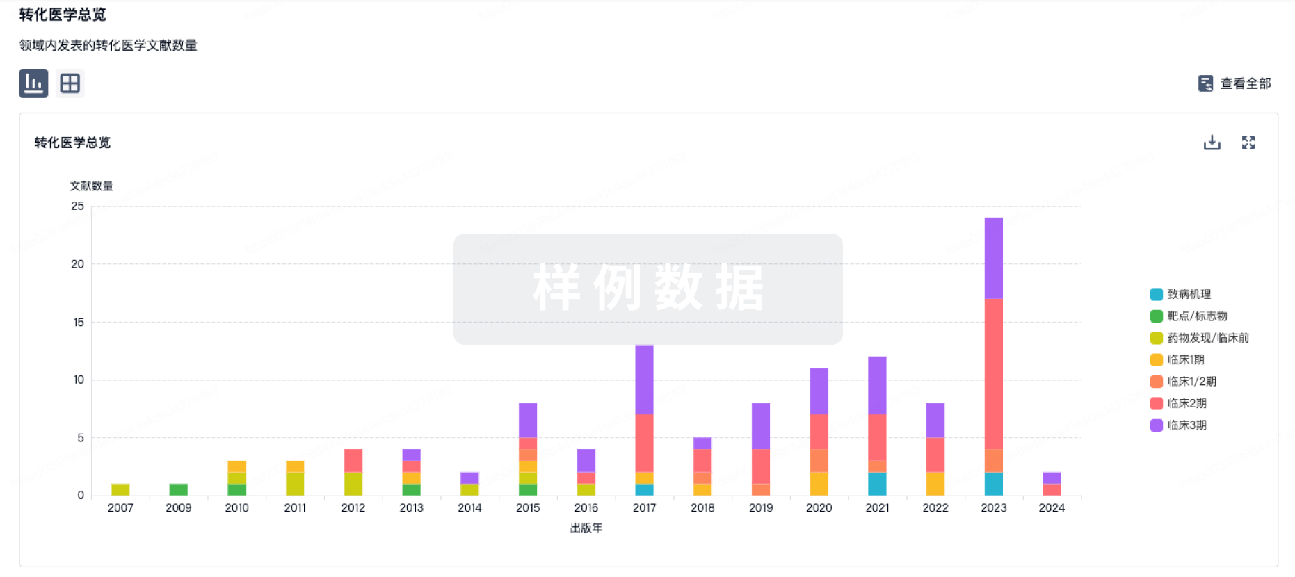
药物交易
使用我们的药物交易数据加速您的研究。
登录
或
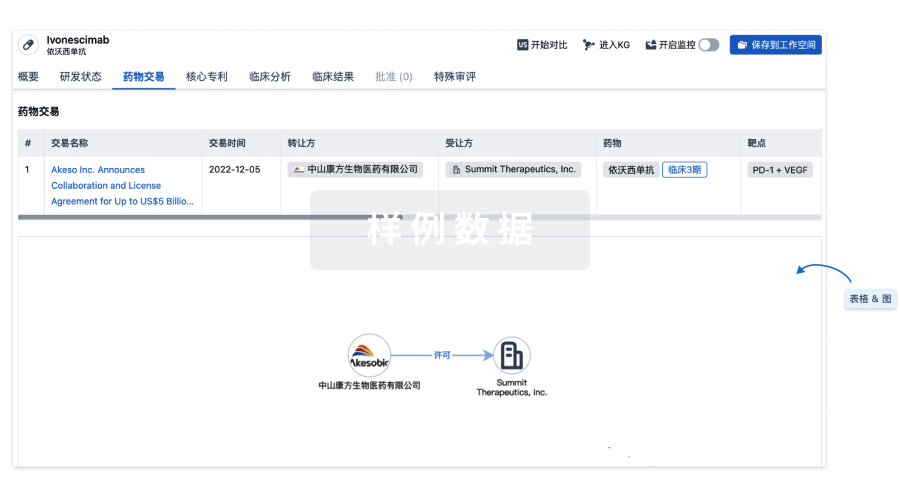
核心专利
使用我们的核心专利数据促进您的研究。
登录
或
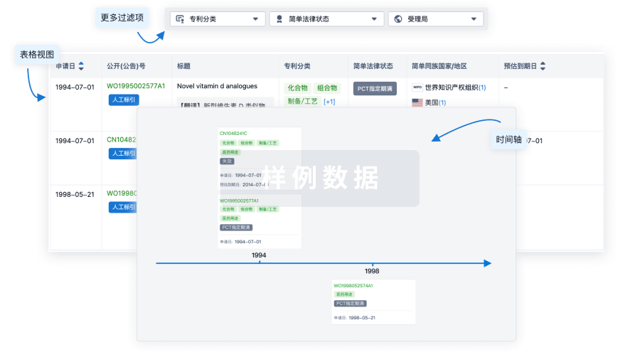
临床分析
紧跟全球注册中心的最新临床试验。
登录
或
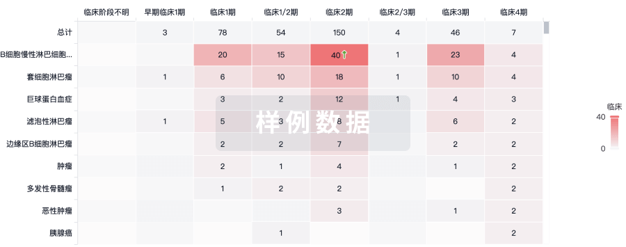
批准
利用最新的监管批准信息加速您的研究。
登录
或
生物类似药
生物类似药在不同国家/地区的竞争态势。请注意临床1/2期并入临床2期,临床2/3期并入临床3期
登录
或
特殊审评
只需点击几下即可了解关键药物信息。
登录
或
生物医药百科问答
全新生物医药AI Agent 覆盖科研全链路,让突破性发现快人一步
立即开始免费试用!
智慧芽新药情报库是智慧芽专为生命科学人士构建的基于AI的创新药情报平台,助您全方位提升您的研发与决策效率。
立即开始数据试用!
智慧芽新药库数据也通过智慧芽数据服务平台,以API或者数据包形式对外开放,助您更加充分利用智慧芽新药情报信息。
生物序列数据库
生物药研发创新
免费使用
化学结构数据库
小分子化药研发创新
免费使用