预约演示
更新于:2026-08-22
Altrakincept
更新于:2026-08-22
概要
基本信息
在研机构- |
非在研机构 |
权益机构- |
最高研发阶段终止临床2期 |
首次获批日期- |
最高研发阶段(中国)- |
特殊审评- |


登录后查看时间轴
关联
100 项与 Altrakincept 相关的临床结果
登录后查看更多信息
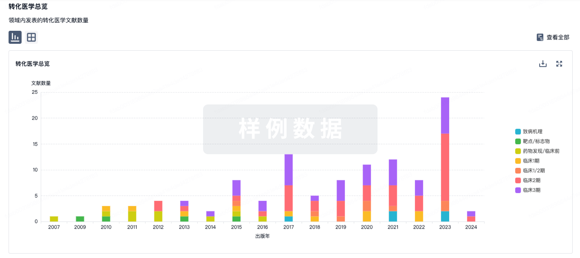
100 项与 Altrakincept 相关的转化医学
登录后查看更多信息
100 项与 Altrakincept 相关的专利(医药)
登录后查看更多信息
30
项与 Altrakincept 相关的文献(医药)2025-03-01·Dermatology and Therapy
Interleukin-4 and Atopic Dermatitis: Why Does it Matter? A Narrative Review
Review
作者: Mendes-Bastos, Pedro ; Torres, Tiago ; Duarte, Bruno ; Filipe, Paulo ; Cruz, Maria J ; Gonçalo, Margarida ; Lopes, Maria J P
Atopic dermatitis (AD) is a common chronic inflammatory skin condition that significantly impairs patients' quality of life as a result of intense itching and persistent eczematous lesions. Although AD has a multifaceted etiology-including genetic predisposition, environmental triggers, barrier dysfunction, and dysregulated immune responses-interleukin-4 (IL-4) has a recognized central role in its pathogenesis. This narrative review explores the role of IL-4 in the pathophysiology of AD, its contribution to the atopic march, and the therapeutic impact of IL-4 inhibition. IL-4 plays a critical role in skin barrier dysfunction, dysbiosis, pruritus, and inflammation, all of which contribute to the debilitating symptoms of AD. Moreover, IL-4 is implicated in other atopic conditions, such as asthma, allergic rhinitis, and food allergies, underscoring its role beyond AD and its importance in the atopic march. Recent advances in targeted therapies, particularly IL-4/IL-13 signaling inhibitors, have changed AD management. Dupilumab, an IL-4 receptor antagonist, has demonstrated significant efficacy in reducing AD symptoms and enhancing patient outcomes in both children and adults. In addition to symptomatic relief, suppressing IL-4 signaling may also offer potential for disease modification, altering AD's progression and possibly preventing the onset of other atopic conditions. This review highlights the crucial role of IL-4 as a therapeutic target in AD. By understanding the role of IL-4 in AD pathogenesis and exploring the therapeutic implications of targeting IL-4 pathways, this work can contribute to guide future research concerning treatment approaches and also emphasize the need for early and targeted interventions to mitigate disease impact and ultimately improve patient quality of life.
2023-10-01·Arthritis & rheumatology (Hoboken, N.J.)
Characterization of a Musculoskeletal Syndrome of Enthesitis and Arthritis in Patients With Atopic Dermatitis Treated With Dupilumab, an Interleukin‐4/13 Inhibitor
Article
作者: Catherine Smith ; Libin Mathew ; Bina Menon ; Andrew E. Pink ; Bruce Kirkham ; Joseph Nathan ; Richard T. Woolf ; Catherine D. Hughes
Objective:
To characterize the presentation and outcomes of patients with atopic dermatitis (AD) who developed musculoskeletal symptoms after treatment with dupilumab, a human IgG4 monoclonal antibody that blocks the functions of interleukin‐4 (IL‐4) and IL‐13, key pathologic pathways in AD.
Methods:
This article reports an observational cohort of patients receiving dupilumab who developed new‐onset musculoskeletal symptoms after dupilumab therapy at our center. All patients had a comprehensive rheumatologic history and examination, with imaging by ultrasonography (US) or magnetic resonance imaging (MRI) in most patients.
Results:
Between October 2018 and February 2021, we recorded 470 patients with AD commencing dupilumab treatment from routine clinical care records. Of 36 patients referred for rheumatologic assessment, we identified 26 patients (14 male, 12 female) with a musculoskeletal syndrome of inflammatory enthesitis, arthritis, and/or tenosynovitis. Clinical findings were confirmed by US and MRI. All patients had very good response to dupilumab treatment, and no specific predictors of musculoskeletal syndrome were noted. Symptoms were mild in 16 patients, moderate in 6 patients, and severe in 4 patients. Receipt of nonsteroidal antiinflammatory drugs or cyclooxygenase 2 inhibitors, reduction of dupilumab dose/frequency, and cessation of dupilumab therapy led to improvement, but moderate or severe symptoms persisted for many months.
Conclusion:
We report a new musculoskeletal syndrome of inflammatory enthesitis/arthritis/tenosynovitis in some patients receiving the IL‐4 receptor antagonist dupilumab. This response to a cytokine‐targeting therapy provides key insights into the pathogenesis of enthesitis.
2022-10-01·Journal of cosmetic dermatology4区 · 医学
Evaluation of adult patients with atopic dermatitis treated with dupilumab: A single‐center real‐life experience
4区 · 医学
Article
作者: Bengisu Güçkan Işik ; Anıl Gülsel Bahali ; Tahsin Çağdaş Akaslan ; Nazan Taşlidere ; Fatma Pelin Özgen ; Begüm Güneş ; Özlem Su Küçük
Abstract:
Background:
Adult atopic dermatitis (AD), especially adult‐onset type appears to have different clinical manifestations. Dupilumab is an IL‐4 receptor antagonist used in patients with moderate and severe atopic dermatitis, aged 12 years and older and it works by inhibiting the IL‐4 and IL‐13 signaling pathway. The purpose of our study is to retrospectively investigate the side effect profile and drug efficacy of thirteen adult patients who received dupilumab treatment and to evaluate the drug use status and the results during the COVID‐19 pandemicour stuAdult atopic dermatitis (AD), especially adult‐onset type appears to have different clinical manifestations. Dupilumab is an IL‐4 receptor antagonist used in patients with moderate and severe atopic dermatitis, aged 12 years and older and it works by inhibiting the IL‐4 and IL‐13 signaling pathway. The purpose of our study is to retrospectively investigate the side effect profile and drug efficacy of thirteen adult patients who received dupilumab treatment and to evaluate the drug use status and the results during the COVID‐19 pandemicAdult atopic dermatitis (AD), especially adult‐onset type appears to have different clinical manifestations. Dupilumab is an IL‐4 receptor antagonist used in patients with moderate and severe atopic dermatitis, aged 12 years and older and it works by inhibiting the IL‐4 and IL‐13 signaling pathway. The purpose of dy is to retrospectively investigate the side effect profile and drug efficacy of thirteen adult patients who received dupilumab treatment and to evaluate the drug use status and the results during the COVID‐19 pandemicAdult atopic dermatitis (AD), especially adult‐onset type appears to have different clinical manifestations. Dupilumab is an IL‐4 receptor antagonist used in patients with moderate and severe atopic dermatitis, aged 12 years and older and it works by inhibiting the IL‐4 and IL‐13 signaling pathway. The purpose of our study is to retrospectively investigate the side effect profile and drug efficacy of thirteen adult patients who received dupilumab treatment and to evaluate the drug use status and the results during the COVID‐19 pandemic.
Materials and Methods:
Thirteen patients with clinical and/or histopathological diagnoses of atopic dermatitis who received dupilumab treatment and were subsequently followed up in Bezmialem Vakif University dermatology outpatient clinic between April 2019 and October 2021 were included in our study.Patient files were reviewed, and patients were interviewed in‐person or by phone to learn about the COVID‐19 contagion.Descriptive statistical analysis was performed with Microsoft Excel, and the data obtained were calculated as mean and percentage.
Results:
All of our patients responded to the treatment after one course of dupilumab injection and also CRP and LDH levels decreased. Conjunctivitis side effect was found at a slightly higher rate than in previous clinical studies. The treatment was continued during the COVID‐19 pandemic in most patients. Meanwhile, four patients had COVID‐19 infection, but one of them was not using dupixent at that time.
Conclusion:
We can conclude that dupilumab is an effective and safe therapy for patients with severe AD also in cases of severe infections.
13
项与 Altrakincept 相关的新闻(医药)2026-08-20
点击上方蓝字 关注我们
虽然大量患者被诊断为不典型病变或小叶原位癌,但是对于这些病变的诊治,缺乏来自研究证据和专家共识的指南。虽然过去几十年里众多高质量数据已经推动乳腺癌诊治的快速发展,但是受限于前瞻研究数据有限和诊断不确定性,不典型良性增生诊治仍然存在诸多争议。虽然影像学、活检技术和放射病理学相关研究进展表明,这些病变仅代表一系列生物学风险,而非一刀切的手术指征,但是传统的常规手术切除模式仍然长期存在。
2026年8月19日,《美国医学会杂志》外科学分册在线发表哈佛大学医学院贝斯以色列女执事医疗中心和达纳法伯癌症研究院、德克萨斯大学MD安德森癌症中心、埃默里大学医学院、纽约大学格罗斯曼医学院、斯坦福大学医学院、纽万斯医疗、纽约大学朗格尼医疗中心、莱格西医疗、妙佑(梅奥)医疗国际、北岸医疗事业埃文斯顿医院的美国乳腺外科学会、乳腺影像学会和美国病理学会乳腺不典型增生和小叶原位癌诊治指南2026年版。
这是三家学会第三次联合制定良性乳腺病变诊治指南。此次,三家学会合作组建指导小组监督制定诊治指南,主要针对不典型病变和小叶原位癌,其中包括不典型扁平上皮增生、不典型导管增生、不典型小叶增生、经典型小叶原位癌以及变异型小叶原位癌,如多形性小叶原位癌和旺炽性小叶原位癌。
不典型导管增生、不典型小叶增生和经典型小叶原位癌的自然病程表明,这些病变与将来乳腺癌风险升高相关,因此建议诊断为这些病变的患者接受全面风险评定和乳腺癌风险降低策略咨询。
多形性小叶原位癌和旺炽性小叶原位癌与将来乳腺癌风险相关程度尚不确定,但是如果确定这些病变为激素受体阳性,那么应该考虑风险降低药物。
目前尚无证据表明不典型扁平上皮增生与将来乳腺癌风险升高相关。
再次病理复核确认:对于不典型导管增生推荐,对于不典型扁平上皮增生应该考虑,对于不典型小叶增生或小叶原位癌不必。
现有证据支持对大多数粗针活检确诊的不典型导管增生、多形性小叶原位癌和旺炽性小叶原位癌病例进行诊断性切除,但是部分符合严格标准并获得多学科共识的不典型导管增生病例可以进行观察。
多形性小叶原位癌和旺炽性小叶原位癌需要切缘阴性,不典型导管增生不需切缘阴性。
如果粗针活检诊断与影像学特征一致,即影像学与病理学一致性得到证实,那么不典型小叶增生和经典型小叶原位癌可以安全地进行观察。
如果影像学与病理学一致性得到证实,那么仅诊断为不典型扁平上皮增生后,通常不必进行诊断性切除。
因此,该指南为乳腺不典型病变(包括不典型导管增生、不典型小叶增生、不典型扁平上皮增生、经典型小叶原位癌以及变异型小叶原位癌,例如多形性小叶原位癌和旺炽性小叶原位癌)诊治提供了来自研究证据或专家共识的推荐意见。临床医生诊治乳腺不典型病变或小叶原位癌患者时,应该考虑将这些指南纳入临床诊治。
对此,洛马琳达大学医学院发表特邀评论:乳腺不典型病变诊治规范更新。
JAMA Surg. 2026 Aug 19. IF: 15.6
American Society of Breast Surgeons, Society of Breast Imaging, and College of American Pathology 2026 Guidelines for the Management of Proliferative Lesions With Atypia and Lobular Carcinoma In Situ.
Nakhlis F, Bedrosian I, King TA, Heller SL, Allison KH, Boolbol S, Pass HA, Johnson NM, Boughey JC, Yao K.
Beth Israel Deaconess Medical Center, Dana-Farber Cancer Institute, Boston, Massachusetts; University of Texas MD Anderson Cancer Center, Houston; Emory University School of Medicine, Atlanta, Georgia; New York University Grossman School of Medicine, New York; Stanford Medicine, Stanford, California; Nuvance Health, Poughkeepsie, New York; NYU Langone Health, New York, New York; Legacy Health, Portland, Oregon; Mayo Clinic, Rochester, Minnesota; NorthShore Endeavor Health, Evanston, Illinois.
IMPORTANCE: Many patients are diagnosed with atypical lesions or lobular carcinoma in situ (LCIS); however, evidence- and consensus-based guidelines for the management of many of these lesions are limited.
OBSERVATIONS: The American Society of Breast Surgeons, in collaboration with the Society of Breast Imaging and College of American Pathology, assembled a steering group to create guidelines for the management of atypical lesions and LCIS, inclusive of flat epithelial atypia (FEA), atypical ductal hyperplasia (ADH), atypical lobular hyperplasia (ALH), classic LCIS (C-LCIS), and the variant LCIS forms pleomorphic LCIS (P-LCIS) and florid LCIS (F-LCIS). The natural history of ADH, ALH, and C-LCIS suggests that these lesions are associated with an elevated future breast cancer risk; therefore, patients diagnosed with these lesions should be recommended to undergo comprehensive risk assessment and counseling about breast cancer risk-reducing strategies. The magnitude of future breast cancer risk associated with P-LCIS and F-LCIS remains uncertain, yet if these lesions are determined to be hormone receptor positive, risk-reducing medications should be considered. There is no evidence that FEA is associated with an elevated future breast cancer risk. A second pathology review confirmation is recommended for ADH, should be considered for FEA, and is not necessary for ALH or LCIS. Currently available evidence supports the need to diagnostically excise most ADH, P-LCIS, and F-LCIS cases identified on core biopsy, although some ADH cases fulfilling strict criteria and multidisciplinary consensus can be observed. P-LCIS and F-LCIS require a negative margin, but ADH does not. ALH and C-LCIS can be safely observed if the core biopsy diagnosis is concordant with imaging features (ie, radiographic-pathologic concordance is established). Provided radiologic-pathologic concordance is confirmed, diagnostic excision is generally not indicated after a diagnosis of FEA alone.
CONCLUSIONS AND RELEVANCE: These guidelines provide evidence-informed, consensus-based recommendations for the management of atypical lesions of the breast, including ADH, ALH, FEA, C-LCIS, P-LCIS, and F-LCIS. Practicing clinicians who treat patients with atypical breast lesions or LCIS should consider integrating these guidelines into clinical management.
PMID: 42616513
DOI: 10.1001/jamasurg.2026.3552
JAMA Surg. 2026 Aug 19. IF: 15.6
Updated Framework for Atypical Breast Lesions.
Vora H, Denham L, Lum S.
Loma Linda University School of Medicine, Loma Linda, California.
PMID: 42616553
DOI: 10.1001/jamasurg.2026.3546
(来源:SIBCS)
声 明
凡署名原创的文章版权属《肿瘤瞭望》所有,欢迎分享、转载。本文仅供医疗卫生专业人士了解最新医药资讯参考使用,不代表本平台观点。该等信息不能以任何方式取代专业的医疗指导,也不应被视为诊疗建议,如果该信息被用于资讯以外的目的,本站及作者不承担相关责任。
2026-08-17
tags: [银屑病, 特应性皮炎, 白癜风, 辉瑞, Tilrekimig, 三抗, 信达生物, IBI3027, 度普利尤, VC005, 索洛奇单抗, 纳米抗体, 三生国健, SSGJ-718, 第十二批集采, 恩适达, 2026医保, 芦可替尼乳膏]皮肤领域医药动态周报(2026.08.10–08.16)
本期要点:辉瑞全球首创IL-4/IL-13/TSLP三抗Tilrekimig启动特应性皮炎III期临床——每月一次皮下注射,同时阻断2型炎症三条关键通路,超越达必妥单靶点机制;信达生物度普利尤单抗生物类似药IBI3027启动III期——IND到III期不到半年,瞄准2026年Q2超50亿欧元的达必妥市场;华东医药/威凯尔VC005片白癜风II期阳性数据公布——24周F-VASI改善43%,国产新一代JAK1抑制剂多适应症全面布局;MoonLake双靶向IL-17纳米抗体索洛奇单抗PsA III期全线阳性——纳米抗体在自免领域关键里程碑;三生国健IL-23/TL1A双抗SSGJ-718递交IND——自免双抗新靶点;第十二批集采正式落地——65品种600亿市场,地奈德软膏剂纳入,10款原研药回归;2026医保谈判进入测算关键期——7/31专家评审结束,8月下旬至9月中旬现场谈判。
---一、本周核心亮点速览
序号
事件
企业/机构
意义
1
**辉瑞Tilrekimig(IL-4/IL-13/TSLP三抗)启动AD III期**(8/14)
辉瑞
全球首个AD三抗进入III期,每月一次给药,超越达必妥单靶点机制
2
**信达IBI3027(度普利尤类似药)启动III期**(8/10)
信达生物
国产首款度普利尤生物类似药冲刺,520例患者,IND到III期不到半年
3
**VC005片白癜风II期阳性数据公布**(8/13)
华东医药/威凯尔
24周F-VASI改善43%,国产新一代JAK1抑制剂白癜风赛道加入
4
**索洛奇单抗PsA III期全线阳性**(8/10)
MoonLake
双靶向IL-17纳米抗体,初治PsA全部主要终点达标
5
**三生国健SSGJ-718(IL-23/TL1A双抗)递交IND**(8/6)
三生国健
自免双抗新靶点,年内第二款1类新药申报
6
**第十二批集采正式落地**(8/6)
国家医保局
65品种600亿,地奈德软膏剂纳入,10款原研药回归
7
**海南省委书记冯飞调研德镁医药**(8/10)
海南省委
百卢妥"乐城研用+海口生产"模式获高度肯定
8
**CSD 2026聚焦银屑病慢病管理**
中华医学会
匹康奇拜单抗本土数据亮相,银屑病迈入系统性慢病管理新阶段
---二、特应性皮炎:三抗时代开启,生物类似药加速入场2.1 🔥🔥🔥 辉瑞Tilrekimig启动AD III期——全球首个IL-4/IL-13/TSLP三抗
2026年8月14日,辉瑞在ClinicalTrials.gov注册了Tilrekimig(研发代码PF-07275315)治疗中重度特应性皮炎的全球III期临床试验(NCT07765888)。
维度
内容
产品
Tilrekimig(PF-07275315)
企业
辉瑞
靶点
**IL-4/IL-13/TSLP三特异性抗体**
给药方案
**每月一次皮下注射**
III期入组
**375例**≥12岁中重度AD患者
预计完成
2028年5月
地位
**全球唯一针对AD等炎症疾病的三抗药物**
#### 核心机制:从"单靶点"到"全链条阻断"
Tilrekimig的三特异性设计可同时阻断与2型炎症密切相关的三条关键信号通路:
靶点
在AD中的作用
**IL-4**
AD发病核心细胞因子,驱动Th2免疫反应,促进IgE生成
**IL-13**
与IL-4共同驱动嗜酸性粒细胞浸润及皮肤屏障破坏
**TSLP**
上游炎症"开关",由角质形成细胞分泌,激活树突状细胞,诱导Th2分化
竞争定位:达必妥(度普利尤单抗)仅通过抑制IL-4/IL-13信号发挥作用。Tilrekimig同时靶向TSLP,实现从炎症启动到下游效应的全链条阻断,理论上比单靶点或双靶点药物更全面地抑制炎症、减少病情反复。全球目前仅4款三抗处于III期,Tilrekimig是其中唯一一款针对炎症疾病的三抗。
#### II期关键数据
指标
低剂量组
中剂量组
高剂量组
EASI-75达标率(安慰剂调整)
38.7%
**51.9%**
49.4%2.2 🔥🔥 信达IBI3027启动III期——国产度普利尤类似药加速冲刺
2026年8月10日,信达生物在ClinicalTrials.gov注册了IBI3027(度普利尤单抗生物类似药)的III期临床试验。
维度
内容
产品
IBI3027
企业
信达生物
参照药
度普利尤单抗(达必妥®)
适应症
中重度特应性皮炎
III期入组
**520例**患者
主要终点
治疗16周EASI-75达标率
预计完成
2028年7月
研发速度
1月IND → 4月I期 → 8月III期,**不到半年**
市场背景:度普利尤单抗2026年Q2销售额达52亿欧元,首次单季突破50亿欧元,是全球自身免疫疾病领域的重磅品种。其化合物专利在美国将于2031年3月到期。信达生物通过生物类似药和创新药并行的方式完善自免产品矩阵,除IBI3027外,还布局了IL-23抗体、TSLP/IL-4R双抗、OX40L抗体、CD40L抗体及IL-1RAP抗体等。2.3 AD赛道竞争格局更新(2026年8月)
赛道
药物
企业
当前进度
差异化优势
**IL-4Rα单抗(原研)**
度普利尤单抗(达必妥)
赛诺菲
已上市+医保
—
**IL-4Rα单抗(国产)**
司普奇拜单抗(康悦达)
康诺亚
已上市+医保
1,039元/支
**IL-4Rα单抗(国产)**
柯美奇拜单抗(MG-K10)
麦济生物
NDA受理
每4周给药,52周瘙痒下降4分+占71.7%
**IL-4Rα单抗(国产)**
泰利奇拜单抗
智翔金泰
NDA受理
国产第三家
**IL-4Rα类似药**
IBI3027
信达生物
**III期启动**
国产首款类似药
**IL-4Rα类似药**
KH816
康弘药业
I期(2026.1获批临床)
—
**IL-4Rα类似药**
SYS6033
石药集团
I期
—
**IL-13单抗**
来瑞奇珠单抗
礼来
NDA受理
16周EASI-75超60%
**JAK1口服**
艾玛昔替尼
恒瑞
已上市+医保
—
**JAK1口服(新一代)**
VC005片
华东医药/威凯尔
AD III期达标,pre-NDA
平衡型高选择性,白癜风II期阳性
**TYK2口服**
Soficitinib(ICP-332)
诺诚健华
AD III期阳性
**无黑框警告潜力**
**三抗**
Tilrekimig
辉瑞
**III期启动**
IL-4/IL-13/TSLP全链条阻断
**T-reg生物制剂**
Rezpegaldesleukin
Nektar
III期启动(7/21)
免疫修复全新机制
趋势判断:AD赛道正从"达必妥独大"向"多靶点、多机制全面竞争"演进。三抗(Tilrekimig)、T-reg生物制剂(Rezpegaldesleukin)等全新机制药物入局,标志着AD治疗从"炎症抑制"向"全链条阻断+免疫修复"升级。同时,度普利尤生物类似药密集推进(信达/康弘/石药),将进一步降低生物制剂用药成本。
---三、白癜风:国产JAK1新药II期数据亮眼,口服赛道持续扩容3.1 🔥 VC005片白癜风II期阳性——国产新一代JAK1抑制剂数据公布
2026年8月13日,华东医药宣布,与江苏威凯尔战略合作的VC005片在口服治疗成人非节段型白癜风的II期临床研究中取得积极顶线结果。
维度
内容
产品
VC005片
企业
江苏威凯尔自主研发,华东医药独家商业化
靶点
**平衡型高选择性新一代JAK1抑制剂**
机制特色
JAK1强效抑制+大幅降低JAK2/JAK3抑制,降低骨髓抑制和感染风险
适应症
成人非节段型白癜风
给药方案
早晚口服,血药浓度平稳可控
#### II期关键数据
指标
结果
24周F-VASI较基线改善
**约43%**
24周F-VASI75应答率
**约24%**
24周面部皮损复色≥50%患者比例
**超过半数**
统计学差异
P<0.001(vs安慰剂)
安全性
整体良好,未发现新的安全信号
临床价值:相较于白癜风主流外用治疗方案,VC005片口服给药能够持续作用于全身,极大解决泛发性白癜风患者大面积涂药困扰,提升治疗依从性。其"平衡型高选择性"设计更适合白癜风患者长期治疗需求。
#### VC005片多适应症布局
适应症
进度
中重度特应性皮炎
**III期已达主要终点**,进入pre-NDA
非节段型白癜风
**II期阳性**,III期即将启动
强直性脊柱炎
III期入组即将结束
斑秃
II期即将启动
轻中度AD(外用凝胶)
III期已获批开展3.2 白癜风口服JAK赛道五强格局(8月更新)
药物
企业
靶点
当前进度
关键数据
安全性标签
**乌帕替尼**
艾伯维
JAK1
**✅ EU已获批(7/29)**
Viti-Up: 48周F-VASI75≈23-25%
FDA黑框警告
**利特昔替尼**
辉瑞
JAK3/TEC
III期双达标→将申报
2,174例,52周F-VASI75+T-VASI50双达标
与斑秃一致
**Povorcitinib**
Incyte
JAK1
III期达标→2027H1申报
52周F-VASI75: 18.9% vs 3.1-6.8%
预期黑框警告
**Soficitinib**
诺诚健华
TYK2
II期阳性→III期推进
24周F-VASI改善41.2% vs 2.2%
**无黑框警告潜力**
**VC005片**
华东医药/威凯尔
JAK1(新一代)
**II期阳性**
24周F-VASI改善43%
未发现新安全信号
格局演变:白癜风口服全身治疗从上周的"四强争霸"扩容为"五强并进"。VC005片作为国产新一代JAK1抑制剂,其F-VASI改善43%的数据在II期阶段表现亮眼,与Soficitinib(41.2%)处于同一量级。五款药物覆盖JAK1、JAK3/TEC、TYK2三种靶点,为不同患者提供差异化选择。3.3 芦可替尼乳膏:海南省委书记调研德镁,医保谈判关键期
2026年8月10日,海南省委书记冯飞一行赴康哲药业皮肤健康子公司海南德镁医药调研指导,实地了解企业发展、创新研发及海南研发生产基地建设情况。
维度
内容
产品
磷酸芦可替尼乳膏(百卢妥®)
企业
Incyte原研/康哲药业/德镁医药商业化
国内获批
2026年1月NMPA批准白癜风适应症
国内AD进展
AD适应症NDA于2026年2月24日获受理
EU AD获批
2026年7月29日获欧盟批准
国内价格
约**5,800-6,800元/支**(100g规格)
医保进程
初审通过→7-8月专家评审→**8-9月价格谈判**
海南模式
**"乐城研用+海口生产"**创新实践成果
关注:冯飞书记对德镁医药聚焦皮肤健康、坚持创新发展的做法给予肯定。8-9月医保谈判是决定百卢妥命运的关键窗口。芦可替尼乳膏EU AD获批将为中国AD NDA审评提供正面催化,同时增强医保谈判筹码。海南研发生产基地即将实现主体结构封顶。3.4 CKBA软膏:全球首创靶点III期推进中
维度
内容
产品
CKBA软膏
企业
江苏博创园(泰恩康子公司)
靶点
**ACC1/MFE-2**(全球首个靶向T细胞脂肪酸代谢通路的免疫调节剂)
成人白癜风III期
2026年4月启动,许爱娥教授牵头
儿童白癜风II期
2026年1月获临床批准,5月完成首例入组
玫瑰痤疮
II/III期无缝试验,3月完成IIa期首例入组
安全性优势
II期显示安全性良好,无皮肤萎缩等严重副作用
---四、银屑病:纳米抗体III期阳性,CSD 2026聚焦慢病管理4.1 🔥 MoonLake索洛奇单抗PsA III期全线阳性——纳米抗体里程碑
2026年8月10日,MoonLake Immunotherapeutics公布核心管线索洛奇单抗(sonelokimab)用于初治银屑病关节炎的全球III期IZAR-1研究顶线阳性结果。
维度
内容
产品
索洛奇单抗(sonelokimab)
企业
MoonLake Immunotherapeutics
靶点
**双靶向IL-17A/IL-17F纳米抗体**
III期研究
IZAR-1(全球多中心、随机、双盲、安慰剂对照)
入组人群
从未接受过生物制剂治疗的活动性PsA成年患者
给药方式
皮下注射
核心节点
第16周多重疗效指标
结果
**所有主要、次要临床终点全部达成**
完整数据
预计2027年上半年试验全部完成后披露
意义:索洛奇单抗是双靶向IL-17纳米抗体,同时结合IL-17A和IL-17F两种促炎亚型,分子量仅约40kDa(传统IgG约150kDa),组织渗透性更强。本次IZAR-1全线阳性标志着纳米抗体在自身免疫炎症领域实现关键里程碑突破,为银屑病关节炎患者提供全新差异化治疗选择。4.2 CSD 2026:银屑病迈入系统性慢病管理新阶段
中华医学会第三十一次皮肤性病学术年会(CSD 2026)传递出明确信号:银屑病正式步入系统性慢病管理的新阶段。
维度
内容
核心转变
从"短期皮损改善"到"长期稳定清除+共病管理+重建生活"
管理框架
患者教育+长期皮损规范管理+共病筛查+特殊人群个体化方案
治疗目标跃升
从"控制症状"到追求皮损长期稳定清除
#### 匹康奇拜单抗CSD 2026本土数据亮相
信达生物自主研发的国产首个IL-23p19单抗匹康奇拜单抗(信美悦®)在CSD 2026上公布多维度临床证据:
维度
数据
靶点
IL-23p19(国产首个)
分子设计
Fc段YTE基因改造→"两增一减"(增强半衰期+降低免疫原性)
维持期给药
**每12周一次**(目前最长间隔)
16周PASI 90
**80.3%**(中国同类药物中唯一突破80%的IL-23抑制剂)
16周sPGA 0/1
93.5%
52周PASI 90
84.9%
52周PASI 100
**超过50%**
难治部位清除率
头皮、生殖器等91.5%
BMI影响
**疗效不受BMI/体重影响**(超重/肥胖占比超60%)
上市时间
2025年11月NMPA获批
医保状态
初审通过,8-9月谈判
价格
6,500元/支
学术价值:CLEAR-1研究问鼎国际顶级期刊《美国皮肤病学杂志》(JAAD)。匹康奇拜单抗的"疗效不受BMI影响"特性,为超重/肥胖银屑病患者的长期慢病管理提供了优选方案。4.3 银屑病治疗药物全景更新(2026年8月)
#### 已上市生物制剂(截至2026年8月)
药物
靶点
企业
获批时间
医保状态
价格参考
司库奇尤单抗(可善挺)
IL-17A
诺华
—
✅医保
~778元/支
依奇珠单抗(拓咨)
IL-17A
礼来
—
✅医保
~1,218元/支
古塞奇尤单抗(特诺雅)
IL-23p19
强生
—
✅医保
3,680元/支
瑞莎珠单抗(利生奇珠)
IL-23p19
艾伯维
—
✅医保
—
乌司奴单抗(喜达诺)
IL-12/23
强生
—
✅医保
—
比奇珠单抗(倍捷乐)
IL-17A/F
优时比
2026.3
初审过
—
赛立奇单抗(金立希)
IL-17A
智翔金泰
2024
✅医保
~756元/支
夫那奇珠单抗(安达静)
IL-17A
恒瑞
2024.8
✅医保
~743.5元/支
匹康奇拜单抗(信美悦)
IL-23p19
信达生物
2025.11
初审过
6,500元/支
安沐奇塔单抗(益赛拓)
IL-17A
三生国健
2026.2
初审过
~1,520元/支
古莫奇单抗(奇佑康)
IL-17A
康方生物
2026.6
待申报
—
瑞西奇拜单抗(华亦净)
IL-36R
华海/华奥泰
2026.7
—
—
佩索利单抗(佩索利)
IL-36R
勃林格殷格翰
—
—
—
#### NDA审评中(预计2026Q4-2027年获批)
药物
靶点
企业
NDA受理
预计获批
莱康奇塔单抗
IL-17A/F
丽珠/鑫康合
2026.1(优先审评)
2026 Q4
偌考奇拜单抗(JS005)
IL-17A
君实/复星万邦
2025.12
2027 H1
HB0017
IL-17A
华海/华奥泰
2026.6
2027 H1
鲁塞奇塔单抗
IL-17A
荃信生物
审评中
2027
#### 口服靶向药
药物
靶点
企业
状态
氘可来昔替尼(颂狄多)
TYK2
BMS
已上市+医保(2026.6新增PsA)
阿普米司特(欧泰乐)
PDE4
安进
已上市+医保
乌帕替尼
JAK1
艾伯维
已上市(PsA)
Icotrokinra(Icotyde)
IL-23R口服多肽
强生
FDA已批(2026.3),中国未上市
#### 外用创新剂型
药物
类别
企业
状态
恩适达(卡泊三醇倍他米松泡沫剂)
VD3+激素泡沫剂
LEO Pharma
2026.4获批,线上预售中
罗氟司特乳膏(Zoryve)
PDE4外用
Arcutis/华东
中国III期阳性,待申报
本维莫德乳膏(泽立美)
AhR
泽德曼
已上市+医保(138元/支)4.4 恩适达泡沫剂线上预售——银屑病外用剂型商业化加速
恩适达(卡泊三醇倍他米松泡沫剂)8月初在阿里健康、京东健康两大平台正式开启预售。
维度
内容
产品
恩适达®(卡泊三醇倍他米松泡沫剂)
企业
LEO Pharma/利奥中国
获批时间
2026年4月NMPA批准
地位
**中国首个且唯一银屑病外用泡沫剂型**
剂型优势
过饱和泡沫剂,透皮渗透提升138%-400%
线上策略
与阿里健康、京东健康合作,打通"最后一公里"
中国III期
604名患者,4周PASI-75约2/3,PASI-90约1/2
行业意义:利奥中国总经理殷晓峰表示,恩适达作为定价较高的自费创新药,入院进程面临客观挑战,线上渠道成为连接药品与患者最直接的桥梁。预计未来3-5年,线上线下药品零售格局将迎来历史性"拐点",线上规模有望扩容至3,500亿元。
---五、自免双抗:三生国健IL-23/TL1A双抗递交IND5.1 SSGJ-718——年内第二款1类新药申报
2026年8月6日,CDE官网显示三生国健1类生物药SSGJ-718递交新药研究申请(IND)。
维度
内容
产品
SSGJ-718
企业
三生国健
靶点
**IL-23/TL1A双抗**
地位
三生年内递交临床的第二款1类新药(首款SSGJ-716用于AD,3月获IND)
布局
"上市产品+NDA阶段+临床阶段+早期管线"多层次梯队
行业意义:IL-23/TL1A双抗是自免领域的新兴方向——IL-23驱动Th17炎症通路(银屑病核心),TL1A则同时参与炎症和纤维化通路(IBD核心),双靶点协同有望覆盖银屑病+IBD的IMID(免疫介导炎症性疾病)共病人群。三生国健继安沐奇塔单抗(IL-17A,2026.2获批)后持续扩容自免管线。
---六、第十二批集采正式落地:皮肤科外用药价格体系重塑6.1 集采核心数据
2026年8月6日,第12批国家组织药品集中带量采购中选结果正式公布。
维度
数据
开标时间
2026年7月31日
中选结果公布
2026年8月6日
采购周期
执行至2029年12月31日
品种数量
**65个**
参与企业
495家、859个产品
中选结果
**327家**企业、**521个**产品
报量机构
全国**4.5万家**医疗机构
市场规模
约**600亿元**(历届之最)
原研药中选
**10款**参比制剂(历届之最)6.2 皮肤科相关品种
品种
中选企业(代表)
皮肤科意义
**地奈德软膏剂**
江苏知原药业、重庆胜凯(华邦)、海南全盈
**首次纳入软膏剂**,皮肤科外用药价格体系重塑
巴瑞替尼口服常释剂型
山东华铂凯盛(泰恩康受托)、杭州民生等
JAK抑制剂,用于斑秃等自免疾病6.3 集采规则迭代要点
规则创新
内容
**"稳临床"**
按厂牌报量+不带量复活+不按省分量,保证临床用药延续性
**"反内卷"**
双锚点机制控制价格离散度,极端低价企业无法获得带量
**"保质量"**
参比制剂"不带量复活"通道,10款原研药回归(历届之最)
**"防围标"**
全流程严格保密和监督
对皮肤科的影响:地奈德乳膏是皮肤科最常用的弱效外用糖皮质激素之一,广泛用于湿疹、皮炎、银屑病等。纳入集采后将重塑外用药价格体系。2025年中国公立医疗机构软膏剂化药销售额逼近90亿元,皮肤病用药占比超83%。集采标志着皮肤科外用药从"分散竞争"走向"规模竞争"。
---七、2026医保谈判进入测算关键期7.1 医保谈判时间线
阶段
时间
状态
准备阶段
2026.4-5月
✅已完成
企业申报+形式审查
2026.6.1-7月上旬
✅已完成
专家综合评审
2026.7-8月
✅7/31公布评审结果
**现场谈判&竞价**
**2026.8下旬-9月中旬**
⏳即将开始
结果公布
2026.10下旬-11月
待定
正式落地
2027.1.1
待定7.2 关键数据
维度
数据
基本医保申报
800份,涉及664个通用名
形式审查通过
**601个**(目录外368+目录内233),通过率91%
商保创新药申报
62份,涉及62个通用名
商保形式审查通过
**58个**(目录外57+目录内1),通过率94%
首次商保→医保衔接
7款商保目录药品通过形式审查7.3 皮肤科重点谈判品种
药物
靶点/类别
企业
当前状态
芦可替尼乳膏(百卢妥)
外用JAK1/2
Incyte/康哲/德镁
初审通过→**8-9月价格谈判**
匹康奇拜单抗(信美悦)
IL-23p19
信达生物
初审通过→谈判
安沐奇塔单抗(益赛拓)
IL-17A
三生国健
初审通过→谈判
比奇珠单抗(倍捷乐)
IL-17A/F
优时比
初审通过→谈判
谈判规则亮点:今年首次引入预申报机制和参照药预沟通办法。药企还没坐上谈判桌,医保已划好三条线——主规格(怎么算)、参照药(跟谁比)、支付范围(给谁付),这三件事在谈判开始前就基本确定。芦可替尼乳膏EU AD获批将增强谈判筹码,但参照药锚点位置将决定其价格空间。
---八、流行病学与发病机制8.1 银屑病
维度
数据
全球患病率
0.5%-3%,约6,400万患者
中国患病率
约0.47%-0.55%,约650-780万患者
共病情况
约73%患者存在至少一种共病,近30%发展为PsA
核心机制
IL-23/Th17轴异常活化,IL-17A/F、TNF-α过度释放
CSD 2026新共识
银屑病正式步入系统性慢病管理,核心挑战为病程迁延、易复发、伴随心血管/代谢共病8.2 白癜风
维度
数据
全球患病率
0.5%-2%
中国患病率
约0.56%,约800万例
发病年龄
约50%在20岁前发病,70%-80%在30岁前
分型
非节段型(NSV)占约84%
核心机制
CD8+ T细胞攻击黑素细胞;IFN-γ→JAK/STAT→CXCL9/10正反馈环路
儿童白癜风
占总数32%-40%,平均发病4-8岁,约25%在10岁前发病,**全球无获批儿童产品**8.3 特应性皮炎
维度
数据
中国1-7岁儿童患病率
12.94%
中国1岁以内婴儿患病率
超过30%
中国成人患病率
3%-8%
发病特点
60%在1岁前发病,半数病程持续至成年
核心机制
皮肤屏障功能障碍+2型炎症(IL-4、IL-13驱动)+皮肤菌群紊乱
三抗机制启示
TSLP为上游"开关",同时靶向IL-4/IL-13/TSLP可实现全链条阻断
---九、竞争格局、医保与价格9.1 主要皮肤科靶向药价格与医保情况(2026年8月更新)
#### 特应性皮炎靶向药
药物
企业
靶点
价格(参考)
医保状态
度普利尤单抗(达必妥)
赛诺菲
IL-4Rα
医保后约数千元/支
✅医保
司普奇拜单抗(康悦达)
康诺亚
IL-4Rα
**1,039元/支**
✅医保
艾玛昔替尼
恒瑞
JAK1
**1,906元/盒**(4mg×28片)
✅医保
乌帕替尼(瑞福)
艾伯维
JAK1
已纳入医保
✅医保
芦可替尼乳膏(百卢妥)
Incyte/康哲
JAK1/2
约**5,800-6,800元/支**
初审通过,谈判中
#### 白癜风药物
药物
企业
靶点/机制
当前状态
价格/备注
芦可替尼乳膏(百卢妥)
Incyte/康哲
外用JAK1/2
国内已上市
5,800-6,800元/支,医保初审通过
乌帕替尼
艾伯维
口服JAK1
EU已获批白癜风
国内已上市AD等适应症
利特昔替尼
辉瑞
口服JAK3/TEC
III期双达标→将申报
国内已上市重度斑秃
Povorcitinib
Incyte
口服JAK1
III期达标→2027H1申报
—
Soficitinib
诺诚健华
口服TYK2
II期阳性→III期推进
—
VC005片
华东医药/威凯尔
口服JAK1
**II期阳性**
—
CKBA软膏
泰恩康/博创园
ACC1/MFE-2
III期推进中
全球首创靶点9.2 度普利尤单抗市场数据
维度
数据
2026年Q2全球销售额
**52亿欧元**(首次单季突破50亿欧元)
美国化合物专利到期
2031年3月
国产生物类似药进度
信达IBI3027(III期)、康弘KH816(I期)、石药SYS6033(I期)十、结语
2026年8月第3周,皮肤领域创新药竞争进入"多机制、多靶点、多剂型"全面爆发阶段:特应性皮炎
:辉瑞三抗Tilrekimig启动III期,从"单靶点抑制"迈向"全链条阻断";信达度普利尤类似药III期启动,生物类似药加速入场。白癜风
:VC005片II期阳性加入口服JAK五强格局;芦可替尼乳膏医保谈判在即,EU AD获批持续催化。银屑病
:纳米抗体索洛奇单抗PsA III期全线阳性;CSD 2026确立慢病管理新范式;恩适达泡沫剂线上预售加速商业化。自免双抗
:三生国健IL-23/TL1A双抗递交IND,双靶点协同覆盖IMID共病人群。政策端
:第12批集采落地,10款原研药回归;2026医保谈判8月下旬至9月中旬现场谈判。
免责声明:本文仅供医药行业学术交流参考,不构成任何诊疗建议或用药指导。具体用药请遵医嘱。
数据来源:CDE官网、NMPA、国家医保局、ClinicalTrials.gov、企业公告、CSD 2026、公开发表的临床研究及权威医学媒体
2026-08-02
·感悟分子
小分子新药靶点深度调研报告 | UniProt P05112 | 细胞因子(IL-4/IL-13 共享 IL-4Rα 轴)
靶点价值:9/10专利拥挤度:9/10小分子起点:❌ 无(抗体/生物药主战场)晶体结构:✅ 有报告日期:2026-07-30一、靶点概述与生物学基础
IL-4(Interleukin-4,基因符号 IL4,UniProt P05112)是经典的 2 型(Th2)炎症核心细胞因子,由 153 个氨基酸的前体经信号肽切除后生成 129 个氨基酸的成熟链,分子量约 17.5 kDa,属分泌型蛋白,归类于 IL-4/IL-13 细胞因子家族。其生物学本质是 Th2 免疫应答的"总开关"——驱动 B 细胞类别转换至高 IgE / IgG1、上调 CD23(低亲和力 IgE 受体)、促进嗜酸性粒细胞募集与替代性活化巨噬细胞分化,并抑制 Th1/Th17 偏向。在病理状态下,IL-4 是过敏、哮喘、特应性皮炎(AD)等 2 型炎症疾病的因果性驱动因子。
属性
内容
蛋白全称
Interleukin-4 / B-cell stimulatory factor 1 (BSF-1)
基因符号
IL4(染色体 5q31.1,与 IL13、IL5、IL3 成簇)
UniProt ID
P05112(次要:Q14630、Q6NZ77)
种属
Homo sapiens
蛋白类型
分泌型细胞因子(四螺旋束折叠,含 3 对二硫键)
分子量
前体 17,492 Da;成熟 129 aa(约 15 kDa)
亚细胞定位
分泌到细胞外(Secreted)
主要分泌细胞
Th2 细胞、肥大细胞、嗜碱性粒细胞、嗜酸性粒细胞、ILC2
受体轴
IL-4Rα(P24394)+ γc(IL2RG)→ Ⅰ型;IL-4Rα + IL-13Rα1(P78552)→ Ⅱ型
关键通路
JAK1 / JAK3 → STAT6 磷酸化 → Th2 转录程序
疾病关联
缺血性卒中遗传易感(MIM 601367,PMID 14681304);核心 2 型炎症驱动
IL-4 的成药逻辑有一个必须首先澄清的关键点:IL-4 本身与 IL-13 共享 IL-4Rα 亚基——这是整个靶点的"枢纽"。Ⅰ型受体(IL-4Rα + γc)主要表达于造血细胞,仅由 IL-4 触发;Ⅱ型受体(IL-4Rα + IL-13Rα1)则表达于造血与非造血细胞,既能被 IL-4 触发、也能被 IL-13 触发。因此阻断 IL-4Rα 能同时沉默 IL-4 和 IL-13 两条信号,而单纯阻断 IL-4 则无法抑制 IL-13 经 IL-13Rα1/α2 的 IL-4Rα 非依赖通路。这一"共享受体"结构决定了临床开发的主战场不在"抗 IL-4 抗体",而在"抗 IL-4Rα 抗体"(双阻断)——下文竞争章节将详述这一历史教训。
组织表达层面,IL-4 不在组织内组成型高表达(HPA 标注为"Not detected"),其本质是诱导型效应分子,在寄生虫感染、过敏原暴露、上皮屏障损伤时被 Th2/ILC2 局部放大。这决定了其靶向策略以"阻断循环/组织间隙中的可溶性细胞因子信号"为主,而非抑制细胞内表达。GWAS 证据链上,IL4/IL13 基因座(5q31)与血清 IgE 水平、特应质(atopy)、哮喘易感性强关联,是 2 型炎症最稳健的遗传信号之一;缺血性卒中关联则提示 IL-4 在脑血管保护/修复中的潜在角色,属次要线索。二、成药性评估
小分子起点:❌ 无。IL-4 是分泌型细胞因子,其生物活性依赖与 IL-4Rα(及共受体)的蛋白-蛋白界面,不存在可被传统小分子占据的酶催化口袋或变构裂隙。全球范围内,IL-4/IL-13 轴的临床在研分子全部为抗体或融合蛋白类生物药,无任何小分子进入临床。这意味着若以"小分子新药靶点"定义严格卡位,IL-4 不属于典型可成药小分子靶点;本报告的"成药性"须放在生物药(单抗 / 双抗 / 长效化 / 吸入化)与 PPI 界面工程的语境下评估。晶体结构与界面可靶向性
IL-4 的结构生物学已相当成熟,为抗体表位设计提供了原子级依据:
PDB ID
内容
方法
分辨率
1RCB
人源游离 IL-4(天然构象)
X-ray
2.25 Å
1IAR
IL-4 / IL-4Rα(胞外域)中间复合物
X-ray
2.30 Å
1ITL / 1IAM
IL-4 突变体(受体结合热点定位)
X-ray
2.0–2.4 Å
1TGW / 1GIG
抗 IL-4 单抗 Fab 复合物(表位解析)
X-ray
~2.0 Å
抗 IL-4Rα 相关
dupilumab 类 Fab / 受体复合体(IL-4Rα 空间构象)
X-ray / EM
—
1IAR(Hage et al., Cell 1999)揭示的界面是理解该轴成药性的核心:IL-4 与 IL-4Rα 形成"马赛克式"结合界面,由三个独立的跨蛋白相互作用簇组成,受体结合诱导 IL-4 发生轻微构象变化(螺旋 C 区重排)。这一界面特征解释了为何不同抗 IL-4Rα 抗体(dupilumab 与其后继者)可通过占据不同表位实现功能等价但序列各异的阻断——也为后继者的"设计绕开"提供了结构空间。需要诚实指出:这些结构中无任何小分子共晶(0 个),因为细胞因子-受体相互作用面大而浅,不适合小分子占据;靶点可成药性完全依赖抗体工程。CRO Assay 可及性
Assay 丰富度评级:🟡 中(生物药 assay 成熟,小分子 panel 缺失)。具体分层如下:
维度
可及性
说明
生化 assay
🟢 成熟
IL-4/IL-13 与 IL-4Rα 或 IL-13Rα1 的 ELISA / 表面等离子共振(SPR)/ 均相时间分辨荧光(HTRF)结合竞争 assay;STAT6 磷酸化(pSTAT6)报告细胞系
细胞 assay
🟢 成熟
TF-1 / THP-1 STAT6 报告基因、原代 PBMC 共培养(CD23 上调、IgE 分泌)、人源化小鼠药效
体内 / PD 标志
🟡 中等
血清 TARC(CCL17)/胸腺活化调节趋化因子、 periostin、IgE、嗜酸性粒细胞计数作为 2 型炎症 PD 生物标志物
小分子定制需求
🔴 不适用
无小分子先导物筛选基础 panel;若开发口服小分子(极早期概念,如细胞因子-受体界面多肽模拟物),需从头建设
对项目启动的影响:若立项为抗体/双抗,CRO 现成 panel 充分,可在数周内启动 lead 筛选与表位竞争分析;若执意开发小分子,则进入"无已知起点"的从头发现,风险与成本陡增,不建议作为独立立项路径。成药性评分理由
以生物药维度评估,IL-4/IL-4Rα 轴的成药性为极高:机制已被"药王"级资产(dupilumab 2025 全球销售约 178 亿美元)临床与商业双重验证,靶点-表型因果关系清晰,生物标志物成熟。但"靶点价值 9/10"已扣除两点:① 这不是可被小分子的靶点(限制了化学空间的创新自由度);② 赛道极度拥挤(见第四章、第五章),新进入者的"增量价值"需靠长效化/双抗/适应症差异化兑现,而非机制新颖性。三、适应症格局
IL-4 的适应症谱系由"阻断 2 型炎症"这一核心机制外推,经 dupilumab 的"Pipeline-in-a-Drug"策略被持续拓宽。以下按证据强度分级。核心适应症(证据强,已获批/Ⅲ期验证)
特应性皮炎(AD):IL-4/IL-13 是 AD 瘙痒与屏障破坏的上游驱动。dupilumab 在 AD 中实现 EASI-75 约 50–60%、IGA 0/1 约 40%,并快速止痒;2026 年真实世界数据显示其短期治疗即可下调皮肤归巢 Th2 细胞(CLA⁺)、恢复角质形成分化蛋白(KRT1/2/10)。中国数据:康诺亚 stapokibart 在 AD Ⅲ期达 EASI-75 约 66%、IGA 0/1 约 44%。AD 是中重度患者约 1960 万(中国,2022)的超大慢病市场,证据等级强。
哮喘(尤其是嗜酸/T2 表型):IL-4 抑制通过降低气道嗜酸性炎症与黏液高分泌改善控制。dupilumab 在儿童哮喘 VOYAGE 试验中(NCT02948959)将年化急性加重降低 47–71%(按病程分层),无论病程长短均获益,且肺功能正常化与临床缓解率更高。证据等级强。
慢性鼻窦炎伴鼻息肉(CRSwNP)与嗜酸性食管炎(EoE):二者均为 IL-4/IL-13 驱动的局部 2 型炎症。dupilumab 在 CRSwNP 显著缩小息肉、改善嗅觉;在 EoE 通过逆转 1,302 基因转录特征(EoE-NES,P<5×10⁻⁸)改善吞咽困难。stapokibart 亦已获批 CRSwNP。证据等级强。拓展适应症(证据中等→强,部分已批)
慢性阻塞性肺疾病(COPD):dupilumab 已成为首个获批用于嗜酸性表型 COPD 的生物制剂(2024–2025),将急性加重率显著降低,是该轴市场放量的关键新引擎。证据等级强(新获批)。
结节性痒疹(PN)、慢性自发性荨麻疹(CSU)、大疱性类天疱疮(BP)、变应性真菌性鼻窦炎(AFRS):均为 dupilumab 近年新获批或 sBLA 在审适应症,体现"同一分子跨疾病复制"的范式。CSU 与 BP 是后继者差异化切入的潜在空白点(如智翔金泰泰利奇拜布局 CSU 这一 dupilumab 已进但竞争未饱和的领域)。证据等级中→强。
过敏性鼻炎(季节性 SAR):stapokibart 于 2025-02 成为全球首个且唯一获批用于 SAR 的 IL-4Rα 抗体(NMPA),在 Nature Medicine 发表的 PHECDA Ⅲ期显示其同时降低鼻分泌物中 cystatin SN、eotaxin-3 等 2 型标志。证据等级强(中国)。罕见病 / 特殊机会
2 型炎症在嗜酸性粒细胞性胃肠炎、结节病、移植物抗宿主病(GVHD)皮肤表现等罕见/难治场景亦有探索价值,但均属 dupilumab 标签外或早期,证据等级弱→中,更适合作为"适应症外溢"的期权而非立项主轴。四、竞争格局深度分析
IL-4 轴的竞争本质是"抗 IL-4Rα 单抗(双阻断)"对"抗 IL-13 单抗(单阻断)"的范式之争,而后者已被前者全面压制。更尖锐的现实是:该赛道已不是"靶点火爆",而是"拥挤红海 + 专利悬崖前的卡位战"。全局态势可概括为——一个药王(dupilumab)垄断跨疾病证据与真实世界数据,一群国产 next-gen 在长效化/双抗/适应症上集群突围,一批 biosimilar 在 2031 窗口前置布局。1. 领跑者:Dupilumab(Dupixent,Sanofi / Regeneron)
全球首个获批 IL-4Rα 抗体,全人源 IgG4,通过结合 IL-4Rα 同时阻断 IL-4 与 IL-13。2025 年全球净销售额约 178 亿美元(同比 +26%,Q4 单季 49 亿美元,同比 +34%),累计获批适应症 11+ 个,覆盖 AD、哮喘、CRSwNP、EoE、PN、CSU、COPD、BP、AFRS、儿童哮喘等 2 型炎症全谱系。其特征点评:护城河来自"跨疾病证据体系 + 真实世界数据 + Pipeline-in-a-Drug 持续拓展",后来者短期内无法在广度上复制;其价格(中国约 1508 元/支)已成为市场基准。它是该靶点的"定义者"与"天花板"。2. 中国首个国产:Stapokibart(CM310,康诺亚 Keymed)
2024-09 获批 AD,成为国内首个、全球第二款 IL-4Rα 抗体;同年 12 月获批 CRSwNP,2025-02 获批季节性 AR(全球首个 SAR 适应症)。2025 上半年销售额约 1.7 亿元,已入 2025 医保(价格 1039 元/300mg,为 dupilumab 的 69%)。其特征点评:以"国产替代 + 价格优势 + 快速适应症扩张"切中市场,但适应症广度与长期安全性数据远落后于 dupilumab;其成功验证了国产 IL-4Rα 的工程可行性与商业可行性。3. IL-13 特异性单抗(机制差异化但被压制)
Lebrikizumab(礼来,抗 IL-13)与 Tralokinumab(LEO Pharma,抗 IL-13)选择"仅阻断 IL-13"路径,理论优势是保留 IL-4 的生理/修复功能、潜在更低结膜炎风险。其特征点评:在 AD 中与 dupilumab 头对头不占优(疗效与便利性的综合劣势),但在特定人群(如 dupilumab 不耐受结膜炎者)保有细分价值;IL-13 单靶点赛道整体被双阻断范式边缘化,但仍有 ~19 项Ⅲ期非原研试验支撑(礼来 19 项、LEO 14 项领跑)。4. 中国 next-gen 集群(长效化 + 双抗突围)
国内已形成"群雄逐鹿"格局,差异化逻辑集中在三方向:
资产(公司)
阶段
差异化特征
SSGJ-611(三生国健)
BLA 已受理
Ⅲ期 EASI-75 维持率 96.9%(Q2W)/95.8%(Q4W),主打长期疗效稳定
AK120 曼多奇(康方生物)
BLA 已受理
"快速止痒"突出;升级双抗 AK139(IL-4R/ST2)已进入Ⅱ期
Rademikibart(康乃德/先声)
BLA 已受理
CBP-201,Q4W 给药直击依从性痛点
MG-K10(麦济生物)
BLA 申报
宣称"全球首个长效",Q4W,一年 13 针 vs dupilumab 26 针
泰利奇拜(智翔金泰)
BLA 已受理
布局 6 适应症含 CSU(dupilumab 已进但未饱和)
SHR-1819(恒瑞)
Ⅱ期
结节性痒疹差异化;探索吸入/自免 ADC(IL-4R-Ab 偶联糖皮质激素)
IL-4R/TSLP 双抗(信达)
早期
双通道协同,瞄准呼吸疾病
AK139(康方,IL-4R/ST2)
Ⅱ期
对标 dupilumab 多靶点协同,覆盖 COPD/哮喘/CSU/AD/PN
其特征点评:国内 10+ 企业扎堆,Ⅲ期占比高达 34.9%,"抗体扎堆"现象显著。突围矛头在于:① 长效化(Q4W/Q8W 提升依从性);② 多靶点协同(IL-4R/ST2、IL-4R/TSLP 双抗突破单靶点天花板);③ 新剂型(吸入/局部,改变安全性特征);④ 差异化适应症(CSU、PN、儿童/青少年)。5. Biosimilar 前置布局(2031 窗口)
Samsung Bioepis、Celltrion、Amgen、Sandoz、Biocon 等已前置投入 dupilumab biosimilar,押注 2031 专利悬崖。其特征点评:对创新药企而言,这是"做 biosimilar 仿制跟随"还是"做 next-gen License-out"的战略岔路——前者拼成本与产能,后者拼差异化价值与出海。康诺亚以 23 项登记试验领跑中国非原研管线,已走在海外布局前列。
IL-4 / IL-4Rα 轴竞争分层图谱(2026)药王层(垄断)Dupilumab (Sanofi/Regeneron) — 11+ 适应症, 2025 $17.8B, 跨疾病证据护城河国产首款Stapokibart (康诺亚) — 价格/替代IL-13 单靶点(被压制)Lebrikizumab(礼来)/Tralokinumab(LEO)国产 next-gen 集群(长效/双抗/差异化适应症)SSGJ-611 · AK120 · Rademikibart · MG-K10 · 泰利奇拜 · SHR-1819 · AK139(IL-4R/ST2) · IL-4R/TSLP 双抗Biosimilar 前置(2031 窗口)Samsung Bioepis · Celltrion · Amgen · Sandoz · Biocon差异化窗口:长效化 / 多靶点双抗 / 新剂型 / 空白适应症 — 而非机制 novelty
图:IL-4/IL-4Rα 轴的竞争分层。新进入者难以在"机制"或"广度"上挑战 dupilumab,价值须来自差异化交付。五、专利态势分析
IL-4Rα 的专利态势高度集中于 Sanofi/Regeneron,对后继者构成严峻 FTO 壁垒,但 2031 年前后将出现系统性松动窗口。
维度
状态
核心专利族数量
Regeneron 在 EoE 单一适应症即持有 >25 条专利记录,覆盖美/欧/澳/加/新/以/星等 10+ 司法辖区(2014–2025);中国登记 5 件(3 个专利族)
中国核心专利
CN102197052B(母案)、CN103739711B、CN106267190B(分案)、CN109963577B、CN109789196B
到期窗口
首批专利到期 2027;主要竞争窗口 2031;最晚保护延伸至 2042
拥挤度评分依据
10 分制给 9 分——原研化合物/用途专利密织,叠加 10+ 国产在研与 biosimilar 前置,FTO 极难绕开
FTO 评估:在 EoE 等已授权适应症上,Regeneron 的专利网"深且广",任何新进入者须谨慎导航。但后继者的现实策略是序列差异化——以全新可变区(如 Akeso 以 IgG4 架构、人源化构建体在美/欧/以/印尼/巴西布局 2024–2025 新申请)实现分子结构层面的绕开;并通过新适应症、新给药方案(儿科专属专利为 2024–2025 新一波)寻求方法专利空间。Biosimilar 玩家则明确押注 2031 化合物专利悬崖后的可互换性路径。六、同源靶点与选择性
IL-4 的"选择性"问题本质是与 IL-13 的纠缠,以及"阻断 IL-4Rα 是否应保留 IL-13 信号"的策略取舍。
家族成员
关系
选择性含义
IL-13(IL13,IL-4/IL-13 家族)
共享 IL-4Rα;可经 IL-13Rα1/α2 独立信号
抗 IL-4 不抑制 IL-13;抗 IL-4Rα 双阻断
IL-4Rα(IL4R,P24394)
共有受体亚基
抗 IL-4Rα 是"广谱"策略,临床赢家
γc(IL2RG)/ IL-13Rα1(IL13RA1)
共受体
决定Ⅰ型/Ⅱ型受体分布与细胞类型选择性
IL-5 / IL-31 / TSLP / ST2
其他 2 型/瘙痒通路
双抗(IL-4R/TSLP、IL-4R/ST2)协同阻断对象
选择性挑战的核心教训:历史上单纯抗 IL-4 单抗(如 pascolizumab)在哮喘临床试验中疗效不足,直接催生了"转攻共享 IL-4Rα 实现 IL-4+IL-13 双阻断"的策略转向。这说明:对 IL-4 轴而言,"选择性 = 仅阻断 IL-4"反而是疗效劣势,而"非选择性 = 双阻断"才是临床赢家。因此后继者的选择性创新不体现在"更纯的 IL-4 阻断",而体现在更优的给药属性(长效)、更广的协同(双抗)、更局部的安全性(吸入/ topical)。脱靶风险上,抗体类分子本身 off-target 低;真正需警惕的是"过度阻断 2 型轴"带来的生理代价(见第七章)。七、安全性评估
IL-4/IL-13 轴的在体阻断总体耐受良好(dupilumab 长期安全性数据库庞大),但存在明确的靶点或类效应风险,须分级管理。On-target 风险:🟡 中
抗寄生虫免疫削弱:IL-4/IL-13 双敲除小鼠(McKenzie et al., JEM 1999)在胃肠道线虫 Nippostrongylus brasiliensis 感染中排虫能力严重受损、IgE 检测不到、嗜酸性粒细胞浸润被消除,并默认向 Th1 表型偏移;单 IL-4 敲除表型较轻但仍有缺陷。这提示全身性阻断该轴会削弱对蠕虫的天然防御——在寄生虫流行区或免疫低下人群中需关注。临床层面 dupilumab 标签含"蠕虫感染"警示。
结膜炎(类效应):dupilumab 在 AD 中结膜炎发生率约 10–20%,是其特征性 AE,机制与眼表 2 型炎症失衡相关。这是"双阻断"无法回避的类效应,后继者(包括 stapokibart)亦须监测,并构成 IL-13 单靶点(保留部分 IL-4 修复功能)的理论差异化点。
注射部位反应与嗜酸性粒细胞升高:常见但多为轻中度;偶见嗜酸性粒细胞显著升高(需排除药物性嗜酸综合征/Churg-Strauss 样事件,极罕见)。Off-target 风险:🟡 中(低分子 off-target,高生理后果)
抗体分子本身 off-target 结合低,但系统性 2 型轴抑制的间接后果需警惕:① 对感染防御的广谱影响(疱疹病毒再激活风险轻微升高、对部分胞内菌/真菌的 Th1/Th17 代偿需求);② IL-4 在脑内参与记忆/学习的高级功能(By similarity 注释),长期中枢效应未知;③ 对代谢/心血管的潜在影响(IL-4 与动脉粥样硬化斑块稳定性存在双向证据,非靶向治疗方向)。安全策略核心是:入组排除活动性感染/蠕虫流行区未治疗者,基线及定期眼科评估,长期随访感染与肿瘤事件。八、前沿动态与机会
2024–2026 年该轴的进展集中在"交付形式创新"而非"新机制":长效化竞赛:
MG-K10 宣称 Q4W、一年 13 针,较 dupilumab 26 针直接减半;恒瑞 SHR-1819 等探索更低频给药。半衰期工程(Fc 改造 / 血清白蛋白融合)是国产集群最务实的突围路径。多靶点双抗:
康方 AK139(IL-4R/ST2)进入Ⅱ期,信达 IL-4R/TSLP 双抗早期,旨在以单分子协同阻断多 2 型/瘙痒通路,对标 dupilumab 的"跨疾病复制"但剂量与覆盖更优。新剂型探索:
恒瑞尝试"可吸入 IL-4R 抗体偶联糖皮质激素"(自免 ADC),若成功将局部给药、改善安全性特征,是真正的给药范式创新。适应症外溢:
COPD 获批为市场注入新增量;CSU、PN、BP、AFRS、儿童人群为后继者提供 dupilumab 已验证但未饱和的切口。Biosimilar 时间窗:
2031 专利悬崖前的"黄金数年",决定国产资产走 License-out 还是仿制跟随。出海(尤其是东南亚、拉美价格敏感市场)是 next-gen 的理性商业化路径。
机会窗口判断:机制已验证、市场高速放量,但增量价值来自"更好的交付"而非"更新的靶点"。对创新药企,最优策略是长效化/双抗/新剂型的差异化资产 + 适应症精准卡位 + 早期 License-out;对 biosimilar 玩家,则是 2031 窗口的产能与可互换性布局。九、高引文献推荐
#
文献
期刊/年
主题
推荐理由
1
Hage T, Sebald W, Reinemer P. Crystal structure of the IL-4/receptor alpha chain complex reveals a mosaic binding interface.
Cell, 1999
结构(1IAR)
IL-4/IL-4Rα 界面原子级基础,高被引(>1000),抗体表位设计基石
2
Wlodawer A, et al. Crystal structure of human recombinant IL-4 at 2.25 Å resolution.
FEBS Lett, 1992
结构(1RCB)
游离 IL-4 四螺旋束折叠首解,细胞因子结构生物学经典
3
McKenzie GJ, et al. Simultaneous disruption of IL-4 and IL-13 defines individual roles in Th2 responses.
J Exp Med, 1999 (PMID 10330435)
体内功能/KO
定义 IL-4/IL-13 冗余与叠加角色,解释阻断轴的安全边界
4
Ong PY, et al. dupilumab (anti-IL-4Rα) for atopic dermatitis.
NEJM, 2016/2018
临床验证
双阻断范式临床奠基,AD 关键Ⅲ期
5
Dupilumab 哮喘/CRSwNP/EoE 关键Ⅲ期(LIBERTY 系列)
NEJM / Lancet / Gastreo, 2018–2023
跨适应症
Pipeline-in-a-Drug 证据体系,适应症拓展范本
6
IL-4/IL-13 in helminth immunity(条件性 KO 系列)
Mucosal Immunol, 2015 (PMID 25336167)
安全/机制
明确先天 ILC2 来源 IL-4/IL-13 的不可替代保护角色
7
Conjunctivitis 类效应机制研究
多个眼科/免疫期刊, 2020–2024
安全性
解释 dupilumab 结膜炎机制,指导后继者差异化
8
Stapokibart (CM310) 临床前与临床
相关药理/临床期刊, 2024
国产资产
国产 IL-4Rα 工程与安全数据范式(PMC11387801)
9
AK139 / IL-4R 双抗临床前
康方/Akeso 披露, 2025
双抗前沿
多靶点协同阻断的代表性 next-gen 方向
10
IL-4Rα 竞争格局 / biosimilar 窗口分析
产业/专利综述, 2024–2026
商业/专利
2031 专利悬崖与出海路径的实务参考
主题分布:结构生物学(2)、体内功能与安全(2)、临床验证(2)、国产/双抗前沿(2)、安全性机制(1)、商业专利(1),覆盖 ≥4 个主题维度。阅读优先级:先读 1、3、4 建立"结构-功能-临床"闭环,再读 5、8、9 把握竞争与 next-gen 走向。十、综合评估与策略建议六维评分
维度
评分
说明
靶点价值
9/10
"药王"级轴,机制与商业双验证,市场高速放量
专利拥挤度
9/10
原研密织 + 10+ 国产 + biosimilar 前置,FTO 极难
小分子起点
❌ 无
细胞因子 PPI,临床全为生物药,无小分子进入临床
晶体结构
✅ 有
~10 个 PDB(1RCB/1IAR/抗体系列),但 0 小分子共晶
Assay 丰富度
🟡 中
生物药 assay 成熟,小分子 panel 缺失
安全可用性
🟡 中
总体耐受好,结膜炎类效应 + 抗寄生虫削弱须管理SWOT 分析
优势(Strengths)
劣势(Weaknesses)
机制因果清晰,被 178 亿美元级资产验证
跨疾病可复制(Pipeline-in-a-Drug)
生物标志物(TARC/periostin/IgE)成熟
抗体工程空间大(表位/序列可绕开)
非小分子靶点,化学创新自由度为零
赛道极度拥挤,机制 novelty 无增量
原研专利密织,FTO 严苛
结膜炎类效应难以规避
机会(Opportunities)
威胁(Threats)
长效化(Q4W/Q8W)提升依从性
双抗(IL-4R/ST2、IL-4R/TSLP)协同
新剂型(吸入/ topical/自免 ADC)
空白适应症(CSU/PN/儿童)+ 2031 biosimilar 窗口
dupilumab 广度与真实世界数据壁垒
国产集群价格战,医保天花板
2031 后 biosimilar 大幅压缩创新药空间
IL-13 单靶点/口服小分子(其他轴)替代竞争立项建议与里程碑
总体立项建议:有条件推荐(针对生物药资产),不推荐(针对小分子资产)。若以"小分子新药"立项,IL-4 直接否决——无起点、无共晶、无临床概念。若立项为抗体/双抗/长效化资产,则应在 dupilumab 已验证的机制上,用"更好的交付"兑现增量价值。
差异化策略优先级(推荐矩阵):机制差异化:
优先布局双抗(IL-4R/ST2、IL-4R/TSLP)或长效化改造,而非重复单靶点单抗;避免"单纯抗 IL-4"(历史失败 pascolizumab 已证伪)。适应症差异化:
避开 AD 头对头,切入 CSU、PN、儿童/青少年、或呼吸(COPD/哮喘)中 dupilumab 未饱和的细分;优先选择有未满足需求且竞品少的切口。分子设计差异化:
以全新可变区实现序列绕开(规避 Regeneron 专利网),并探索吸入/ topical 新剂型改善安全性特征。临床策略差异化:
早期规划儿科/长期给药方案专利;评估 2031 前的 License-out(东南亚/拉美价格敏感市场)而非独自商业化。
关键里程碑建议:Phase 1 证明长效 PK(Q4W 达标)→ Phase 2 头对头安慰剂 + 与 dupilumab 间接比较(止痒/皮损速度)→ 注册性Ⅲ期选 CSU/PN 等空白适应症 → 同步布局双抗与儿科专利 → 商业端在 2028–2030 窗口完成 License-out 或 biosimilar 联动。
一句话结论:机制已封神的 2 型炎症"药王轴",但小分子不可成药、赛道红海;增量只来自更好的交付(长效/双抗/新剂型/空白适应症),而非更新的机制。
数据截至 2026-07-30 | 基于公开披露(UniProt / RCSB PDB / 公司管线与财报 / Phase 3 顶线 / 专利文献)
100 项与 Altrakincept 相关的药物交易
登录后查看更多信息
研发状态
10 条进展最快的记录, 后查看更多信息
登录
| 适应症 | 最高研发状态 | 国家/地区 | 公司 | 日期 |
|---|---|---|---|---|
| 过敏性哮喘 | 临床2期 | 美国 | 1999-05-01 |
登录后查看更多信息
临床结果
临床结果
适应症
分期
评价
查看全部结果
| 研究 | 分期 | 人群特征 | 评价人数 | 分组 | 结果 | 评价 | 发布日期 |
|---|
No Data | |||||||
登录后查看更多信息
转化医学
使用我们的转化医学数据加速您的研究。
登录
或
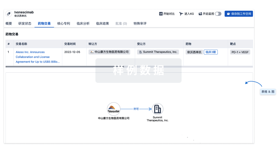
药物交易
使用我们的药物交易数据加速您的研究。
登录
或
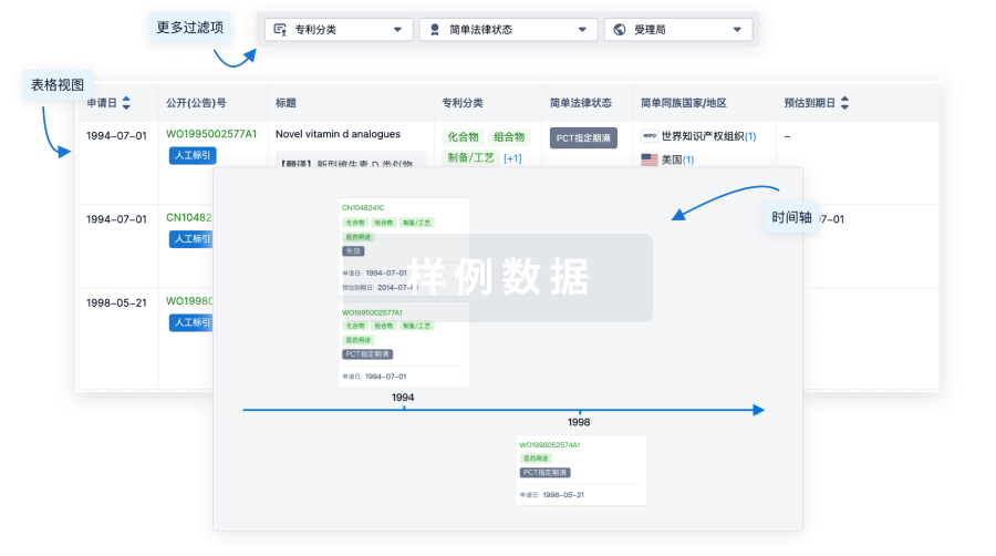
核心专利
使用我们的核心专利数据促进您的研究。
登录
或
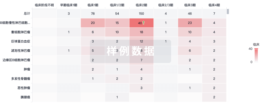
临床分析
紧跟全球注册中心的最新临床试验。
登录
或
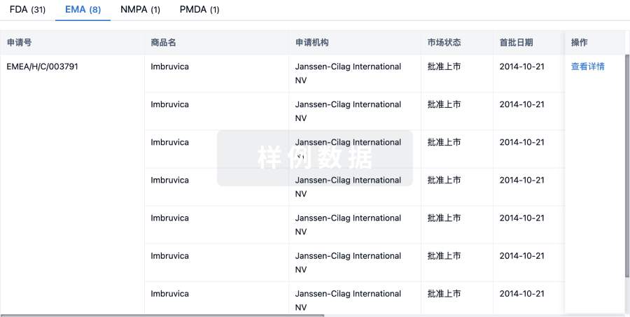
批准
利用最新的监管批准信息加速您的研究。
登录
或
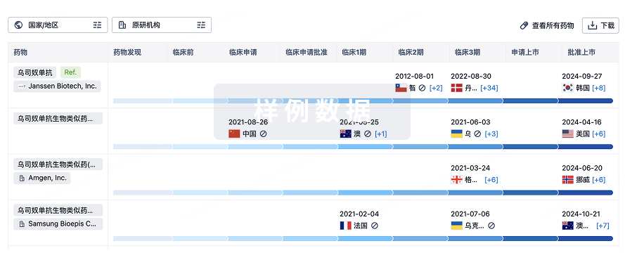
生物类似药
生物类似药在不同国家/地区的竞争态势。请注意临床1/2期并入临床2期,临床2/3期并入临床3期
登录
或
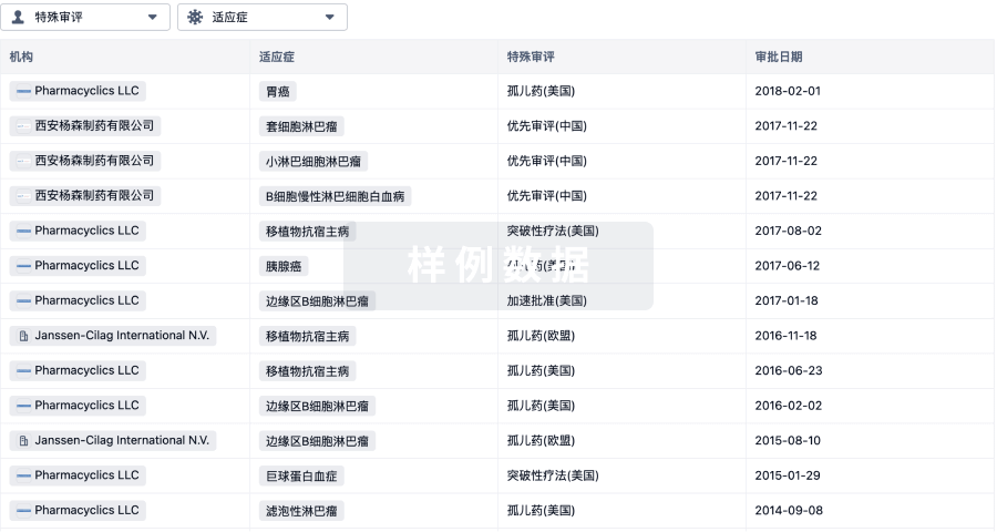
特殊审评
只需点击几下即可了解关键药物信息。
登录
或
提出任何生物医药研究问题
让每一步生物医药决策都精准制胜
基于十亿级生物数据与全球情报的精准 AI,赋能早期药物发现
立即开始免费试用!
智慧芽新药情报库是智慧芽专为生命科学人士构建的基于AI的创新药情报平台,助您全方位提升您的研发与决策效率。
立即接入生物医药数据包
智慧芽新药库数据也通过智慧芽开放平台,以MCP或API服务形式对外开放。立即使用我们的MCP服务定制您的LLM智能体。
生物序列数据库
生物药研发创新
免费使用
化学结构数据库
小分子化药研发创新
免费使用