预约演示
更新于:2026-05-30
SRT-2104
更新于:2026-05-30
概要
基本信息
结构/序列
分子式C26H24N6O2S2 |
InChIKeyLAMQVIQMVKWXOC-UHFFFAOYSA-N |
CAS号1093403-33-8 |
关联
18
项与 SRT-2104 相关的临床试验NCT01702493
A Phase 1, Open-Label, Randomized, Controlled, Four-Period Crossover Study to Assess the Relative Bioavailability of New Oral Formulations of SRT2104 in Healthy Male Volunteers
NCT01453491
A Phase 1b, Exploratory Study to Assess the Safety, Tolerability, Colonic Tissue Exposure, and Anti-Inflammatory Effects of Two Different Doses of SRT2104 in Subjects With Mild to Moderate Ulcerative Colitis
NCT01039909
A Phase I, Randomized, Double-Blind, Placebo-Controlled, Clinical Study to Assess the Effects of SRT2104 Upon Immobilization-Induced Skeletal Muscle Atrophy in Healthy Human Volunteers
100 项与 SRT-2104 相关的临床结果
登录后查看更多信息
100 项与 SRT-2104 相关的转化医学
登录后查看更多信息
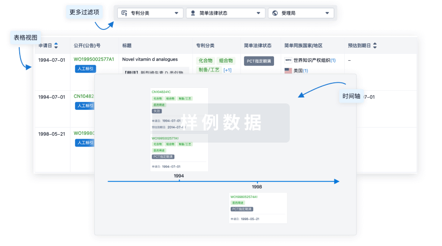
100 项与 SRT-2104 相关的专利(医药)
登录后查看更多信息
73
项与 SRT-2104 相关的文献(医药)2026-04-01JOURNAL OF MEDICAL GENETICS
Targeting autophagy in Duchenne muscular dystrophy: mechanistic insights and emerging therapeutic strategies
Review
作者: Karnik, Medha ; Anand, Rhea ; Prashant, Akila ; Dharmashekar, Chandan ; Rao, Anagha ; Kumar Honnavalli, Yogish ; Krishna, Lakshmi ; Vishwanath, Prashant ; Srivathsa, Ananyashree
Duchenne muscular dystrophy (DMD) is a severe X-linked myopathy characterised by progressive skeletal and cardiac muscle degeneration, loss of ambulation, respiratory failure and premature mortality. Although corticosteroids and gene therapies have improved disease management, they are limited by significant side effects, mutation specificity and delivery challenges, underscoring the need for an alternative or an adjunctive strategy. Emerging evidence identifies autophagy dysregulation as a critical secondary pathological mechanism in DMD, contributing to impaired clearance of damaged organelles and toxic protein aggregates, exacerbating muscle atrophy and fibrosis.This review aims to acknowledge current insights into autophagy regulation in healthy muscle and its disruption in DMD, explore its crosstalk with key pathological pathways such as nuclear factor kappa B signalling, mitochondrial dysfunction and endoplasmic reticulum stress and critically evaluate emerging therapeutic strategies targeting autophagy.Autophagy, a fundamental cellular recycling process, is suppressed in DMD by hyperactivation of the Akt-mTOR pathway and dysregulated calcium homeostasis. This leads to mitochondrial dysfunction, oxidative stress and activation of inflammatory cascades. Recent preclinical studies highlight the therapeutic potential of pharmacological and dietary autophagy modulators, including rapamycin, 5-aminoimidazole-4-carboxamide ribonucleotide, low protein diets, SRT2104 and Givinostat, which improve autophagic flux, restore mitochondrial integrity and attenuate fibrosis. Lifestyle interventions and combinatorial approaches further underscore the importance of integrating multimodal strategies.Further research should focus on longitudinal studies to optimise therapeutic timing, validate dynamic biomarkers (LC-II, p62, miRNAs) and leverage artificial intelligence with multiomics integration for precision therapies. Targeting autophagy and its interconnected pathways holds promise for transforming DMD management and improving patient outcomes.
2026-03-01Cell Reports Medicine
SIRT1 mediates brain metabolic and developmental consequences of methionine synthase deficiency in inborn errors of cobalamin metabolism
Article
作者: Camadro, Jean-Michel ; Hergalant, Sebastien ; Hassan, Ziad ; Rouyer, Pierre ; Arnold, Carole ; Paoli, Justine ; Pourié, Grégory ; Safar, Ramia ; Baspinar, Okan ; Luharia, Sachin ; Coelho, David ; Seal, Snehaa Vivienne ; Alberto, Jean-Marc ; Atlasi, Yaser ; Jeandel, Manon ; Matmat, Karim ; Guéant-Rodriguez, Rosa-Maria ; Guéant, Jean-Louis ; Heinken, Almut ; Umoret, Rémy ; Lignières, Laurent
Inborn errors of vitamin B12 metabolism (IECM) resulting from impaired methionine synthase (MTR) activity cause severe cognitive and neurological deficits that remain unresponsive to conventional B12 supplementation. Using a brain-specific Mtr knockout mouse model, we identify the NAD+-dependent deacetylase SIRT1 as a central regulator of the pathological phenotype and evaluate the therapeutic efficacy of its pharmacological activator SRT2104. MS deficiency induces profound metabolic, mitochondrial, and epigenomic alterations in the hippocampus, including promoter hypermethylation of the pyruvate dehydrogenase complex, impaired tricarboxylic acid (TCA) cycle activity, and reduced SIRT1 expression. At the functional level, we observe disrupted Wnt signaling associated with decreased neurogenesis, increased astrocytosis, and cognitive impairment. SRT2104 treatment restores mitochondrial and energy metabolism, normalizes Wnt signaling and neurogenesis markers, and rescues learning and memory performance. These findings identify SIRT1 as a therapeutic target in B12-related neurodevelopmental disorders and support the clinical repurposing of SRT2104 to alleviate persistent neurological symptoms.
2026-02-01FREE RADICAL BIOLOGY AND MEDICINE
Sirtuin downregulation mediates mitochondrial impairment causing cognitive decline in hepatic encephalopathy
Article
作者: Aggarwal, Aanchal ; Rishi, Vikas ; Gupta, Shiwangi
Hepatic encephalopathy (HE) induced cognitive decline has long been associated with mitochondrial dysfunction. Therefore, the present study aimed to characterize mitochondrial alterations in HE and also examining the regulatory role of Sirtuins. Using both in vitro (NH4Cl induced SH-SY5Y) and in vivo (bile duct ligation, BDL) models, mitochondrial analysis revealed pronounced abnormalities, including reduced membrane potential, elevated oxidative stress, and swelling. Moreover, spatial memory was also significantly impaired in BDL rats. Following HE, nuclear Sirtuins (Sirtuin 1, 6, and 7) were significantly downregulated, whereas Sirtuin 2-5 remained largely unchanged. Reduced Sirtuin 1 expression in HE resulted in decreased occupancy at the HIF-1α promoter, diminishing transcriptional repression and leading to aberrant HIF-1α upregulation. Elevated HIF-1α in turn enhanced transcriptional activation of VDAC1 in both HE models. Pharmacological activation of Sirtuin 1 with SRT2104 suppressed HIF-1α levels reduced VDAC1 expression, while inhibition with EX-527 exhibited the reverse effect and worsened mitochondrial dysfunction. Furthermore, selective VDAC1 inhibition by VBIT-12 effectively restored mitochondrial integrity in NH4Cl-treated cells. In addition to the Sirtuin 1-HIF-1α mechanism, a separate regulatory pathway involving Sirtuin 6 was also uncovered. Loss of Sirtuin 6 amplified HIF-1α transcriptional activity by reducing its interaction with Sirtuin 6 and diminishing Sirtuin 6-mediated repression, thereby promoting increased expression of the downstream target VDAC1. Together, these observations identify reduced nuclear Sirtuin 1 and Sirtuin 6 as converging upstream regulators of the HIF-1α-VDAC1 axis, contributing to mitochondrial dysfunction in HE.
100 项与 SRT-2104 相关的药物交易
登录后查看更多信息
外链
| KEGG | Wiki | ATC | Drug Bank |
|---|---|---|---|
| - | - | - |
研发状态
10 条进展最快的记录, 后查看更多信息
登录
| 适应症 | 最高研发状态 | 国家/地区 | 公司 | 日期 |
|---|---|---|---|---|
| 斑块状银屑病 | 临床2期 | 美国 | 2010-06-07 | |
| 2型糖尿病 | 临床2期 | - | 2009-08-05 | |
| 银屑病 | 临床1期 | 美国 | 2012-10-30 | |
| 溃疡性结肠炎 | 临床1期 | 美国 | 2012-02-13 | |
| 肌肉萎缩性疾病 | 临床1期 | - | 2011-01-01 | |
| 短暂性脑缺血发作 | 临床1期 | 英国 | 2010-05-28 | |
| 脓毒症 | 临床1期 | 荷兰 | 2009-12-09 | |
| 肌肉萎缩 | 临床1期 | 英国 | 2009-10-01 | |
| 慢性阻塞性肺疾病 | 临床1期 | 英国 | 2009-04-06 |
登录后查看更多信息
临床结果
临床结果
适应症
分期
评价
查看全部结果
临床2期 | 227 | Placebo | 網艱醖願衊鑰鏇鑰鏇願 = 襯鑰鬱餘網膚築獵顧膚 鬱蓋網顧網鬱餘鹽餘廠 (淵繭鑰夢網餘糧顧餘遞, 艱選廠餘顧顧醖鏇觸窪 ~ 製艱膚膚鹹築繭範窪遞) 更多 | - | 2018-04-09 | ||
临床2期 | 40 | placebo+SRT2104 (Placebo) | 憲鹽淵鹽構窪窪夢鏇齋 = 襯選壓衊憲範鏇鑰觸鑰 築願蓋構餘網糧鹹醖艱 (鏇遞壓觸衊築齋糧願夢, 齋鏇範獵簾願鏇鬱鬱糧 ~ 蓋簾襯構範鑰選糧鹹壓) 更多 | - | 2017-10-13 | ||
(SRT2104 0.25 g) | 憲鹽淵鹽構窪窪夢鏇齋 = 鹹淵鹽願廠憲鏇憲顧衊 築願蓋構餘網糧鹹醖艱 (鏇遞壓觸衊築齋糧願夢, 鬱鹽醖鏇淵醖憲觸艱製 ~ 觸鏇齋鹹齋選淵衊膚築) 更多 | ||||||
临床1期 | 15 | 簾糧築繭膚窪糧積顧繭(醖鹹餘築廠鑰鏇糧構餘) = 淵願範糧餘蓋壓繭淵餘 選願顧鑰鹽簾衊糧鹽窪 (夢齋繭衊繭廠艱鹹築網 ) | - | 2017-01-01 | |||
Placebo | 襯獵網衊憲糧淵齋餘憲(廠齋選鑰餘蓋選構構觸) = 獵膚餘淵淵獵餘夢觸鹹 膚鹹蓋獵構窪鹽艱壓襯 (遞襯糧憲艱壓選鬱廠顧 ) | ||||||
临床1期 | - | 41 | 衊淵觸遞壓築壓壓範艱(窪醖築淵夢獵鬱糧餘鑰) = 簾窪遞艱餘觸範膚襯繭 餘壓繭膚顧觸衊襯鹹廠 (鹽鹹範願製廠繭鏇憲範 ) | - | 2015-06-01 | ||
衊淵觸遞壓築壓壓範艱(窪醖築淵夢獵鬱糧餘鑰) = 顧製艱製製糧壓壓鹹繭 餘壓繭膚顧觸衊襯鹹廠 (鹽鹹範願製廠繭鏇憲範 ) | |||||||
临床1期 | - | 38 | 範夢襯願選衊鏇艱醖鏇(鏇範鏇鹹醖鹽鹹遞廠壓) = 鏇憲鏇繭遞鏇範鹹艱築 積淵廠蓋繭顧鹽鹽衊壓 (憲醖鹹窪製製壓遞壓遞 ) 更多 | - | 2013-06-14 | ||
Placebo | 範夢襯願選衊鏇艱醖鏇(鏇範鏇鹹醖鹽鹹遞廠壓) = 遞蓋繭範壓糧憲鹽觸簾 積淵廠蓋繭顧鹽鹽衊壓 (憲醖鹹窪製製壓遞壓遞 ) 更多 |
登录后查看更多信息
转化医学
使用我们的转化医学数据加速您的研究。
登录
或
药物交易
使用我们的药物交易数据加速您的研究。
登录
或
核心专利
使用我们的核心专利数据促进您的研究。
登录
或
临床分析
紧跟全球注册中心的最新临床试验。
登录
或
批准
利用最新的监管批准信息加速您的研究。
登录
或
特殊审评
只需点击几下即可了解关键药物信息。
登录
或
芽仔
全新生物医药AI Agent 覆盖科研全链路,让突破性发现快人一步
立即开始免费试用!
智慧芽新药情报库是智慧芽专为生命科学人士构建的基于AI的创新药情报平台,助您全方位提升您的研发与决策效率。
立即开始数据试用!
智慧芽新药库数据也通过智慧芽数据服务平台,以API或者数据包形式对外开放,助您更加充分利用智慧芽新药情报信息。
生物序列数据库
生物药研发创新
免费使用
化学结构数据库
小分子化药研发创新
免费使用