预约演示
更新于:2026-07-09
Central South University of Forestry & Technology
更新于:2026-07-09
概览
标签
其他疾病
呼吸系统疾病
小分子化药
疾病领域得分
一眼洞穿机构专注的疾病领域

技术平台
公司药物应用最多的技术
靶点
公司最常开发的靶点
关联
靶点 |
作用机制 |
在研机构 |
原研机构 |
在研适应症 |
非在研适应症 |
最高研发阶段 |
首次获批国家/地区 |
首次获批日期 |

100 项与 中南林业科技大学 相关的临床结果
登录后查看更多信息
登录后查看更多信息
2026-12-31Virulence
The interaction between CfAda3 and CfGcn5 impacts growth, development, and virulence of
Colletotrichum fructicola
Article
作者: Li, He ; Zhang, Shengpei ; Chen, Yan ; Luo, Jing ; Wang, Yiling
Camellia oleifera is an important economic woody oil plant in many Asian countries, and the anthracnose caused by Colletotrichum fructicola is prevalent in its cultivation regions, causing significant losses annually. We previously found that CfGcn5-mediated H3 acetylation governs virulence of C. fructicola. To further elucidate the regulatory mechanism of CfGcn5, we carried out mass spectrometry analysis for CfGcn5-interacting proteins and identified CfAda3 protein for functional analysis. We found that CfAda3 was mainly localized in nucleus and cooperated with CfGcn5 to acetylate H3K18 for global gene transcription. Targeted gene deletion revealed that CfAda3 is involved in growth and conidiation. Similar to ΔCfgcn5 mutant, the ΔCfada3 mutant is defective in conidial germination, appressorial formation, autophagy, and in the response to environmental stresses. These combined effects result in its non-virulence on C. oleifera. In addition, we provided evidence showing that both NLS region and ADA3 domain are required for the localization and function of CfAda3. Moreover, we indicated that the interaction with CfGcn5 is essential but not sufficient for the normal localization and full function of CfAda3. Taken together, our studies not only illustrate the prominent roles of CfAda3 in growth, development, and virulence but also highlight how CfAda3 functions together with CfGcn5 in C. fructicola.
2026-12-31MECHANICS OF ADVANCED MATERIALS AND STRUCTURES
Numerical simulation study on mechanical properties of multi-stranded steel wire ropes with different contact modes
作者: Luo, Wusheng ; Liu, Yihua ; Li, Qiao ; Chen, Yimin ; Yu, Shengfei ; Jiang, Feng
In order to elucidate the mech. properties of wire ropes in different contact modes, this paper, based on ANSYS finite element software, investigated the stress and deformation distributions, adjacent wire contact stresses and rope strand slip characteristics of wire ropes with three contact types, namely point (PCWR), line (LCWR) and face (FCWR), resp.The results show that under the same tensile and torsional loads, the deformation results of FCWR are the smallest, which are 0.1680% and 0.3822%, resp., while the overall stress performance of PCWR is the largest, and its maximum principal stresses mainly exist in the contact position between the strands, which indicates that rupture is more likely to occur here due to stress overloading.Meanwhile, the simulation results were compared with the theor. calculations of Costello, and it was found that the fitting effect was better, and the reliability of the model was further investigated.In addition, the interlayer contact stresses of the wire ropes all showed a linear growth trend with the increase of axial load, in which the overall contact stresses and slips of the PCWR were the largest, especially the interstrand slips accounted for 92.69% of the overall.
2026-12-01Nano-Micro Letters
Bioinspired Precision Peeling of Ultrathin Bamboo Green Cellulose Frameworks for Light Management in Optoelectronics
Article
作者: Zhang, Yuan ; Wu, Yiqiang ; Zhao, Dawei ; Wang, Yan ; Zuo, Yingfeng
Abstract:
Cellulose frameworks have emerged as promising materials for light management due to their exceptional light-scattering capabilities and sustainable nature. Conventional biomass-derived cellulose frameworks face a fundamental trade-off between haze and transparency, coupled with impractical thicknesses (≥ 1 mm). Inspired by squid’s skin-peeling mechanism, this work develops a peroxyformic acid (HCOOOH)-enabled precision peeling strategy to isolate intact 10-µm-thick bamboo green (BG) frameworks—100 × thinner than wood-based counterparts while achieving an unprecedented optical performance (88% haze with 80% transparency). This performance surpasses delignified biomass (transparency < 40% at 1 mm) and matches engineered cellulose composites, yet requires no energy-intensive nanofibrillation. The preserved native cellulose I crystalline structure (64.76% crystallinity) and wax-coated uniaxial fibril alignment (Hermans factor: 0.23) contribute to high mechanical strength (903 MPa modulus) and broadband light scattering. As a light-management layer in polycrystalline silicon solar cells, the BG framework boosts photoelectric conversion efficiency by 0.41% absolute (18.74% → 19.15%), outperforming synthetic anti-reflective coatings. The work establishes a scalable, waste-to-wealth route for optical-grade cellulose materials in next-generation optoelectronics.
2026-07-08
2026-06-24
·荒糖学术
100 项与 中南林业科技大学 相关的药物交易
登录后查看更多信息
100 项与 中南林业科技大学 相关的转化医学
登录后查看更多信息
组织架构
使用我们的机构树数据加速您的研究。
登录
或
管线布局
2026年07月21日管线快照
管线布局中药物为当前组织机构及其子机构作为药物机构进行统计,早期临床1期并入临床1期,临床1/2期并入临床2期,临床2/3期并入临床3期
临床前
1
登录后查看更多信息
当前项目
登录后查看更多信息
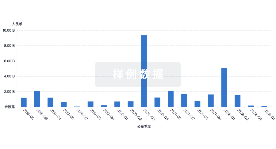
药物交易
使用我们的药物交易数据加速您的研究。
登录
或
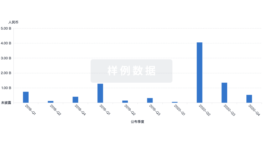
转化医学
使用我们的转化医学数据加速您的研究。
登录
或
营收
使用 Synapse 探索超过 36 万个组织的财务状况。
登录
或
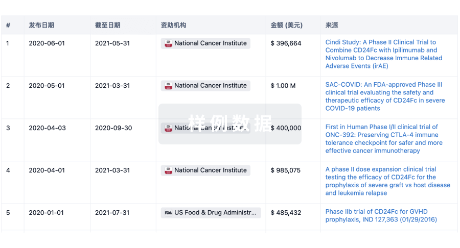
科研基金(NIH)
访问超过 200 万项资助和基金信息,以提升您的研究之旅。
登录
或
投资
深入了解从初创企业到成熟企业的最新公司投资动态。
登录
或
融资
发掘融资趋势以验证和推进您的投资机会。
登录
或
芽仔
全新生物医药AI Agent 覆盖科研全链路,让突破性发现快人一步
立即开始免费试用!
智慧芽新药情报库是智慧芽专为生命科学人士构建的基于AI的创新药情报平台,助您全方位提升您的研发与决策效率。
立即开始数据试用!
智慧芽新药库数据也通过智慧芽数据服务平台,以API或者数据包形式对外开放,助您更加充分利用智慧芽新药情报信息。
生物序列数据库
生物药研发创新
免费使用
化学结构数据库
小分子化药研发创新
免费使用