预约演示
更新于:2026-03-05
AntiCancer, Inc.
更新于:2026-03-05
概览
标签
肿瘤
皮肤和肌肉骨骼疾病
其他疾病
生物药
毒素
腺相关病毒基因治疗
疾病领域得分
一眼洞穿机构专注的疾病领域

技术平台
公司药物应用最多的技术
靶点
公司最常开发的靶点
关联
靶点 |
作用机制 |
在研机构 |
原研机构 |
在研适应症 |
非在研适应症 |
最高研发阶段 |
首次获批国家/地区 |
首次获批日期 |

靶点 |
作用机制 |
在研机构 |
原研机构 |
在研适应症 |
非在研适应症 |
最高研发阶段 |
首次获批国家/地区 |
首次获批日期 |
靶点 |
作用机制 |
在研机构 |
原研机构 |
在研适应症 |
非在研适应症 |
最高研发阶段 |
首次获批国家/地区 |
首次获批日期 |
100 项与 AntiCancer, Inc. 相关的临床结果
登录后查看更多信息
登录后查看更多信息
2026-02-01ANTICANCER RESEARCH
Glutamine Dependence Is Not a Cancer-specific Vulnerability in Contrast to Methionine Dependence
Article
作者: Li, Shukuan ; Han, Qinghong ; Miyashi, Yuta ; Hoffman, Robert M ; Nishida, Kotaro ; Kang, Byung Mo ; Bouvet, Michael ; Kim, Jin Soo ; Mizuta, Kohei ; Tome, Yasunori ; Asano, Yohei
BACKGROUND/AIM:
Glutamine (GLN) addiction has been proposed as a cancer vulnerability and a therapeutic target. However, the glutamine requirement of normal cells is poorly understood. In the present study, we used a unique co-culture model to study the glutamine requirement of cancer cells compared to normal cells co-cultured together.
MATERIALS AND METHODS:
The human fibrosarcoma cell line HT1080 and normal human fibroblasts HS27 were co-cultured in 12-well dishes seeded with equal numbers of cells of each type. Additionally, HS27 cells were cultured alone in 6-well plates. The cells were grown in Dulbecco's Modified Eagle's Medium (DMEM) which did not contain GLN, methionine (MET), or cystine (CYS). 150 μM L-cystine 2HCl was added to all media. Co- and mono- cultures were grown under the following conditions: Complete medium (GLN 4 mM and MET 100 μM); MET restriction [Methionine restriction (MR), GLN 4 mM and MET 0 μM]; GLN restriction [Glutamine restriction (GR), GLN 0 mM and MET 100 μM] and MR+GR (GLN 0 mM and MET 0 μM). Cells were observed under phase-contrast and fluorescence microscopy for seven days. ImageJ was used to compare the three groups: MR, GR and MR+GR.
RESULTS:
In complete DMEM, HT1080 fibrosarcoma cells dominated HS27 normal fibroblasts in co-culture. Under MR, HT1080 cells became mostly non viable, but HS27 cells remained viable. Under GR and MR+GR, both HT1080 and HS27 cells became mostly non-viable. Monoculture experiments showed that normal cells survived under MR but not GR.
CONCLUSION:
GR is not a cancer-specific vulnerability, while MR is. Therefore, GR is not a promising cancer-therapy strategy.
2026-02-01ANTICANCER RESEARCH
Lack of Cancer Specificity of Methionine Adenosyltransferase 2A (MAT2A) Inhibitor AG-270 in Combination With Recombinant Methioninase In Vitro.
Article
作者: Han, Qinghong ; Kang, Byung Mo ; Hoffman, Robert M ; Bouvet, Michael ; Miyashi, Yuta ; Kim, Jinsoo ; Asano, Yohei ; Mizuta, Kohei ; Li, Shukuan
BACKGROUND/AIM:
Methionine addiction is a fundamental and general hallmark of cancer termed the Hoffman effect. Methionine restriction using recombinant methioninase (rMETase) has shown synergistic efficacy with numerous types of chemotherapeutic agents against cancer cells and not normal cells. Methionine adenosyltransferase 2A (MAT2A) is a crucial enzyme converting methionine to S-adenosylmethionine (SAM). A MAT2A inhibitor, AG-270, has been proposed as a potential anti-cancer drug. The present study evaluated whether AG-270 is a cancer-specific agent by comparing its efficacy in combination with rMETase on cancer and normal cells.
MATERIALS AND METHODS:
The half-maximal inhibitory concentrations (IC50) of rMETase and AG-270 were determined on HCT116 human colon-cancer cells and Hs-27 human normal fibroblasts in vitro. The efficacy of rMETase combined with AG-270, at their respective IC50 values, on HCT116 and Hs-27 was also determined. Cell viability was evaluated using the WST-8 reagent.
RESULTS:
The IC50 values of rMETase were 0.35 U/ml for HCT116 and 1.14 U/ml for Hs-27. The IC50 values of AG-270 were 4.38 μM for HCT116 and 6.55 μM for Hs-27. The combination of rMETase and AG-270, at their respective IC50, had synergistic efficacy on both cancer and normal cells, reducing viability to approximately 20% in both cell lines (p<0.05).
CONCLUSION:
AG-270 showed lack of cancer specificity in combination with rMETase when tested on both cancer and normal cells. The present results contrast with numerous chemotherapy agents, which in combination with rMETase are synergistic on cancer cells but not on normal cells. The present findings suggest that MAT2A inhibition affects crucial metabolic pathways in normal as well as cancer cell types and thus AG-270 may not be suitable as a cancer-specific therapeutic strategy.
2026-01-01JOURNAL OF GASTROINTESTINAL SURGERY
Enhanced visualization of colonic polyps using a fluorophore-conjugated claudin-1 antibody in a CPC-APC mouse model
Article
作者: Primeaux, Mark ; Hoffman, Robert M ; Liu, Shanglei ; Jaiswal, Sunidhi ; Batra, Surinder K ; Bouvet, Michael ; Talib, Sumbal ; Amirfakhri, Siamak ; Pisegna, Joseph R ; Mohs, Aaron M ; Jih, Lily J ; Singh, Satish K ; Dhawan, Punita ; Cox, Kristin E
BACKGROUND:
Early detection and removal of polyps for primary colon cancer prevention during colonoscopy are still fraught with significant miss rates. In the current study, we determined the ability of claudin-1 (CLDN1) antibodies conjugated to near-infrared fluorophores to target and visualize colonic polyps with high resolution in the CPC-APC mouse colonic polyp model.
METHODS:
CPC-APC mice that developed distal colon polyps at 12 weeks of age were used in the present study. The formation of polyps was confirmed by mouse colonoscopy using the ColoView system (Karl Stortz). For polyp labeling, anti-CLDN1 and control immunoglobulin G (IgG) antibodies were conjugated to IRDye800 and 50, 100, and 150 μg doses were administered intravenouly to CPC-APC mice. Mice were sacrificed at different time points, and colons were harvested for imaging using the LI-COR Pearl Small Animal Imaging System. Polyp-to-background fluorescence intensity ratios (PBRs) were calculated for each polyp. Histology and immunohistochemistry (IHC) were performed on the polyps.
RESULTS:
A total of 9 mice were used in the experiment, with a total of 45 polyps analyzed. The presence of observed polyps on gross examination was confirmed via histology. CLDN1-IRDye800 had a PBR of 7.6 ± 3.9, which was significantly higher than the nonspecific IgG-IRDye800 control (P =.0428). CLDN1-IRDye800 was further analyzed and overall had a similar range of PBR with 50, 100, and 150 μg injections at 72 h and had a statistically significant higher PBR of 8.9 ± 4.2 than the IgG-IRDye800 control (P =.0184) at 150 μg. Adenomatous polyps as small as 1 mm could be identified using CLDN1-IRDye800. Hematoxylin and eosin staining confirmed adenomatous polyps with high-grade dysplasia, and IHC confirmed the expression of CLDN1.
CONCLUSION:
CLDN1-IRDye800 enabled polyp visualization with ultrahigh-resolution fluorescence imaging technology in CPC-APC mice. Polyps as small as 1 mm could be visualized, indicating clinical potential to overcome the current limitations of colonic polyp detection.
100 项与 AntiCancer, Inc. 相关的药物交易
登录后查看更多信息
100 项与 AntiCancer, Inc. 相关的转化医学
登录后查看更多信息
组织架构
使用我们的机构树数据加速您的研究。
登录
或
管线布局
2026年07月21日管线快照
管线布局中药物为当前组织机构及其子机构作为药物机构进行统计,早期临床1期并入临床1期,临床1/2期并入临床2期,临床2/3期并入临床3期
药物发现
1
2
临床前
其他
7
登录后查看更多信息
当前项目
登录后查看更多信息
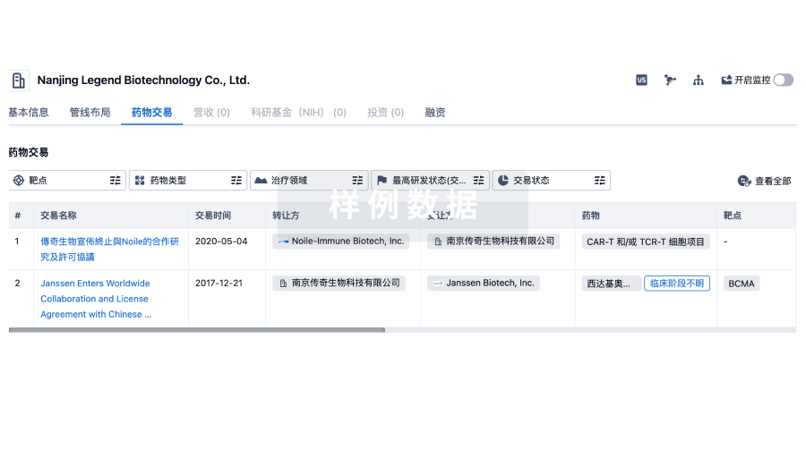
药物交易
使用我们的药物交易数据加速您的研究。
登录
或
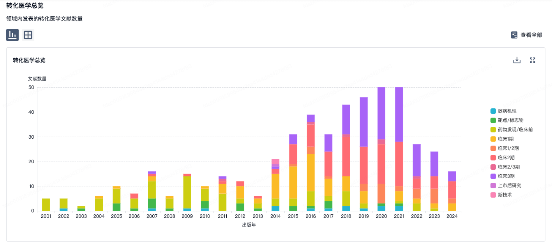
转化医学
使用我们的转化医学数据加速您的研究。
登录
或
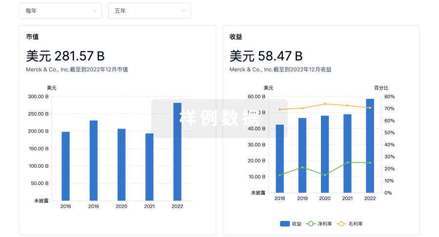
营收
使用 Synapse 探索超过 36 万个组织的财务状况。
登录
或
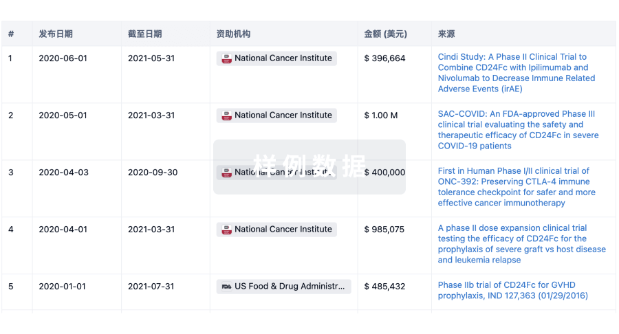
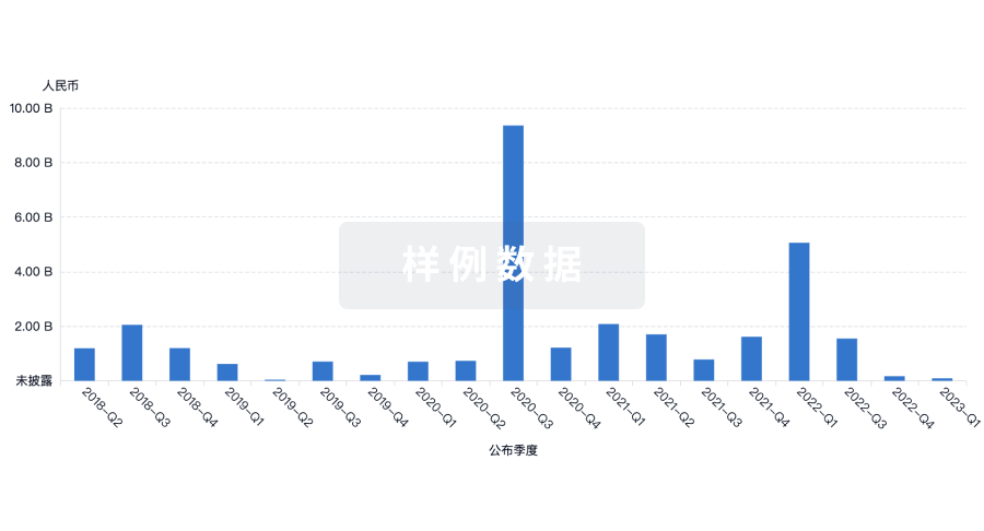
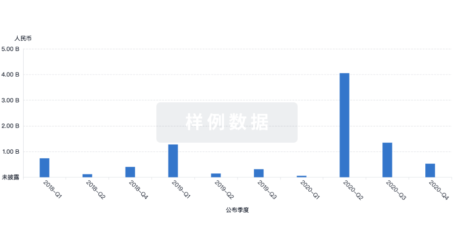
科研基金(NIH)
访问超过 200 万项资助和基金信息,以提升您的研究之旅。
登录
或
投资
深入了解从初创企业到成熟企业的最新公司投资动态。
登录
或
融资
发掘融资趋势以验证和推进您的投资机会。
登录
或
芽仔
全新生物医药AI Agent 覆盖科研全链路,让突破性发现快人一步
立即开始免费试用!
智慧芽新药情报库是智慧芽专为生命科学人士构建的基于AI的创新药情报平台,助您全方位提升您的研发与决策效率。
立即开始数据试用!
智慧芽新药库数据也通过智慧芽数据服务平台,以API或者数据包形式对外开放,助您更加充分利用智慧芽新药情报信息。
生物序列数据库
生物药研发创新
免费使用
化学结构数据库
小分子化药研发创新
免费使用