预约演示
更新于:2026-02-02

Laboratoires Expanscience SA
更新于:2026-02-02
概览
标签
皮肤和肌肉骨骼疾病
免疫系统疾病
肿瘤
小分子化药
疾病领域得分
一眼洞穿机构专注的疾病领域
技术平台
公司药物应用最多的技术
靶点
公司最常开发的靶点

暂无数据
关联
靶点 |
作用机制 |
在研机构 |
原研机构 |
在研适应症 |
非在研适应症 |
最高研发阶段 |
首次获批国家/地区 |
首次获批日期 |
EUCTR2007-004973-25-PL
Prospective multicentric open clinical study comparing the efficacy and safety of piascledine 300 plus standard treatment versus standard treatment only in patients with osteoarthritis of the knee
EUCTR2005-004457-10-BE
Clinical and biological activity of Piascledine® 300 in patients with chronic periodontitis
EUCTR2004-000979-33-IT
A multinational multicentre randomised paralle group study on therapeutic efficacy and tolerability of PIASCLEDINE capsules 300 mg in comparison with placebo in the treatment of hip osteoarthritis
100 项与 Laboratoires Expanscience SA 相关的临床结果
登录后查看更多信息
登录后查看更多信息
2019-02-01The Journal of investigative dermatology
Age-Dependent Transformation of Skin Biomechanical Properties and Micromorphology during Infancy and Childhood
Letter
作者: Darlenski, Razvigor ; De Belilovsky, Clarence ; Fluhr, Joachim W ; Boyer, Gaëtan ; McGuckin, Colin P ; Lachmann, Nadège ; Pellacani, Giovanni ; Ferrari, Chiara ; Chadoutaud, Bernard ; Forraz, Nico ; Msika, Philippe ; Bellemère, Gaëlle ; Baudouin, Caroline
It is well known that skin continues to undergo structural and functional changes after birth. These include skin surface acidification (Fluhr et al., 2012Fluhr J.W. Darlenski R. Lachmann N. Baudouin C. Msika P. De Belilovsky C. et al.Infant epidermal skin physiology: adaptation after birth.Br J Dermatol. 2012; 166: 483-490Crossref PubMed Scopus (116) Google Scholar); increased hydration over the first few weeks of life, followed by decreased hydration (Nikolovski et al., 2008Nikolovski J. Stamatas G.N. Kollias N. Wiegand B.C. Barrier function and water-holding and transport properties of infant stratum corneum are different from adult and continue to develop through the first year of life.J Invest Dermatol. 2008; 128: 1728-1736Abstract Full Text Full Text PDF PubMed Scopus (263) Google Scholar); and changes in cell organization and structure (Bensaci et al., 2015Bensaci J. Chen Z.Y. Mack M.C. Guillaud M. Stamatas G.N. Geometrical and topological analysis of in vivo confocal microscopy images reveals dynamic maturation of epidermal structures during the first years of life.J Biomed Optics. 2015; 20: 095004Crossref PubMed Scopus (7) Google Scholar, Fluhr et al., 2014Fluhr J.W. Lachmann N. Baudouin C. Msika P. Darlenski R. De Belilovsky C. et al.Development and organization of human stratum corneum after birth: electron microscopy isotropy score and immunocytochemical corneocyte labelling as epidermal maturation's markers in infancy.Br J Dermatol. 2014; 171: 978-986Crossref PubMed Scopus (21) Google Scholar). In terms of biomechanical properties, skin elasticity decreases over the first decade of life (Sugihara et al., 1991Sugihara T. Ohura T. Homma K. Igawa H.H. The extensibility in human skin: variation according to age and site.Br J Plast Surg. 1991; 44: 418-422Abstract Full Text PDF PubMed Scopus (41) Google Scholar) and collagen and elastin fiber density increases for several decades (Vitellaro-Zuccarello et al., 1994Vitellaro-Zuccarello L. Cappelletti S. Dal Pozzo Rossi V. Sari-Gorla M. Stereological analysis of collagen and elastic fibers in the normal human dermis: variability with age, sex, and body region.Anat Rec. 1994; 238: 153-162Crossref PubMed Scopus (72) Google Scholar). Studies of the biomechanical maturation of the skin have focused primarily on changes that occur in adults during aging, and most studies of skin maturation in children have focused on the maturation of the epidermis, which increases in thickness with age and has high cell turnover in the first months of life (Stamatas et al., 2010Stamatas G.N. Nikolovski J. Luedtke M.A. Kollias N. Wiegand B.C. Infant skin microstructure assessed in vivo differs from adult skin in organization and at the cellular level.Pediatr Dermatol. 2010; 27: 125-131Crossref PubMed Scopus (174) Google Scholar). One recent study showed that skin biomechanical properties evolve throughout infancy (Visscher et al., 2017Visscher M.O. Burkes S.A. Adams D.M. Hammill A.M. Wickett R.R. Infant skin maturation: preliminary outcomes for color and biomechanical properties.Skin Res Technol. 2017; 23: 545-551Crossref PubMed Scopus (13) Google Scholar), but such studies remain rare. Here, we investigated the correlation between the maturation of biomechanical properties of the skin and the evolution of skin topography and micromorphology from infancy to early adulthood (study approved by Provincial Ethical Committee of Modena, University Hospital of Modena). We recruited a cohort of 70 subjects in seven age groups: 1–15 days, 5 weeks, 5–7 months, 2 years, 4–5 years, 7–8 years, and 20–35 years (Supplementary Table S1 online); all patients or their parent or guardian gave their informed written consent. Skin properties were examined by cutometry and reflectance confocal microscopy in vivo, and by immunohistochemistry in a limited number of foreskin biopsy samples (see Supplementary Material online for detailed methods). Cutometry showed that skin elasticity, as measured by the ratio of immediate retraction to maximum distention (Ur/Uf), increased from infancy to 2 years of age and then plateaued (Figure 1a). The viscoelastic component, calculated as the ratio of immediate to delayed distension (Uv/Ue), decreased from infancy to adulthood (Figure 1b). Total recovery (Ua) was slightly higher at older ages (Figure 1c) and total deformation (Uf) did not vary between age groups (Figure 1d). The parameters related to skin elasticity and recovery (Ur/Uf, Ua/Uf, Ur/Ue, and Ua) were positively correlated with age and body surface area, whereas Uv/Ue was negatively correlated with both of these and Uf did not have any significant correlations (Figure 1e). None of the parameters were correlated with stratum corneum hydration. The viscoelastic properties of the skin are related to the presence of interstitial fluid in the dermal extracellular matrix, and thus changes in Uv/Ue may reflect the water content of the epidermis and the dermis (Dobrev, 2002Dobrev H.P. A study of human skin mechanical properties by means of Cutometer.Folia Med. 2002; 44: 5-10Google Scholar). Although we cannot completely rule out this possibility, none of the parameters examined were significantly (ie, P < 0.01) correlated with stratum corneum hydration as measured by cutometer (Figure 1e), suggesting that the changes in these skin biomechanical properties are related to structural maturation rather than hydration. This is consistent with reports that skin biomechanical properties are related to the structure of the extracellular matrix in adults, specifically elastin fibers, fibrillin microfibrils, rete ridges, and, to a lesser extent, collagen fibrils (Langton et al., 2017Langton A.K. Graham H.K. McConnell J.C. Sherratt M.J. Griffiths C.E.M. Watson R.E.B. Organization of the dermal matrix impacts the biomechanical properties of skin.BrJ Dermatol. 2017; 177: 818-827Crossref PubMed Scopus (37) Google Scholar). Skin microstructure was examined by reflectance confocal microscopy, which allows fast, in vivo imaging of the cyto-architectural aspects of the skin. The thickness of the stratum corneum and the supra papillary epidermis increased with age (Figure 2a), consistent with previous findings (Stamatas et al., 2010Stamatas G.N. Nikolovski J. Luedtke M.A. Kollias N. Wiegand B.C. Infant skin microstructure assessed in vivo differs from adult skin in organization and at the cellular level.Pediatr Dermatol. 2010; 27: 125-131Crossref PubMed Scopus (174) Google Scholar). The homogeneity and furrow architecture of the stratum corneum changed dramatically between infants and older children (Supplementary Figure S1a–S1d). Reflecting spheroids were observed throughout childhood and were most prevalent during infancy (Supplementary Figure S1e–S1f), and the nature of these structures warrants further investigation. The observed evolution from poorly defined to well-defined keratinocyte outline (Supplementary Table S2 online) might reflect the fact that keratinocyte proliferation is high during the first months of life (Stamatas et al., 2010Stamatas G.N. Nikolovski J. Luedtke M.A. Kollias N. Wiegand B.C. Infant skin microstructure assessed in vivo differs from adult skin in organization and at the cellular level.Pediatr Dermatol. 2010; 27: 125-131Crossref PubMed Scopus (174) Google Scholar). Dermal papillae increased in number with age, consistent with a previous study by Miyauchi et al., 2016Miyauchi Y. Shimaoka Y. Fujimura T. Koike Y. Yatabe M. Nishikawa M. et al.Developmental changes in neonatal and infant skin structures during the first 6 months: in vivo observation.Pediat Dermatol. 2016; 33: 289-295Crossref PubMed Scopus (15) Google Scholar, but contrary to this previous study, rete ridge thickness was stable across age groups (Figure 2a). This contradiction could be explained by a difference in age group stratification in the studies. Interestingly, collagen fibers were fibrillar and showed a parallel orientation in newborns, whereas they were thicker, coarse, and multidirectional in older infants through adults (Figure 2a–2c). Mechanical forces are known to increase the diameter of collagen fibrils (Sanders and Goldstein, 2001Sanders J.E. Goldstein B.S. Collagen fibril diameters increase and fibril densities decrease in skin subjected to repetitive compressive and shear stresses.J Biomech. 2001; 34: 1581-1587Crossref PubMed Scopus (63) Google Scholar). Our data support a contribution of mechanical forces to dermal maturation postnatally, including the reorganization of the collagen matrix and increased collagen fiber thickness (Sanders and Goldstein, 2001Sanders J.E. Goldstein B.S. Collagen fibril diameters increase and fibril densities decrease in skin subjected to repetitive compressive and shear stresses.J Biomech. 2001; 34: 1581-1587Crossref PubMed Scopus (63) Google Scholar). We also noticed a structural pattern that has not been reported previously. Circular "cuffing" of the follicle by collagen fibers was observed in newborns only, and was very rare or completely absent in all other age groups (Figure 2a, 2d, 2e). This interesting feature may reflect the unique properties of postnatal hair follicle growth (Zhou et al., 2016Zhou L. Yang K. Wickett R.R. Andl T. Zhang Y. Dermal sheath cells contribute to postnatal hair follicle growth and cycling.J Dermatol Sci. 2016; 82: 129-131Abstract Full Text Full Text PDF PubMed Scopus (8) Google Scholar). The elastin component of the skin cannot be observed by reflectance confocal microscopy. Therefore, we analyzed its structural maturation by immunohistochemistry on foreskin samples from patients of various ages. Both fibrillin and elastin fibers increased in length and intensity with age, especially at the dermal–epidermal junction (Supplementary Figure S2 online). The strength of the present study is the analysis of both structural and biomechanical properties of the skin in the same subjects over a wide range of age groups. In addition to evidence supporting the relationship between structural and biomechanical properties during skin maturation, we introduce the observation of collagen fiber cuffing around hair follicles in newborns. Collectively, these data demonstrate the biochemical and structural evolution of the dermis during postnatal development, as well as the translation of these changes into maturation of the biomechanical properties of the skin. GB, CDB, GB, NL, PM, and CB are current or former employees of Expanscience Laboratories; JF and RD provided consulting services for Expanscience; CPMG, NF, and BC provided services on a commercial basis for this study; GP and CM received a research grant; GP was investigator of the clinical study. This study was funded by Expanscience Laboratories. The authors thank BioScienceWriters for diligence in the full review and editing of the manuscript. Download .pdf (1.09 MB) Help with pdf files Supplementary Data
2014-02-01Annals of the rheumatic diseases1区 · 医学
Randomised, controlled trial of avocado–soybean unsaponifiable (Piascledine) effect on structure modification in hip osteoarthritis: the ERADIAS study
1区 · 医学
Article
作者: Bernard Mazières ; Christian Cadet ; Maxime Dougados ; Dominique Moyse ; Emmanuel Maheu ; Philippe Coste ; Isabelle Kerloch ; Hafid Halhol ; Tim D Spector ; Jean-Marie Grouin ; Michel Lequesne ; Marc Marty
OBJECTIVE:
To assess the ability of avocado-soybean unsaponifiable-Expanscience (ASU-E) to slow radiographic progression in symptomatic hip osteoarthritis (OA).
METHODS:
Prospective, randomised, double blind, parallel group, placebo controlled 3 year trial. Patients with symptomatic (painful ≥1 year, Lequesne Index between 3 and 10) hip OA (American College of Rheumatology criteria) and a minimum joint space width (JSW) of the target hip between 1 and 4 mm on a pelvic radiograph were randomly assigned to 300 mg/day ASU-E or placebo. Standing pelvis, target hip anteroposterior (AP) and oblique views were taken annually. The primary outcome was JSW change at year 3, measured at the narrowest point on pelvic or target hip AP view (manual measure using a 0.1 mm graduated magnifying glass). The full analysis dataset (FAS) included all patients having at least two successive radiographs. An analysis of covariance Mixed Model for Repeated Measurements with Missing at Random (for missing data) was performed to compare adjusted 3 year JSW changes (primary outcome) and the percentages of 'progressors' (JSW loss≥0.5 mm) between groups.
RESULTS:
399 patients were randomised (345 kept in the FAS), aged 62 (35-84) years, 54% women, mean body mass index 27 (SD 4) kg/m(2), mean symptom duration 4 (SD 5) years, 0-100 normalised Lequesne Index 30 (SD 9) and global pain visual analogue scale 37 (SD 23) mm. Mean baseline JSW was 2.8 (0.9) mm. There was no significant difference on mean JSW loss (-0.638 mm vs -0.672 mm, p=0.72, in the ASU-E and placebo groups, respectively) but there were 20% less progressors in the ASU-E than in the placebo group (40% vs 50%, respectively, p=0.040). No difference was observed on clinical outcomes. Safety was excellent.
CONCLUSIONS:
3 year treatment with ASU-E reduces the percentage of JSW progressors, indicating a potential structure modifying effect in hip OA to be confirmed, and the clinical relevance requires further assessment.
2012-10-01Archives of dermatological research3区 · 医学
Patented natural avocado sugar modulates the HBD-2 and HBD-3 expression in human keratinocytes through Toll-like receptor-2 and ERK/MAPK activation
3区 · 医学
Article
作者: Adone Baroni ; Philippe Msika ; Giovanna Donnarumma ; Maria Antonietta Tufano ; Caroline Baudouin ; Iole Paoletti ; Alessandra Fusco ; Elisabetta Buommino
Keratinocytes stimulated by microbial organisms secrete not only a variety of cytokines, chemokines and growth factors, but also antimicrobial peptides such as beta-defensins (HBDs) such as HBD-2 and HBD-3. AV119, a patented blend of avocado sugar, triggers the up-regulation of HBD-2 in skin epithelia upon contact with AV119, but the signalling mechanisms involved are not completely understood. The purpose of this study was to determine if AV119 was able to induce also the expression of HBD-3 in human keratinocytes. In addition, the receptor and intracellular pathways involved in the AV119 up-regulation of HBD-2 and HBD-3 were investigated. Our results demonstrated that AV119 induces a significantly increase of the expression of HBD-3. In addition, the HBD-2 and HBD-3 AV119-induced gene expression and release are TLR-2 dependent. Finally, we demonstrated that AV119 induced ERK/MAPK phosphorylation in human keratinocytes, thus providing evidence that HBD-2 and HBD-3 secretion is through the same transductional pathway. The ability of AV119 to induce also HBD-3 may amplify its therapeutic potential against a broader spectrum of bacterial and yeast strains responsible for human skin disorders.
2025-10-24
·百度百家
临床3期专利到期并购
100 项与 Laboratoires Expanscience SA 相关的药物交易
登录后查看更多信息
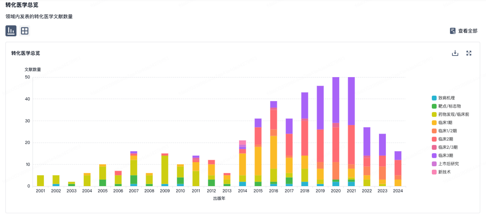
100 项与 Laboratoires Expanscience SA 相关的转化医学
登录后查看更多信息
组织架构
使用我们的机构树数据加速您的研究。
登录
或

管线布局
2026年03月16日管线快照
管线布局中药物为当前组织机构及其子机构作为药物机构进行统计,早期临床1期并入临床1期,临床1/2期并入临床2期,临床2/3期并入临床3期
批准上市
1
登录后查看更多信息
当前项目
登录后查看更多信息
药物交易
使用我们的药物交易数据加速您的研究。
登录
或

转化医学
使用我们的转化医学数据加速您的研究。
登录
或
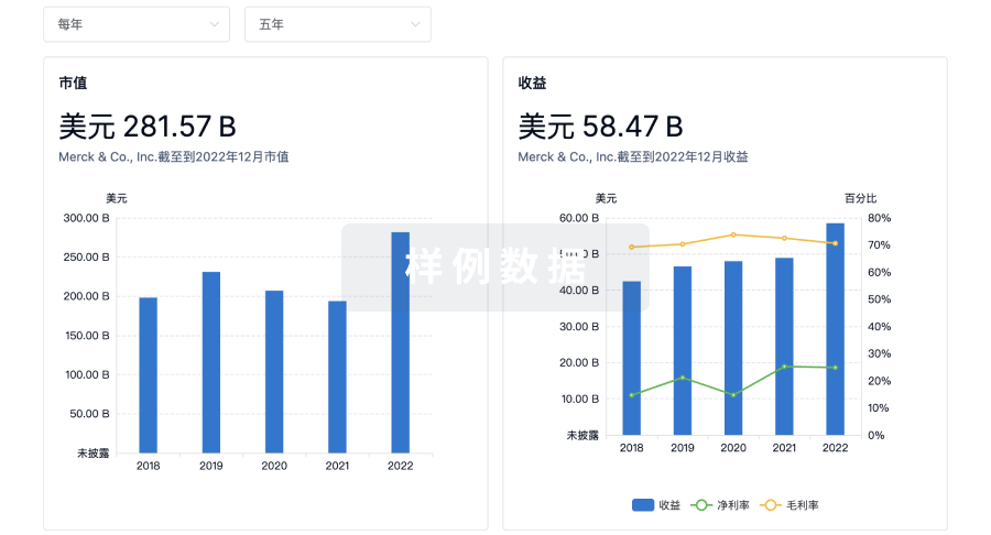
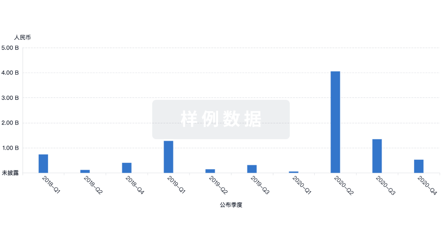
营收
使用 Synapse 探索超过 36 万个组织的财务状况。
登录
或
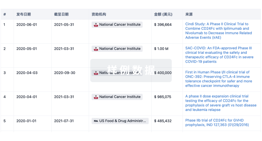
科研基金(NIH)
访问超过 200 万项资助和基金信息,以提升您的研究之旅。
登录
或
投资
深入了解从初创企业到成熟企业的最新公司投资动态。
登录
或
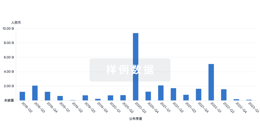
融资
发掘融资趋势以验证和推进您的投资机会。
登录
或
生物医药百科问答
全新生物医药AI Agent 覆盖科研全链路,让突破性发现快人一步
立即开始免费试用!
智慧芽新药情报库是智慧芽专为生命科学人士构建的基于AI的创新药情报平台,助您全方位提升您的研发与决策效率。
立即开始数据试用!
智慧芽新药库数据也通过智慧芽数据服务平台,以API或者数据包形式对外开放,助您更加充分利用智慧芽新药情报信息。
生物序列数据库
生物药研发创新
免费使用
化学结构数据库
小分子化药研发创新
免费使用