预约演示
更新于:2025-12-04
PF-06688992
更新于:2025-12-04
概要
基本信息
原研机构 |
非在研机构- |
权益机构- |
最高研发阶段临床1期 |
首次获批日期- |
最高研发阶段(中国)- |
特殊审评- |
登录后查看时间轴
结构/序列
分子式C11H22N4O4 |
InChIKeyAGGWFDNPHKLBBV-YUMQZZPRSA-N |
CAS号159858-33-0 |
查看全部结构式(2)
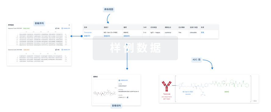
使用我们的ADC技术数据为新药研发加速。
登录
或

关联
1
项与 PF-06688992 相关的临床试验NCT03159117
A Phase I Open-Label Dose Escalation of GD3 ADC (Pfizer PF-06688992) in Subjects With Unresectable Stage III or Stage IV Malignant Melanoma (B802WI209568)
The purpose of this research study is to learn about the safety and effectiveness of the study drug, PF-06688992. Before this study, PF-06688992 has never been given to people.
PF-06688992 is a targeted therapy for people with cancer. The investigators linked a chemotherapy drug to an antibody (protein found in the blood). The antibody will connect to GD3 which is found on most melanomas but on very few other cells in the body. The investigators hope that in this way, it will deliver this chemotherapy directly to the melanoma and not to normal tissues.
PF-06688992 is a targeted therapy for people with cancer. The investigators linked a chemotherapy drug to an antibody (protein found in the blood). The antibody will connect to GD3 which is found on most melanomas but on very few other cells in the body. The investigators hope that in this way, it will deliver this chemotherapy directly to the melanoma and not to normal tissues.
开始日期2017-05-16 |
申办/合作机构 |
100 项与 PF-06688992 相关的临床结果
登录后查看更多信息
100 项与 PF-06688992 相关的转化医学
登录后查看更多信息
100 项与 PF-06688992 相关的专利(医药)
登录后查看更多信息
3
项与 PF-06688992 相关的新闻(医药)2025-06-14
·抗体圈
摘要:抗体-药物偶联物(ADC)领域经历了复兴,在过去的6年里,最近的大量开发投资和随后的药物批准。2022年11月,ElahereTM成为最新获得美国食品药品监督管理局(FDA)批准的ADC。迄今为止,已在临床上针对各种肿瘤适应症测试了260多种ADC。本文回顾了目前已获得FDA批准的ADC(11)、目前正在临床试验但尚未批准的药物(164)以及临床试验后停产的候选药物(92)。这些经过临床测试的ADC通过其靶向肿瘤抗原、接头、有效载荷选择和达到的最高临床阶段进行进一步分析,突出了与已停产候选药物相关的局限性。最后,本文讨论了临床前证明可以提高治疗指数的生物工程修饰,如果掺入,可能会增加成功过渡到监管批准的分子比例。【NO.1】ADC作为一类新型靶向治疗药物2000年,随着美国食品和药物管理局(FDA)批准MylotargTM用于治疗急性髓性白血病(AML),一类新的精准药物抗体-药物偶联物(ADC)被引入肿瘤学临床实践。ADC分子将抗体介导的肿瘤抗原靶向的精确性与强效细胞毒性药物相结合,从而为恶性肿瘤创造了靶向递送载体。通过这种方式,ADC提供了一种通过限制正常组织中的有效载荷暴露来降低肿瘤外毒性的方法。虽然大多数ADC临床候选药物利用细胞毒性化疗有效载荷,但最近的ADC候选药物也加入了靶向小分子1和免疫调节剂。自MylotargTM首次注册以来的23年里,经过临床测试的267种ADC中只有12种获得了监管部门的批准;过去6年内发生了10次[图1]。对生物工程的见解和对效力较弱的接头有效载荷(例如EnhertuTM)的利用为该领域重新注入了活力,并迎来了新一轮的药物审批浪潮。图1:FDA批准的时间表。迄今为止,已有12款ADC获得FDA批准(绿框)。由于在批准后试验中未能满足必要的终点,两名候选药物MylotargTM和BlenrepTM的批准被撤销(红框)。MylotargTM随后以较低剂量与化疗联合使用重新获得批准。目前有11种ADC疗法已获得FDA批准。【NO.2】影响ADC活性的因素与标准化疗相比,ADC具有多项优势,特别是:(1)将细胞毒性有效载荷精确递送至表达所选靶抗原的细胞,(2)能够实现比全身给药更有效的细胞毒性有效载荷利用,(3)可能最小化靶向/脱靶肿瘤毒性。ADC设计成功后,其前景是能够扩大治疗指数,优于全身化疗。通过将细胞毒性有效载荷直接递送到肿瘤组织,最小有效剂量(MED)降低,靶向/脱靶肿瘤不良事件相应减少。对临床测试的ADC分子进行有效分析需要从根本上了解调节其生物活性的因素。ADC细胞毒性有效载荷递送的基本细胞过程有三个关键部分。首先,抗体与抗原阳性细胞表面的靶抗原结合。其次,抗原-ADC复合物通过受体介导的内吞作用内化到靶细胞中。第三,抗原-ADC复合物被溶酶体酶消化,释放出触发细胞死亡的细胞毒性有效载荷。如图2所示,这些基础ADC临床活性的基本细胞过程的有效性受到各种因素的进一步调节,特别是靶抗原、所产生抗体的功能属性、偶联化学、接头属性以及所选肿瘤适应症的有效载荷效力和有效性。图2.控制ADC活动的因素。灰色箭头表示ADC进入信元的路径。抗体与细胞表面的靶抗原结合,抗原-ADC复合物通过内吞作用内化,抗原-ADC复合物要么循环回细胞表面,要么过渡到溶酶体区室。溶酶体加工释放细胞毒性有效载荷(红点),最终触发细胞死亡。控制这一过程的因素包括靶抗原、抗体、将有效载荷连接到生物制剂的偶联方法、接头、有效载荷和选定的肿瘤适应症。【NO.3】已进入临床试验的肿瘤ADC分析在这里,我们回顾了截至2023年1月1日注册的至少一项针对肿瘤适应症的人体临床试验的ADC,这些ADC包含在Beacon靶向治疗临床试验和管道数据库(beacon-intelligence.com)中。我们纳入了具有以下两个要素的ADC:(1)包含抗体、抗体融合或抗体片段的靶向部分,以及(2)有效载荷。利用的有效载荷来自常规化疗类别或靶向小分子和/或免疫调节剂。放射性同位素ADC被排除在该分析之外。自1997年首次ADC临床试验以来的26年里,又有266种ADC在1200多项临床试验中进行了测试。在此期间,54个ADC项目已正式终止,38个ADC已从公司管道中删除。本综述涵盖的ADC分为(1)批准(由FDA),(2)活性(未获得FDA批准,但目前正在进行≥1项临床试验),以及(3)停产(不再列入公司的临床管道,无论是否宣布停产)[图3]。应该注意的是,所有已批准的ADC目前也参与了几项临床试验,尽管它们未被归入“活性”类别(以消除重复计算)。此外,除美国外,所有FDA批准的ADC都在其他国家获得批准。图3:经过临床测试的ADC。此条形图捕获了已进行临床测试的267种ADC,其中:11种已获得FDA批准(绿色扇区),164种处于活跃临床试验中(蓝色扇区),92种已停产(红色扇区)。此外,对于有源ADC,它们已被细分以突出其最高开发阶段(第1阶段-第4阶段,P1-P4)。第4阶段(P4)中列出的此类候选药物disitamabvedotin已在中国获得批准,但尚未获得FDA的批准。3.1经临床测试的ADC靶向的肿瘤抗原总结图4说明了肿瘤抗原靶标和临床测试的最先进阶段。迄今为止,ADC候选药物共靶向了106种肿瘤抗原。获批的11种ADC靶向10种独特的癌症抗原:5种ADC靶向血液癌抗原,6种靶向实体瘤[图5,表1]。精选抗原是多种ADC的靶标,包括HER2(41个候选抗原)、Trop-2(14)、CLDN18.2(11)和EGFR(11)。不到2%的临床ADC候选药物靶向选定癌症抗原的1个以上表位:本综述包括4个双特异性ADC和1个双旁位ADC。图4:经临床测试的ADC的抗原靶标。在临床测试的267种ADC中,260种具有已知抗原(7种未披露)。在临床试验的各个阶段(1期-4期,P1-P4)中靶向给定肿瘤抗原的ADC数量显示在FDA批准的ADC(绿色扇区,绿色文本)、有源ADC(蓝色扇区,蓝色文本)和已停产的ADC(红色扇区,红色文本)类别中。双抗原靶向ADC以斜体显示。紫色文本所示的4期HER2候选药物是disitamabvedotin,该药物已在中国获得批准,但尚未获得FDA的批准。图5:按有效载荷类别和恶性肿瘤设置分类的获批ADC。提供了批准的ADC药物名称和有效载荷。ADC根据所利用有效载荷的效力从上到下列出,其中PBD有效载荷最有效,SN-38有效载荷最弱。表1.FDA批准的ADC和批准适应症的属性3.2经临床测试的ADC使用的接头总结接头分为两大类:可切割和不可切割[图6]。在临床ADC中,54%使用可切割接头,这是最常用的接头类别。11种临床批准的ADC中有10种使用蛋白酶可切割的接头。在经过临床测试的ADC中,16%使用不可切割的接头,包括临床活性ADCBlenrepTM。只有一种获批的ADCKadcylaTM使用不可切割的接头。31%的临床测试的ADC未披露接头类别。图6:用于临床测试的ADC的接头。使用FDA批准的ADC(绿色)、有源ADC(蓝色)和已停产的ADC(红色)的外圈中显示了使用不同连接子类别的ADC的数量。FDA批准的ADC与其各自的连接子一起显示。3.3经临床测试的ADC使用的有效载荷总结有效载荷分为四大类:(1)微管抑制剂,(2)DNA损伤剂,(3)拓扑异构酶I抑制剂,以及(4)靶向小分子(SM)[图7]。微管破坏剂是经过临床试验的最大有效载荷类别(57%)。11种获批的ADC中有7种使用微管抑制剂有效载荷。DNA损伤剂是ADC的第二大有效载荷类别(17%)。在这个亚组中,45个分子中有26个使用高效的PBD有效载荷,其中只有一个获得了FDA批准。另外两种获批的ADC通过利用calicheamicin有效载荷来采用DNA损伤类。拓扑异构酶I抑制剂包含在7%的临床测试的ADC中。在获批的11种ADC中,有2种使用拓扑异构酶I抑制剂有效载荷。除了这些传统的化疗有效载荷类别外,大约5%的ADC还包含靶向小分子(如Bcl-xL抑制剂)以及免疫调节剂(如TLR和STING激动剂)。该非化疗有效载荷类别中的候选药物尚未获得FDA批准。15%的临床测试的ADC的有效载荷未披露。图7:用于临床测试的ADC的有效载荷。与FDA批准的ADC(绿色)、有源ADC(蓝色)和已停产的ADC(红色)扇区的外环中显示了与有效载荷类型相对应的ADC数量3.4经临床测试的ADC使用的偶联方法总结在267个临床ADC中,111个候选药物使用非特异性氨基酸偶联,72个候选药物使用位点特异性偶联,84个候选药物未披露用于创建ADC的偶联方法。在使用位点特异性ADC偶联的ADC候选药物中,2种获得批准(EnhertuTM和TrodelvyTM)、50种活性ADC和26种已停产ADC接受了临床试验。除了DAR=8ADC(例如,EnhertuTM和TrodelvyTM)利用所有天然二硫键进行偶联外,其余ADC使用位点特异性偶联方法,这些方法要么保留四个链间二硫键,要么用化学共价键(例如,二硫键再桥接)代替它们。【NO.4】批准的ADC迄今为止,FDA已批准了12种ADC[图1、图5、图8和表1],血液系统和实体瘤恶性肿瘤各6种[图5、表1]。12个已获批准的ADC中有9个获得了加速附条件批准。12种ADC中的2种(MylotargTM和BlenrepTM)的批准被撤销[图1]。由于安全性与临床益处问题,MylotargTM于2010年被撤回,但在2017年以较低剂量与化疗联合使用重新获得批准。BlenrepTM于2022年撤回,当时验证性试验未达到必要的批准后疗效终点。图8:FDA批准的ADC,按有效载荷类别分类。显示了ADC药物名称、靶抗原以及有效载荷的名称和化学结构。箭头标记有效载荷与抗体的附着点在目前FDA批准的11种ADC中,有6种使用微管抑制剂有效载荷。3个获批的ADC使用破坏DNA的有效载荷,而2个携带抑制拓扑异构酶I的有效载荷[图5、图8、]。这些有效载荷涵盖从高效DNA损伤剂PBD(IC50~pM)到低效拓扑异构酶I抑制剂SN-38(IC50~nM)的效力范围。尽管样本量小,但经批准的ADC在靶向血液系统恶性肿瘤时使用更有效率的有效载荷,而在针对实体瘤的ADC中使用较低效力的有效载荷。在实体瘤环境中疗效所需的较高药物暴露可能会限制更有效有效载荷的利用,据报道,在首选生物剂量下全身毒性增加。【NO.5】有源ADC在164个有源ADC中,~7%处于3期临床试验中。这些活性晚期ADC靶向以下肿瘤抗原:BCMA(belantamabmafodotin)、CEACAM5(tusamitamabravtansine)、c-Met(telisotuzumabvedotin)、HER2(曲妥珠单抗duocarmazine和曲妥珠单抗rezetecan)、HER3(patritumabderuxtecan)、NaPi-2b(upifitamabrilsodotin)和Trop-2(datopotamabderuxtecan和SKB264)。活性ADC组中的大多数ADC(~54%)都使用微管抑制剂有效载荷,其次是DNA损伤(10%)和拓扑异构酶I抑制剂(~9%)有效载荷。~22%的有源ADC的有效载荷未披露[图9]。在微管抑制剂ADC中,auristatins含量最高,其次是maytansines。在DNA损伤有效载荷类别中,PBD占临床活性ADC的~50%。图9:按有效载荷类分类的有源ADC。在临床试验中的活性ADC中,大多数使用微管抑制剂有效载荷,其次是DNA损伤剂、拓扑异构酶I抑制剂(Topo-I)和靶向小分子(SM)。~22%的有源ADC尚未披露所使用的有效载荷(未披露)。在临床活性ADC靶向的癌症抗原中,~16%靶向血液肿瘤抗原,~80%靶向实体瘤抗原,~4%靶向在血液肿瘤和实体瘤恶性肿瘤中表达的癌症抗原。活性ADC类别中最常靶向的肿瘤抗原包括HER2(32个候选)、Trop-2(11)、CLDN18.2(11)和EGFR(8)。【NO.6】已停产的ADC停用ADC可归因于以下三个原因中的一个或多个:1)由于无法耐受的毒性导致治疗效果不足,2)由于疗效不足,治疗效果不如目前的护理标准,和/或3)商业/商业考虑。表2显示了所有已停产ADC的详细信息。表2.按有效载荷类别和恶性肿瘤设置停产的ADC【由于表格内容较多,请参考原文】由于无法耐受的毒性而导致治疗获益不足的潜在因素包括(1)靶向/脱离肿瘤毒性,(2)将非常高效的有效载荷用于需要更高生物暴露的抗原,(3)不稳定的连接子导致有效载荷的非肿瘤释放,(4)脱靶毒性,可能是由于ADC的胞饮作用,以及(5)有效载荷代谢转化为毒性更强的代谢物。大约29%的临床测试的ADC表示无法耐受的毒性是终止计划的原因。部分可能由于靶向/脱靶肿瘤毒性而具有无法耐受毒性的ADC的例子包括bivatuzumabmertansine(CD44v6,在皮肤角质形成细胞中表达)——致命的脱屑,MEDI-547(EphA2)——出血和凝血不良反应(通常与MMAE有效载荷无关的不良事件),和PF-06664178–皮疹不良事件(Trop-2,在包括皮肤在内的正常上皮表面表达)。对于PF-06664178的后一个例子,导致皮肤毒性严重程度的另一个潜在因素是有效的auristatin有效载荷与这种靶向Trop-2的ADC配对。事实上,PF-06664178的皮肤毒性严重程度与已批准的靶向Trop-2的ADCTrodelvyTM明显不同,后者使用较低效力的拓扑异构酶I抑制剂有效载荷。此外,还注意到另一种针对Nectin-4(也在皮肤中表达)的auristatinADCPadcevTM的皮肤毒性。微管抑制剂有效载荷ADC占已停产候选药物的63%,其次是DNA损伤(~27%)有效载荷。拓扑异构酶I抑制剂、靶向小分子和未公开的有效载荷合计占已停产ADC的10%[图10]。对需要更高生物制剂暴露量的抗原使用高效有效载荷可能是导致几种已停产的ADC候选药物出现无法耐受毒性的一个因素。双旁位四价HER2定向ADC的有效载荷选择MEDI4276可能导致剂量为>0.3mg/kg时出现无法耐受的毒性。事实上,所选的微管溶血素类似物有效载荷(IC50~低pM)在PBD有效载荷的效力范围内。临床批准的实体瘤ADC(包括2个靶向HER2抗原的ADC)均未使用该效力范围内的有效载荷——其中最活跃的是采用效力较弱的有效载荷(EnhertuTM)的ADC。安全性被认为是PBD偶联ADCADCT-502和DHES0815A终止HER2的原因。图10:按有效载荷类别分类的已停产ADC。已停产的ADC中使用的主要有效载荷类别是微管抑制剂和DNA损伤剂。拓扑异构酶I抑制剂(Topo-1)、靶向小分子(SM)和未公开的候选药物合计占已停产ADC的~9%。靶向已批准ADC的6种肿瘤抗原(CD19、CD22、CD33、CD79b、HER2和Trop-2)的ADC也已停产,其中一些是由于无法耐受的毒性。TrodelvyTM是经批准的Trop-2ADC,使用较低效力的拓扑异构酶I有效载荷SN-38(IC50~nM),需要高生物暴露才能达到所需的疗效益处(21天治疗周期的第1天和第8天10mg/kg)。两种靶向Trop-2的ADC已停产,很可能是由于有效载荷选择与需要更高生物暴露的肿瘤抗原靶标配对太强。PF-06664178使用高效的auristatin类似物有效载荷(IC50~低pM),在每3周接受高达4.8mg/kg的剂量治疗的患者(剂量≥3.6mg/kg由于皮疹、粘膜炎和中性粒细胞减少症的剂量限制性毒性被认为无法耐受)中产生剂量限制性毒性,而没有任何部分和/或完全反应。BAT8003,尚未发表关于高效美坦新有效载荷ADC的临床试验数据,尽管怀疑存在剂量限制性毒性。CD79b是经批准的ADCPolivyTM的靶标。与利妥昔单抗联合测试了后续位点特异性靶向CD79b的ADC,iladatuzumabvedotin。Iladatuzumabvedotin最终停药,因为由于较高剂量的眼毒性,没有注意到治疗指数(vsPolivyTM)的改善。三种靶向MylotargTM靶标CD33的ADC也已停产。AVE9633(DM4有效载荷)显示低于毒性剂量没有临床活性;IMGN779(吲哚基-苯二氮卓类二聚体有效载荷)未报告疗效;和vadastuximabtalirine(PBD有效载荷)在与低甲基化药物的联合研究后停止使用,理由是存在包括致命感染在内的安全问题。一种靶向CD33且具有管溶素有效载荷的ADCDXC007目前处于1期(注册号CTR20221074),但安全性和有效性数据尚未公布。停用LOP628(c-KIT)和losatuxizumabvedotin(EGFR)引用了输液相关不良事件。此外,在测试剂量下DCLL9718S(CLL-1)的耐受性差和缺乏客观反应并不能证明其进一步开发的合理性。在一些已停产的ADC中,临床毒性特征与临床前观察结果不符,例如靶向CDH6的ADCHKT288在临床前模型中未观察到的患者中出现神经毒性。同样,aprutumabixadotin(FGFR2)的临床MTD低于临床前估计的治疗阈值。后两个例子突出表明需要更好的预测模型来指导ADC临床开发。除了无法耐受的毒性外,疗效不足也是ADC停药的一个原因。导致疗效不足的因素包括(1)肿瘤靶抗原密度低和/或停产ADC的内化特性差,(2)有效载荷效力不足,(3)异质性DARADC产品导致有效载荷剂量不理想,(4)肿瘤中肿瘤外有效载荷释放和/或药物释放不完全,(5)由于PK特性差导致ADC快速清除,(6)未能证明疗效优于标准护理,以及(7)通过肿瘤中药物外排转运蛋白升高介导的多药耐药性。在有数据可获得的已停产ADC候选药物中,疗效不足可能是~47%病例的促成因素。据报道,疗效不足以保证进一步临床测试的候选药物包括但不限于tamrintamabpamozirine(DPEP3)、PF-06647263(EFNA4)、和PCA062(P-Cadherin)。其中一些ADC靶标可能具有异质性肿瘤表达和/或肿瘤抗原密度不足,无法诱导有效的ADC内化。利用效力不足的有效载荷,导致疗效不足,是导致HER2靶向免疫调节ADCNJH395和SBT6050停止的可能因素。在18例接受NJH395治疗(TLR7激动剂有效载荷)的患者中未观察到客观反应。同样,14名患者中只有1名实现了SBT6050的部分缓解(TLR8激动剂有效载荷)。对于这些TLR激动剂ADC,临床活性的缺乏也可能与抗肿瘤免疫反应的次优激活有关。临床HER2美登素类ADCBAT800190已停用,可能是为了推进效果较弱的拓扑异构酶I抑制剂有效载荷ADC(BAT8010)。这一停产/推进决定与两种已批准的HER2ADCKadcylaTM和EnhertuTM的临床经验一致,其中采用较低有效载荷的ADC(EnhertuTM)表现出更强的临床活性。与特异性半胱氨酸(THIOMABTM)偶联物ADC相比,非特异性半胱氨酸偶联物MUC16ADCSOFITUZUMABVEDOTIN,52观察到的疗效较低的可能原因可能是非特异性半胱氨酸偶联物(THIOMABTM)偶联物ADCDMUC4064A的疗效较低的原因。53CMB-401(MUC1)是由于疗效不足而停产的ADC的一个例子,部分原因可能是接头选择不当导致非肿瘤有效载荷释放。有人认为,这种卡利希霉素ADC未能引起单个部分缓解是由于使用了不稳定的amid接头。MEDI4267是由于PK性能差(和无法耐受的毒性)而停产的ADC的一个例子。值得注意的是,这种靶向HER2的管溶素ADC在MTD时,相对于HER2靶向的ADCKadcylaTM,在MTD时具有非常短的半衰期和高清除率。由于未能证明优于标准化疗对照组,7种ADC停药:rovalpituzumabtesirine(DLL3),depatuxizumabmafodotin(EGFRvIII),AMG595(EGFRvIII),AGS16F(ENPP3),glembatumumabvedotin(gpNMB),和lifastuzumabvedotin(NaPi-2b)。用lorvotuzumabmertansine(CD56)补充标准化疗会增加不良事件的发生率,而不会提高疗效。 关于92种已停产ADC中其余22种的临床信息仍未发表(AbGn-107、AGS67E、BAT8003、BIIB015、cantuzumabravtansine、IMGN388、milatuzumabdoxorubicin、laprituximabemtansine、lupartumabamadotin、MEDI2228、MEDI7247、PF-06688992、SAR428926、SBT6290、SC-005、SC-006、SGN-CD19B、SGN-CD48A、SGN-CD123A、SGN-CD352A、sirtratumabvedotin和XMT-1592)。在这22家公司中,分别有48%和2%的公司提到了投资组合优先级/战略考虑和缺乏应计项目,但其余50%的公司没有给出停产的原因。【NO.7】对未来ADC药物设计的影响开发具有提高治疗指数潜力的下一代ADC可分为ADC的三个主要组成部分(抗体、接头、有效载荷)和用于将抗体与有效载荷连接的偶联技术。此外,需要考虑需要将适当的有效载荷与给定的肿瘤适应症相匹配,同时注意靶向肿瘤的生物制剂的癌症抗原密度。7.1生物制剂的改进抗体设计的改进包括结合剂的选择和工程设计,以(1)选择促进最大内化的表位/亲和力,(2)优化/降低结合剂对关注正常组织中表达较高的靶标的亲和力,以及(3)微调ADC的净电荷以减轻靶标非依赖性毒性。靶向促进受体介导的快速内化表位的生物制剂比靶向非内化抗原表位的生物制剂显示出更大的活性。此外,据报道,双旁位和双特异性ADC生物制剂可改善ADC内化,提高靶抗原密度较低的肿瘤中的ADC有效性。目前正在测试的双旁生和双特异性ADC包括REGN5093-M114(c-MET,c-MET)、zanidatamabzovodotin(HER2,HER2)、IMGN151(FRαFRα)、BL-B01D1(EGFR,HER3)、M1231(EGFR,MUC1)和ORM-5029(HER2,HER3)。除了选择内化表位和/或双旁位/双特异性抗原靶向外,还需要针对所选抗原定制ADC生物制剂的生物亲和力优化。事实上,亲和力较低的生物制剂在较低的靶抗原密度下可能表现出结合和/或内化不足,而细胞亲和力过高的生物制剂可能导致受体占有率降低和/或内化。生物亲和力调节也可能有助于减轻对正常组织中表达的抗原的靶标/非肿瘤毒性。亲和力失谐已被证明可以降低正常组织中的靶标依赖性毒性,同时保持对靶抗原表达较高的肿瘤细胞的活性。最后,优化ADC的净电荷已被证明可以减轻与靶标无关的毒性。这方面的一个例子是通过将单个Lys到Asp突变引入ADC的生物制剂AGS-16C3F来降低眼毒性。这些结果表明,在ADC上产生净负表面电荷可以抑制不依赖靶标的毒性。7.2对链接器的改进接头不仅仅是抗体和有效载荷之间的惰性桥梁;它们会影响给定ADC的稳定性和PK。一些早期ADC的性能不佳,如CMB-401,被归因于不稳定的接头。ADC接头的改进已被证明可以减少全身有效载荷释放并改善PK特性。按照这些思路,接头开发的改进可能包括(1)有效载荷掩蔽接头,(2)亲水接头,(3)增加药物负荷的支链接头,(4)串联切割接头,以及(5)双切割特异性接头。修饰接头以掩盖疏水有效载荷可以提高治疗指数。一般来说,降低ADC的疏水性可以提高PK和治疗活性,至少部分是由于减少了微胞饮作用诱导的脱靶毒性。事实上,在ADC中加入亲水性大环以掩盖疏水有效载荷提高了AdcetrisTM样ADC的体内活性。修饰接头以增加药物负荷是提高包含低有效载荷的ADC有效性的另一种策略。创建具有较高DAR负载量的传统细胞毒性ADC的一个挑战是ADC分子的疏水性增加,这是由于疏水有效载荷数量的增加,这既增加了聚集的可能性又加快了ADC从生物体中的清除。在抗体和接头之间或从传统接头内的某个位置分支,创建聚合物接头,例如FleximerTM接头或PEG链添加物,可以增加ADC分子上的药物负荷,而不会产生生物降解和/或清除的相关责任。使用这种方法,可以在不增加整体ADC疏水性的情况下增加DAR。此外,多肽由亲水性中性或带负电荷的氨基酸(Ala、Gly、Pro、Ser、Thr、Glu;基于XTENTM肽的平台)可以产生DAR高达18的ADC,而不会影响增加接头亲水性可以通过减少MDR1泵对有效载荷代谢物的排斥来调节旁观者效应,从而改变ADC的毒性特征。然而,这种方法可能并不适用于所有ADC。最后,修饰可切割接头以最大限度地减少全身释放,同时仍保持肿瘤旁观者效应可以提高后续ADC分子的治疗指数。需要仅在溶酶体内发现的酶连续切割的工程接头可以实现这一特性。这样一个例子是针对葡萄糖醛酸酶可切割的接头,当切割时发现了一个组织蛋白酶切割位点,该位点能够释放有效载荷——确保两个切割步骤都只发生在溶酶体内部。在大鼠毒性模型中发现这种串联切割接头可以提高ADC的稳定性和耐受性。7.3对有效负载的改进可以提高后续ADC治疗效果的有效载荷的修饰包括(1)基于前药的有效载荷以减轻非肿瘤毒性,(2)产生亲水性细胞毒性有效载荷,以及(3)产生双功能有效载荷以提高肿瘤疗效。前药有效载荷利用酸性、低氧、高唾液酸化和富含蛋白酶的TME来触发肿瘤中的活性有效载荷释放。前药可能涉及通过“加帽”掩盖有毒、疏水的有效载荷,例如PBD。前药帽被TME酶(如β-葡萄糖醛酸酶)切割,以最大限度地减少肿瘤外有效载荷的释放。鉴定用于有效载荷释放的其他内体运输调节剂和溶酶体途径调节因子可能有助于设计下一代前药有效载荷。亲水性细胞毒性有效载荷的产生是开发DAR升高的ADC的另一项潜在进展,这些ADC可保持生物完整性和良好的PK属性。这方面的一个例子是亲水性有效载荷auristatinβ-D-葡萄糖醛酸苷MMAU。这种糖苷有效载荷还有一个额外的好处,即在其未共轭的游离形式中相对惰性。溶酶体酶促加工成去糖基化状态会激活有效载荷的细胞毒性和旁观者活性。ADC的效力也可以通过创建双有效载荷来提高肿瘤疗效。与给定生物制剂的两种或多种不同有效载荷的偶联已被证明比携带单个有效载荷的ADC混合物具有更大的抗肿瘤活性。探索双有效载荷ADC的临床前研究包括两种不同的微管抑制剂有效载荷MMAE和以及微管抑制剂有效载荷与DNA损伤剂(如MMAE和PBD)偶联的微管抑制剂有效载荷或MMAF和PNU-159682。所有这些双有效载荷ADC已被证明比单有效载荷ADC的混合物更能提高抗肿瘤活性。此外,发现这些双有效载荷ADC在健康小鼠中的耐受性与通过体重减轻和肝脏临床化学测量的单有效载荷ADC相似。7.4有效载荷偶联的改进有效载荷的位点特异性附着产生具有受控和确定的DAR的ADC制备。第一种通过半胱氨酸氨基酸工程生产此类ADC的方法给出了均相制剂,与随机偶联的ADC相比,表现出优异的临床前PK特性和安全性。这些发现激发了该领域的热情,并导致了其他位点特异性偶联方法的开发。迄今为止,位点选择性偶联方法分为八类:半胱氨酸工程、非天然氨基酸工程、与天然半胱氨酸偶联、肽标签、聚糖修饰、酶促修饰、二硫键桥接和天然赖氨酸偶联。迄今为止,尚未证明用于位点特异性偶联的方法对FcRn回收有直接影响,从而改变ADCPK、疗效和安全性。目前正在探索通过非天然氨基酸方法进行的接头-有效载荷偶联。然而,已经注意到,尽管稳定性和PK相当,但非天然氨基酸偶联位置对接头-有效载荷附着的影响对肿瘤杀伤有显着影响。使用肽标签技术的位点特异性偶联示例包括SMARTagTM和谷氨酰胺标签。SMARTagTM通过使用醛标签将接头有效载荷连接到甲酰甘氨酸来实现位点特异性偶联。谷氨酰胺标签技术利用转谷氨酰胺酶连接接头有效载荷。两种技术都被证明可以改善PK和疗效。GlycoConnectTM是位点特异性聚糖修饰偶联方法的一个例子。在这里,抗体在Asparagine-297位点进行聚糖重塑后,通过连接子-有效载荷的附着实现位点特异性偶联。然而,由于天冬酰胺-297聚糖对抗体Fcγ受体效应子功能很重要,因此该方法需要与Fc效应子功能的丧失相平衡,否则可能会为开发的ADC提供疗效益处。位点特异性技术的一项显着进步是AJICAPTM方法,该方法利用天然赖氨酸进行位点特异性接头有效载荷连接。该方法不需要抗体工程或酶促反应。在临床前模型中,这样生产的ADC被证明具有更好的治疗指数。在临床上,与非特异性半胱氨酸偶联的sofituzumabvedotin(MUC16)相比,位点特异性ADCDMUC4064A(MUC16)可以以更高的生物剂量给药,具有更高的总体反应率。虽然前景广阔,但位点特异性有效载荷偶联并不总是导致治疗改善。例如,位点特异性偶联ADCiladatuzumabvedotin(CD79b)和SC-002(DLL3)未显示出临床反应/治疗指数优于非特异性半胱氨酸偶联ADCPolivyTM和rovalpituzumabtesirine。【NO.8】总结在测试肿瘤适应症的267种ADC中,11种已获得FDA批准;92已停产。分析与已停产候选药物相关的局限性有助于为下一个系列分子的设计和选择提供信息。重要的是,新的生物工程修饰已在临床前被证明可以提高治疗指数。采用谨慎选择靶标的综合多因素方法,同时优化抗体、接头和有效载荷(与感兴趣的适应症相匹配),有望迎来下一波新的ADC批准。识别微信二维码,添加抗体圈小编,符合条件者即可加入抗体圈微信群!请注明:姓名+研究方向!本公众号所有转载文章系出于传递更多信息之目的,且明确注明来源和作者,不希望被转载的媒体或个人可与我们联系(cbplib@163.com),我们将立即进行删除处理。所有文章仅代表作者观点,不代表本站立场。
抗体药物偶联物申请上市临床结果
2023-03-07
人人都在聊ADC,一时风头无二。MNC药企在该领域找寻新的重磅炸弹,Biotech希望借此成为“进击的巨人”。但当场子热起来时,要辨认谁才是ADC领域的硬核玩家,越来越难。是靠“买买买”来布局ADC的Big Pharma吗?还是在技术不成熟年代就在耕耘的Biotech?当中国的ADC公司频频出现在MNC药企的视野,能否证明他们所代表的ADC中国实力已经能与全球同台PK?最直观的,谁是最流行ADC技术的开拓者、拥有者、良好继承者,谁就是先锋。细胞毒素药物作为ADC的重要部分,被行业认为是相较于抗体和linker部分最有希望突破的。在该领域,现下应用最为广泛的细胞毒素药物都被ADC技术先驱们牢牢掌握,后来的ADC参与者们都需要得到授权,或是另择它路。整体来说,中国能见面貌的ADC大多仍聚焦于应用层面的改良创新上,靶点同质化不可避免。但ADC药物与其他不同,需要抗体、linker、细胞毒素药物都刚刚好的组合。所以很多人认为改良创新也是有机会的,寄望再创一个新的DS-8201,而这也正是MNC们考虑中国ADC公司的一大原因。国内ADC第一梯队基本上都布局者7款以上的在研ADC产品,数量直追全球的技术先驱们,不过在差异化的靶点和进度上仍有相当的差距。比如拥有国产首个ADC产品的荣昌生物,还有6款在研ADC产品,靶点覆盖Her2、CLDN18.2、CD19等。再比如多禧生物,作为ADC产品布局最多的企业之一,创始人均来自美国的ADC技术先驱企业,掌握着ADC密码,披露了多达20个的候选产品,除Trop-2、Her-2、Muc-1,大多在临床前阶段。技术先驱:把握ADC要塞ADC似乎成了如今医药界最热的词,但谁又最懂ADC?问题或许没有一个清晰的答案,但至少时间、历史沉淀、管线潜力、ADC相关专利数量、已上市产品的根源、技术先驱,能够给予所有奔赴ADC战场的企业们一定启示。在全球尚无一款ADC药物上市的年代,Seagen、ImmunoGen和Spirogen的出现,在某种程度上勾勒出了ADC有着金戈铁马般气势的未来。或是其他在2000前后成立的ADC“先驱”型企业,遑论他们如今有多大的市值或知名度,都持续在给押注ADC赛道的各方较大的期待。后面的ADC产品,或是ADC方面的BD交易,或多或少,都能见到他们的身影。举个例子,从当前已上市ADC产品情况来看,超过30%是与Seagen合作开发而出,该公司也成了人们口中要被默沙东、辉瑞以重金收购的“香饽饽”,ImmunoGen也是近两年引起业界最多关注的生物技术公司之一;另外在2002年成立的Mersana Therapeutics,其市值虽未超过10亿美元,但仅在过去一年时间里,该公司便与GSK、德国默克和杨森分别达成了ADC新药的重磅交易。不过,能够最直观感受到这几家企业深刻影响了ADC未来的,是他们的技术与专利布局。花了半个世纪来研究的ADC领域,其在技术层面上的难有目共睹,全球首款与第二款ADC药物上市中间,隔着漫长的11年。在做ADC药物构建时,不得不提细胞毒药物的选择,其开发也是ADC构建壁垒的关键点。据了解,目前大部分ADC药物所携带的细胞毒药物为靶向DNA和微管抑制剂的药物。其中,奥瑞斯他汀衍生物、美登木素生物碱这两大细胞毒药物,具备在成为ADC药物后保持较好水溶性、稳定性等优势,占到了所有ADC药物研发的60%以上。奥瑞斯他汀衍生物是ADC药物研发中最多的一类,其技术拥有者即为Seagen,相关技术后授权给了艾伯维、拜耳、辉瑞、罗氏、武田、GSK等多家大药企;美登木素生物碱衍生物是ADC药物中第二大类研发最多的药物,技术拥有者为ImmunoGen,相关技术授权给了拜耳、安进、诺华、罗氏、赛诺菲等公司。此外,吡咯并苯二氮卓类化合物(PBDs)是一类源自链霉菌属的天然产物,具有很好的抗肿瘤活性,也被不少大药企青睐。基于PBD的ADC研究的早期创新者是Spirogen,后将其技术授权给了Seagen,罗氏等企业,有数据显示,自2013年起,有10个以上携带PBDs的ADCs药物进入临床,这成为继奥瑞斯他汀和美登木素生物碱之后最具前景之一的ADC药物。而作为Spirogen于2011年分拆出来的子公司,ADC Therapeutic现布局多款有PBD药物。以技术为基础,数十年积累沉淀,如今,三者在研ADC管线已是潜力深厚,被多家大药企虎视眈眈。截至目前,三家公司均有至少10项以上的ADC项目在研,Seagen的在研ADC项目更是超30项,其中近10项已到临床III期。Seagen与ImmunoGen均与不少跨国大药企达成了ADC技术协议,其技术平台是许多高价值合作伙伴关系的基础。其中,Seagen全球技术或商业化合作伙伴超过10家,目前有四大上市产品,2022年四款药物的商业收入共计17亿美元,同比增长23%。除了技术之外,近十年来ADC药物专利量快速增长,这或能预示着未来ADC市场更为激烈的竞争。这其中,Seagen依靠ADC核心技术平台突围的同时,是拥有超100项专利的大户,其专利类型覆盖化合物、序列、医药用途和组合物等,靶点丰富。总体来看,如今的ADC领域,无论是看上市ADC药物的表现,还是在研管线的潜力或“专而精”程度,说来说去,都难以绕开这几家企业。当然,其余被跨国大药企看中、来自那些目前“小而优”的企业们,也在以自己的方式共同推动ADC的发展,国外如开发新一代 ADC 和 ROR1双特异性抗体临床前管道的VelosBio,国内如科伦博泰等。跨国药企:ADC“摘果者”ADC曲折发展的历史长河,完美诠释了做药的机遇和挑战。跨国药企在新型疗法方向的布局向来都是“摘果者”的逻辑,ADC也不例外。最为典型的“摘果者”——阿斯利康,通过收购Spirogen、投资ADC Therapeutics、合作第一三共,后来者追上,跻身第一梯队;之后也有艾伯维、吉利德通过大额并购占有一席之地。当然也有更加风险主义的“摘果者”,仅仅通过合作。罗氏是该类型中最为典型的代表,他没有开发自己的ADC平台,但通过与ImmunoGen合作获得了如今市场最畅销的ADC产品Kadcyla;以及通过与Seagen合作推出了Polivy,也有不错销售表现。后来者有赛诺菲、默沙东。也有不那么典型的“摘果者”——辉瑞,在不以补充ADC产品为目的的收购中打开大门,成为了早期开发上的“胜利者”。而后经历技术周期,放弃,欲重新上路的故事。以及潜心研究,后来惊艳四座的第一三共。辉瑞在ADC领域的布局可以总结为“起了个大早,赶了个晚集”。他是最先摘果的MNC药企。早在2009年,辉瑞以680亿美元的交易金额并购惠氏制药(Wyeth)时,全球首款ADC(吉妥珠单抗,CD33 ADC)作为不显著资产被其收入囊中。该药经撤市后于2017年重新获批上市,暂未显出优异的市场表现。同年,辉瑞推出了Besponsa(奥加伊妥珠单抗,CD22 ADC),但销售表现糟糕。后受多因素推动,从2018年起,辉瑞(当时手握ADC产品数量最多)开始削减对ADC的关注,先后终止了与CytomX Therapeutics关于ADC的长期合作,以及ADC药物PF-06688992的开发,并将自主研发的两款ADC PYX-201和PYX-203的全球权益授权出去。究其原因,辉瑞对ADC的重视程度和其ADC药物的市场表现互为因果。如今在辉瑞现有研发管线中,相较于双抗、基因疗法、mRNA等新疗法,ADC几乎可说是被忽略的存在。随着ADC药物不断被验证,辉瑞再次将锚头对准了ADC领域。近日据外媒报道,Seagen已成为辉瑞的潜在收购目标,正在展开初步谈判。SVB证券评论道,这笔交易可能会为辉瑞2030年的营收增加80亿至100亿美元。而且凭借此交易,辉瑞或将再次跻身全球ADC第一梯队。阿斯利康是典型的胜利“摘果者”。与辉瑞不同,阿斯利康的ADC布局可以归结为“搭上好船,后来追上”。阿斯利康2013年才开始正式布局ADC产品,当时辉瑞、武田、罗氏都有上市的ADC药物了。尽管来得晚了些,但不得不佩服阿斯利康的眼光。第一步决定了成功的一半,当年10月,阿斯利康全球生物药研发部门MedImmune斥资4.4亿美元收购了PBD技术领域的先驱Spirogen公司,由此涉足ADC领域。之后,以PBD为基础的ADCs药物源源不断进入临床,由此苯二氮平类药物成为颇有前景的负载药物,而此药正是由Spirogen开发,并授权给Seagen、罗氏、艾伯维、ADC Therapeutics等多家。“长驱直入、抓住要害”的阿斯利康在尝到甜头后,又做了第二个决定——加码ADC Therapeutics,并与其多款在研ADC管线达成商业合作。值得关注的是,ADC Therapeutics是从Spirogen分拆而来。尽管获得了10多个早期的ADC管线,但想要“摘果”仍依赖后期管线。当时市场上仍大有选择,阿斯利康挑中了DS-8201(Her2 ADC)。尽管DS-8201已经小有名气,可当阿斯利康以69亿美元的交易额获得该产品的非日本权益时,其股价当日下跌了5.9%。后来之事诸君皆知。阿斯利康对DS-8201的押注成为其在ADC领域战略布局的关键一棋,凭此AZ直接跃升至全球ADC的第一梯队。总体而言,MNC对于ADC的重视程度和他们的收获互为因果,当然也会存在一些偶然性。第一梯队的MNC基本上是有1~2个成熟产品+小有规模(7个以上)的研发管线构成;而第二梯队的MNC则是有个别产品上市,管线围绕展开。参考资料:https://mp.weixin.qq.com/s/z2torpj3z61qNJED93vFoQ登记邮箱信息订阅E药经理人信息服务扫描二维码精彩推荐集采 | 国谈 | 医保动态 | 药审 | 人才 | 薪资 | 榜单 | CAR-T | PD-1 | mRNA | 单抗 | 商业化 | 国际化 | 猎药人系列专题启思会 | 声音·责任 | 创百汇 | E药经理人理事会 | 微解药直播 | 大国新药 | 营销硬观点 | 投资人去哪儿 | 分析师看赛道 | 药事每周谈 | 医药界·E药经理人 | 中国医药手册创新100强榜单 | 恒瑞 | 中国生物制药 | 百济 | 石药 | 信达 | 君实 | 复宏汉霖 |翰森 | 康方生物 | 上海医药 | 和黄医药 | 东阳光药 | 荣昌 | 亚盛医药 | 齐鲁制药 | 康宁杰瑞 | 贝达药业 | 微芯生物 | 复星医药 |再鼎医药跨国药企50强榜单 | 辉瑞 | 艾伯维 | 诺华 | 强生 | 罗氏 | BMS | 默克 | 赛诺菲 | AZ | GSK | 武田 | 吉利德科学 | 礼来 | 安进 | 诺和诺德 | 拜耳 | 莫德纳 | BI | 晖致 | 再生元
抗体药物偶联物并购
2021-03-18
辉瑞开始从
ADC领域“撤退”了?
3月18日,辉瑞与美国生物技术公司Pyxis Oncology(以下简称“Pyxis”)共同宣布,双方已针对两款ADC ( antibody-drug conjugate,抗体偶联药物)候选产品达成全球许可协议,但具体交易金额未透露。
根据许可协议,辉瑞将为Pyxis提供两种创新ADC候选药物PYX-201和PYX-203的全球范围内许可权、开发及商业化权利。同时,辉瑞还将为Pyxis提供其ADC技术平台的许可,包括各种有效载荷类别、链接器技术和针对特定ADC的缀合技术,以用于将来开发其他ADC药物。
而辉瑞也将获得Pyxis支付的预付款和股权,并有资格获得基于开发和销售的里程碑付款以及潜在销售的分层特许权使用费。
01 边研发,边放弃
此次交易中的PYX-201是辉瑞研发的一款全球首创非内化ADC药物,通过靶向实体瘤中过表达的肿瘤限制性抗原,选择性地杀死肿瘤细胞,同时增强抗癌免疫应答。与内化ADC药物相比,非内化ADC药物可以不用进入癌症细胞内,而在胞外释放细胞毒药物,在局部微环境中发挥作用。而PYX-203是一款靶向血液瘤的ADC药物,通过高效DNA破坏剂来减少肿瘤耐药性和防止肿瘤复发。
Pyxis在获得这两款药物后将会进一步开发并扩充自己ADC药物管线。目前,除了PYX-201和PYX-203外,Pyxis还拥有ADC药物PYX-202和4款单抗药物。
作为一家2019年才成立的公司,Pyxis却与辉瑞颇有渊源。其CEO Lara Sullivan(沙利文)曾在辉瑞工作多年,并在2017年成为辉瑞分拆公司SpringWorks的总裁,于2019年离开并创立Pyxis。这也让双方的合作也充满情分。
Pyxis首席科学官Ronald Herbst博士说:“早期ADC药物表现出了巨大的潜力,但仍有很大创新空间来研发安全性更高的ADC药物,PYX-201和PYX-203代表着使用创新缀合技术的下一代ADC。我们很高兴将辉瑞进行的临床前研究推进临床试验阶段。”
作为研发出全球首个上市ADC药物的公司,辉瑞的ADC药物研发之路也是十分曲折。
2000年5月,美国FDA批准了辉瑞旗下ADC药物Mylotarg(gemtuzumab ozogamicin)上市,用于治疗首次复发的CD33阳性急性髓系白血病(AML)的60岁以上患者。
但在随后的试验中,Mylotarg不但未显示出明显的临床益处,还存在着严重的安全问题,Mylotarg治疗组出现严重致命性肝损伤,联合用药组的死亡率高于单独使用化疗组。2010年,辉瑞选择主动将Mylotarg退市。直到2017年9月,Mylotarg再次在美国批准上市,用于治疗新确诊的CD33阳性AML成人患者,及2岁及以上复发/难治的CD33阳性AML患者。
如今,已有多款上市ADC药物在手的辉瑞,却走着一边研发一边放弃的路。
2018年,辉瑞公司终止了与CytomX Therapeutics公司的药物Probody 用于癌症的几种ADC药物开发平台开发和商业化的五年合作关系。辉瑞在终止信中表示,决定不追求之前选择的两个ADC药物开发目标。
在2019年第四季度营收下滑9%的同时,辉瑞宣布终止对ADC药物PF-06688992的研发。
PF-06688992是一款由抗神经节苷脂GD3单抗组成的药物,研发用于治疗黑素瘤。
PYX-201和PYX-203的出售,又是辉瑞在ADC领域的“新举动”。
02 火热的ADC会重蹈PD-1覆辙吗?
相比于2000年Mylotarg上市后ADC市场的十年寂寥,如今的ADC领域已是十分火热。
随着小分子毒素、连接子以及偶联技术的发展和成熟,ADC技术平台经过多次迭代,安全性、有效性大大增加,治疗窗口大大拓宽。有数据分析,2026年全球ADC药物市场容量将达100亿美元,到2030年将突破150亿美元。ADC领域因而成为各大药企争相布局的重要赛道。
目前,全球已有11 款ADC 药物上市。仅2019年一年就有三款ADC药物在美国获批上市,显然,“生物导弹”ADC 药物研发被推向高潮。
而国内制药企业对于ADC的研发热情丝毫不亚于国外。
据ClinicalTrials.gov显示,截止2021年3月19日,全球有155项ADC药物临床试验正在进行,其中国内有29项。据不完全统计,国内研发ADC药物的公司已超过20家。
部分公司ADC药物研发一览表
目前,荣昌生物的RC48(纬迪西妥单抗)是国内进展最快的自主研发HER2抗体-药物偶联 (ADC)药物,已于2020年8月向中国国家药品监督管理局提交胃癌适应症的新药上市申请,并被纳入优先审评审批程序。除了胃癌已经申报新药上市外,纬迪西妥单抗还在国内进行尿路上皮癌Ⅱ期关键性临床研究、HER2低表达乳腺癌Ⅲ期临床研究,以及肺癌和胆管癌的I期临床研究,并在美国获得尿路上皮癌的Ⅱ期临床试验许可。
处于国内ADC药企第一梯队的东曜药业,其候选核心产品TAA013已进入III期临床阶段。TAA013是一种含有曲妥珠单抗-美坦新衍生物(曲妥珠-MCC-DM1)的在研抗体偶联药物,旨在成为Kadcyla的实惠替代药物,用于治疗HER2阳性乳腺癌。
作为国内创新药研发龙头企业,恒瑞医药不仅加入了ADC大战,并且是多管齐下。
目前, 恒瑞医药已披露4款ADC药物研发进展。靶向c-MET的ADC药物SHR-A1403、靶向HER2的SHR-A1201和SHR-A1811都处于临床阶段。今年1月,恒瑞医药第四款ADC药物SHR-A1904的临床实验申请也获得NMPA受理。
2020年4月,Immunomedic研发的Trop-2 ADC药物Trodelvy在美获批上市。Trodelvy凭借着远高于传统治疗方法的效果,一鸣惊人,成为备受关注的治疗三阴性乳腺癌新药。而云顶新耀早在2019年便获得了Trodelvy在大中华区、韩国、及部分东南亚国家和地区,针对所有癌症适应症开发、注册和商业化的独家授权。目前,云顶新耀正在国内进行Trodelvy用于治疗接受过至少两线既往治疗的转移性三阴性乳腺癌IIb期临床试验。
从科伦药业最近公布的调研活动会议纪要可以看到,
科伦药业2021年将重点布局ADC药物。
靶向HER2的A166已向 CDE 提交关键Ⅱ期申请;靶向Trop-2 的SKB264已获得一期临床数据;靶向Claudin 18.2的ADC药物将在2021年进入临床。
而石药集团自主研发的Claudin 18.2靶向ADC药物SYS1801是国内药企首个Claudin 18.2靶向的ADC药物,去年11月获得FDA的孤儿药资格认定。石药集团也计划今年递交中国、美国的临床试验申请。
但,如此火热的ADC赛道或许将面临“拥挤”且“入不敷出”的问题。
今年3月初,百奥泰宣布放弃其ADC药物BAT8003和单抗BAT1306的临床开发。而在2月份,百奥泰已宣布另一款ADC药物BAT8001 III期临床试验失败。根据百奥泰公布的2020年报计算,三款药物让百奥泰损失研发投入费用3.4亿元。
百奥泰的BAT8001,曾是国内首个进入临床III期靶向HER2的ADC药物,一直被寄予厚望,BAT8003也是一款靶向热门靶点Trop2的ADC药物。然而,两款热门药物相继退场,无疑是给百奥泰,甚至是ADC药物浇了一盆“冷水”。
对于暂停BAT8003的研发,百奥泰方面表示,是为了合理配置该公司研发资源,聚焦研发管线中的优势项目。
百奥泰的“及时止损”也显示出投入与收入的严重失衡。火热赛道的背后意味着研发成本的升高,也暗藏着未来竞品颇多之下的“价格战”。
而如今“拥挤”的PD-1单抗赛道是否会成为ADC的“明天”?

免疫疗法抗体药物偶联物抗体合作创新药
100 项与 PF-06688992 相关的药物交易
登录后查看更多信息
研发状态
10 条进展最快的记录, 后查看更多信息
登录
| 适应症 | 最高研发状态 | 国家/地区 | 公司 | 日期 |
|---|---|---|---|---|
| 不可切除的黑色素瘤 | 临床1期 | 美国 | 2017-05-16 | |
| 不可切除的黑色素瘤 | 临床1期 | 美国 | 2017-05-16 | |
| 多发性骨髓瘤 | 临床1期 | - | - | |
| 多发性骨髓瘤 | 临床1期 | - | - |
登录后查看更多信息
临床结果
临床结果
适应症
分期
评价
查看全部结果
| 研究 | 分期 | 人群特征 | 评价人数 | 分组 | 结果 | 评价 | 发布日期 |
|---|
No Data | |||||||
登录后查看更多信息
转化医学
使用我们的转化医学数据加速您的研究。
登录
或

药物交易
使用我们的药物交易数据加速您的研究。
登录
或

核心专利
使用我们的核心专利数据促进您的研究。
登录
或

临床分析
紧跟全球注册中心的最新临床试验。
登录
或

批准
利用最新的监管批准信息加速您的研究。
登录
或

生物类似药
生物类似药在不同国家/地区的竞争态势。请注意临床1/2期并入临床2期,临床2/3期并入临床3期
登录
或

特殊审评
只需点击几下即可了解关键药物信息。
登录
或

生物医药百科问答
全新生物医药AI Agent 覆盖科研全链路,让突破性发现快人一步
立即开始免费试用!
智慧芽新药情报库是智慧芽专为生命科学人士构建的基于AI的创新药情报平台,助您全方位提升您的研发与决策效率。
立即开始数据试用!
智慧芽新药库数据也通过智慧芽数据服务平台,以API或者数据包形式对外开放,助您更加充分利用智慧芽新药情报信息。
生物序列数据库
生物药研发创新
免费使用
化学结构数据库
小分子化药研发创新
免费使用

