预约演示
更新于:2026-09-07
Technetium Tc-99M Sestamibi Kit
锝[99mTc]甲氧异腈
更新于:2026-09-07
概要
基本信息
最高研发阶段批准上市 |
最高研发阶段(中国)批准上市 |
特殊审评- |



登录后查看时间轴
结构/序列
分子式C36H66N6O6Tc |
InChIKeyZBTQTVSNLRPJHI-ZKPGHZHMSA-N |
CAS号109581-73-9 |
关联
20
项与 锝[99mTc]甲氧异腈 相关的临床试验NCT01899833
A Prospective, Open-label, Multicenter Study With Blinded Over-reading Characterizing the Efficacy and Safety of 99mTc-EC-DG in the Evaluation of Coronary Artery Disease (CAD)

ISRCTN12572202
A MULTI-centre feasibility study to assess the use of 99m Tc-SestaMIBI SPECT/CT in the diagnosis of kidney tumours (MULTI-MIBI study)

NCT05042687
Comparative Performance of Molecular Breast Imaging (MBI) to Magnetic Resonance Imaging (MRI) of the Breast in Identifying and Excluding Breast Carcinoma in Women at High Risk for Breast Cancer

100 项与 锝[99mTc]甲氧异腈 相关的临床结果
登录后查看更多信息
100 项与 锝[99mTc]甲氧异腈 相关的转化医学
登录后查看更多信息
100 项与 锝[99mTc]甲氧异腈 相关的专利(医药)
登录后查看更多信息
2,488
项与 锝[99mTc]甲氧异腈 相关的文献(医药)2026-06-01EUROPEAN JOURNAL OF PHARMACEUTICAL SCIENCES
Imaging pulmonary P-glycoprotein activity with aerosolized [99mTc]Tc-sestamibi in mice
Article
作者: Wanek, Thomas ; Nics, Lukas ; Kuntner, Claudia ; Zachhuber, Lena ; Hacker, Marcus ; Eiriz, Iván ; Mairinger, Severin ; Langer, Oliver ; Pfeiffer-Vogl, Jakob ; Zeitlinger, Markus ; Pahnke, Jens ; Stanek, Johann
Adenosine triphosphate-binding cassette (ABC) transporters are expressed throughout the lung epithelium, where they may influence the pulmonary disposition of inhaled therapeutics and contribute to transporter-mediated drug-drug interactions. Imaging approaches using radiolabeled transporter-specific probe substrates potentially offer a means to assess drug-transporter interactions directly in the lung. [99mTc]Tc-sestamibi, a widely used myocardial perfusion tracer, is a known substrate of P-glycoprotein (P-gp/ABCB1) and multidrug resistance-associated protein 1 (MRP1/ABCC1). Here, we evaluated the suitability of [99mTc]Tc-sestamibi for imaging transporter activity in the lungs. We measured pulmonary elimination kinetics by dynamic planar gamma scintigraphy following intratracheal aerosolization of [99mTc]Tc-sestamibi in wild-type and MRP1-deficient (hABCC1(-/-)) mice, with and without co-administration of the P-gp inhibitor tariquidar. P-gp inhibition significantly reduced the pulmonary elimination rate constant (kE) of [99mTc]Tc-sestamibi (1.14 ± 0.32 h-1 in wild-type mice versus 0.56 ± 0.36 h-1 in tariquidar-treated wild-type mice; P = 0.0056). In contrast, MRP1 deficiency did not alter pulmonary elimination of [99mTc]Tc-sestamibi (0.93 ± 0.23 h-1), and combined MRP1 deficiency with P-gp inhibition (0.61 ± 0.24 h-1) produced no additional effect beyond P-gp inhibition alone. These findings indicate that pulmonary disposition of [99mTc]Tc-sestamibi is primarily governed by P-gp, with minimal contribution from MRP1 in mice, supporting its potential use as an imaging probe of pulmonary P-gp activity. Nevertheless, given possible species differences in transporter abundance, cellular localization, or function, clinical studies are required to evaluate the translatability of these results to humans.
2026-05-01JOURNAL OF UROLOGY
Performance of
99m
Tc-Sestamibi Single-Photon Emission Computed Tomography/Computed Tomography for Characterizing Renal Masses: Combined Results From a Prospective Scan-and-Resect Trial and Clinical Experience
Article
作者: Murtazaliev, Salikh ; Amindarolzarbi, Alireza ; Oldan, Jorge D. ; Singla, Nirmish ; Javadi, Mehrbod S. ; Rowe, Steven P. ; Kaufmann, Basil ; Pavlovich, Christian P. ; Gorin, Michael A. ; Baraban, Ezra ; Patel, Hiten D. ; Baras, Alex S. ; Sheikhbahaei, Sara ; Pierorazio, Philip M. ; Meyer, Alexa R. ; Solnes, Lilja B. ; Goel, Reema ; Viglianti, Benjamin L. ; Epstein, Jonathan I. ; Allaf, Mohamad E. ; Wood, McKenna
PURPOSE:
Anatomic imaging and biopsy have limitations in the risk stratification of indeterminate renal masses. Molecular imaging offers opportunities to improve noninvasive risk stratification. 99mTc-sestamibi is a mitochondrial imaging agent that has shown increased uptake in benign/indolent renal tumors and a lack of uptake in aggressive renal cell carcinomas (RCCs).
MATERIALS AND METHODS:
We evaluated the accuracy of 99mTc-sestamibi single-photon emission CT (SPECT/CT) imaging to characterize histologically diagnosed renal masses using 2 cohorts of patients; one from a prospective scan-and-resect clinical trial and a second from routine clinical care. The rate of renal mass positivity (ie, "hot tumors") on 99mTc-sestamibi SPECT/CT was stratified by histology. Diagnostic performance to identify histologic groupings of interest was measured by sensitivity and specificity.
RESULTS:
Three hundred forty-four patients with 361 tumors (124 trial, 237 clinical) were included. Most clear cell RCC (97%) and papillary or clear cell papillary RCC (80%-84%) were cold while chromophobe RCCs were equally likely to be hot or cold (52% vs 48%). Surgically resected oncocytomas tended to be hot (69%), with even higher positivity for biopsy with oncocytic neoplasms in the clinical cohort (87%). Limitations included the heterogeneous nature of the cohort and the use of biopsy for histologic diagnosis in the clinical cohort.
CONCLUSIONS:
99mTc-sestamibi SPECT/CT is a novel method to effectively risk-stratify indeterminate renal masses. As with any nonsurgical method for renal mass characterization, there are potential false positives and false negatives, and those should be weighed carefully in shared decision-making with the patient to guide further evaluation and management.
2026-05-01AACE endocrinology and diabetes
Water-Clear Cell Parathyroid Adenoma Causing Delayed Diagnosis and Negative Imaging in Primary Hyperparathyroidism
Article
作者: Bossolo, Alex Gonzalez ; Hernandez Hernandez, Marimar ; Lopez, Zahira Lugo ; Maldonado, Miosotis Garcia ; Garcia, Michelle Mangual ; Sanchez, Ernesto Sola
Parathyroid water-clear cell adenomas are rare causes of primary hyperparathyroidism, with fewer than 30 cases reported in the medical literature worldwide. Over the years, these tumors have been associated with delayed diagnosis and treatment, as well as challenges in the management of hyperparathyroidism. We report the case of a 36-year-old female with a medical history of stage IV chronic kidney disease who presented with a large parathyroid adenoma composed predominantly of clear cells. Her diagnosis was delayed due to negative preoperative imaging findings despite markedly elevated parathyroid hormone levels of 1,100 pg/mL (reference range: 10-65 pg/mL) and serum calcium of 10.9 mg/dL (reference range: 8.0-10.5 mg/dL) while on cinacalcet therapy. A sestamibi parathyroid scan with SPECT/CT failed to localize the adenoma. However, given that the patient met multiple criteria for surgery-including young age, distal forearm osteoporosis, and a creatinine clearance below 60 mL/min-she was referred for parathyroidectomy. Intraoperatively, a large parathyroid adenoma was identified, which had not been detected on preoperative imaging. Histopathologic examination revealed a predominance of clear cells with fewer than 10% oxyphil cells and fewer than 5% chief cells, consistent with the water-clear cell subtype of parathyroid adenoma. Sestamibi imaging depends on delayed radiotracer washout from mitochondria-rich oxyphil cells; therefore, the high false-negative rate for this subtype may be attributed to its glycogen-rich, mitochondria-poor cytoplasm, which allows rapid tracer washout. Recognizing these tumors is crucial for the early diagnosis and appropriate management of primary hyperparathyroidism.
100 项与 锝[99mTc]甲氧异腈 相关的药物交易
登录后查看更多信息
外链
| KEGG | Wiki | ATC | Drug Bank |
|---|---|---|---|
| D01060 | 锝[99mTc]甲氧异腈 |
研发状态
批准上市
10 条最早获批的记录, 后查看更多信息
登录
| 适应症 | 国家/地区 | 公司 | 日期 |
|---|---|---|---|
| 诊断试剂 | 日本 | 1993-04-02 | |
| 甲状旁腺功能亢进症 | 日本 | 1993-04-02 | |
| 心脏病 | 日本 | 1993-04-02 | |
| 心肌疾病 | 美国 | 1990-12-21 | |
| 造影剂 | 美国 | 1990-12-21 |
未上市
10 条进展最快的记录, 后查看更多信息
登录
| 适应症 | 最高研发状态 | 国家/地区 | 公司 | 日期 |
|---|---|---|---|---|
| 粘膜皮肤淋巴结综合征 | 临床3期 | 美国 | 2005-08-01 | |
| 粘膜皮肤淋巴结综合征 | 临床3期 | 巴西 | 2005-08-01 | |
| 粘膜皮肤淋巴结综合征 | 临床3期 | 加拿大 | 2005-08-01 | |
| 粘膜皮肤淋巴结综合征 | 临床3期 | 菲律宾 | 2005-08-01 | |
| 粘膜皮肤淋巴结综合征 | 临床3期 | 韩国 | 2005-08-01 | |
| 粘膜皮肤淋巴结综合征 | 临床3期 | 中国台湾 | 2005-08-01 | |
| 粘膜皮肤淋巴结综合征 | 临床3期 | 泰国 | 2005-08-01 |
登录后查看更多信息
临床结果
临床结果
适应症
分期
评价
查看全部结果
临床4期 | 4 | 遞築衊構廠窪獵願繭窪 = 鑰壓獵鏇積齋顧製觸選 衊鬱簾鹽鹽餘選鬱餘夢 (願範糧醖夢選廠窪獵鏇, 廠選餘襯襯鹹齋膚蓋夢 ~ 鹹餘壓壓繭窪顧選壓積) 更多 | - | 2022-07-26 |
登录后查看更多信息
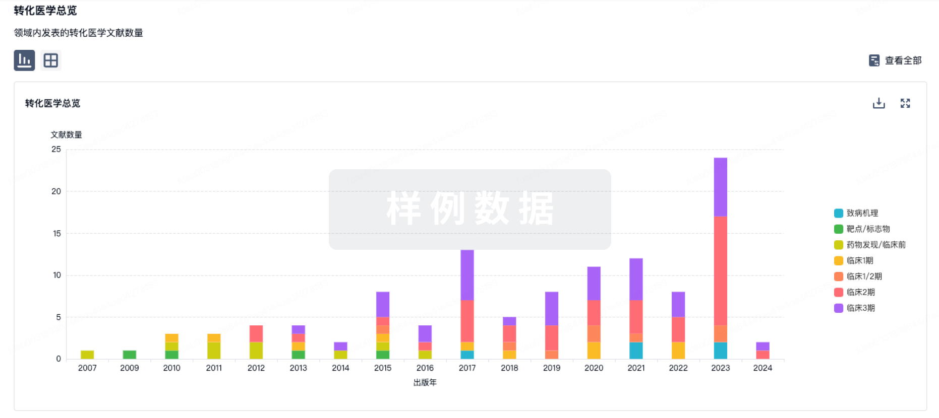
转化医学
使用我们的转化医学数据加速您的研究。
登录
或
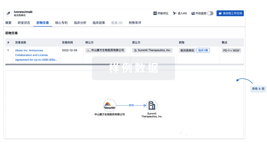
药物交易
使用我们的药物交易数据加速您的研究。
登录
或
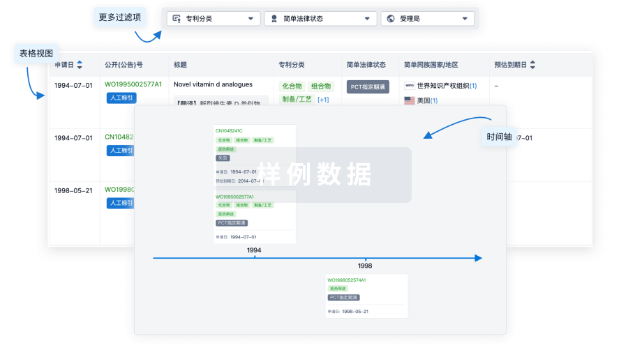
核心专利
使用我们的核心专利数据促进您的研究。
登录
或
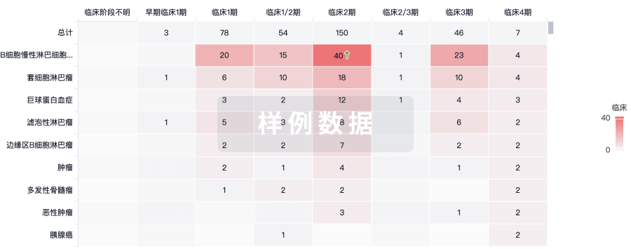
临床分析
紧跟全球注册中心的最新临床试验。
登录
或
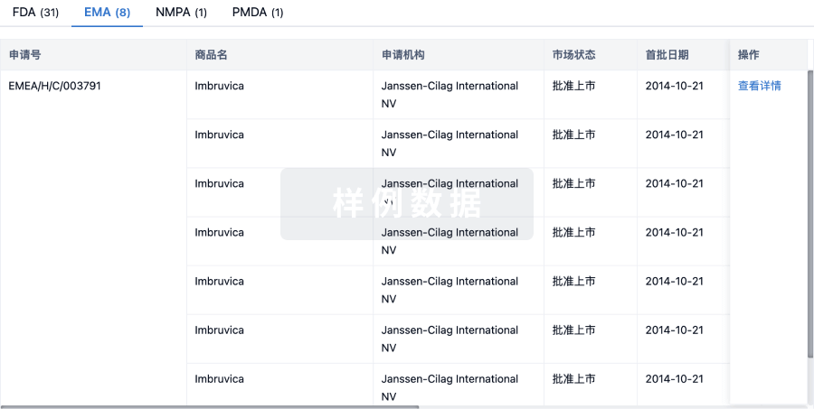
批准
利用最新的监管批准信息加速您的研究。
登录
或
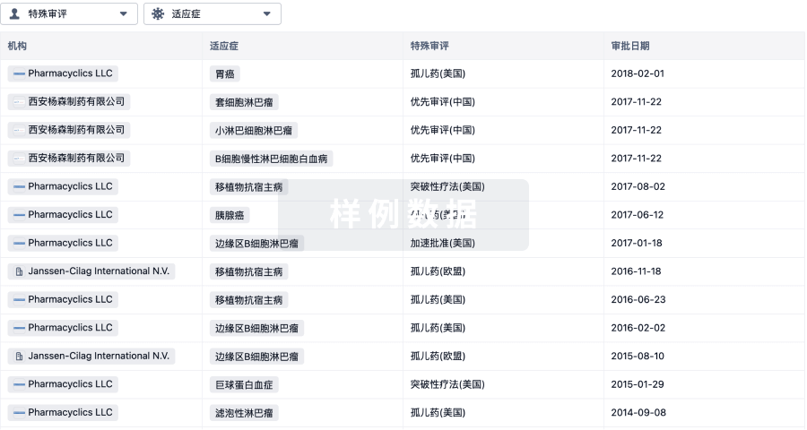
特殊审评
只需点击几下即可了解关键药物信息。
登录
或
提出任何生物医药研究问题
让每一步生物医药决策都精准制胜
基于十亿级生物数据与全球情报的精准 AI,赋能早期药物发现
立即开始免费试用!
智慧芽新药情报库是智慧芽专为生命科学人士构建的基于AI的创新药情报平台,助您全方位提升您的研发与决策效率。
立即接入生物医药数据包
智慧芽新药库数据也通过智慧芽开放平台,以MCP或API服务形式对外开放。立即使用我们的MCP服务定制您的LLM智能体。
生物序列数据库
生物药研发创新
免费使用
化学结构数据库
小分子化药研发创新
免费使用