预约演示
更新于:2026-03-17

Singapore Health Services Pte Ltd.
更新于:2026-03-17
概览
标签
心血管疾病
感染
其他疾病
单克隆抗体
疾病领域得分
一眼洞穿机构专注的疾病领域

技术平台
公司药物应用最多的技术
靶点
公司最常开发的靶点
关联
靶点 |
作用机制 |
在研机构 |
原研机构 |
在研适应症 |
非在研适应症 |
最高研发阶段 |
首次获批国家/地区 |
首次获批日期 |

靶点 |
作用机制 |
在研机构 |
原研机构 |
在研适应症 |
非在研适应症 |
最高研发阶段 |
首次获批国家/地区 |
首次获批日期 |

靶点 |
作用机制 |
在研机构 |
原研机构 |
在研适应症 |
非在研适应症 |
最高研发阶段 |
首次获批国家/地区 |
首次获批日期 |

NCT07452133
AI-Powered Closed-Loop Multielectrode Transcutaneous Spinal Cord Stimulation: Real-Time Adjustments for Enhanced Motor Recovery in Spinal Cord Injury (AIM RECOVER)
NCT07438353
Home-Based Digital Rehabilitation Program Optimized With Transcutaneous Spinal Cord Stimulation for Upper Limb Functional Enhancement in Tetraplegia (HOPE): A Safety, Efficacy, and Feasibility Study
NCT07452159
Diabetes in Asians at Risk in Youth
100 项与 Singapore Health Services Pte Ltd. 相关的临床结果
登录后查看更多信息
登录后查看更多信息
2026-12-31INTERNATIONAL JOURNAL OF HYPERTHERMIA
2024 International expert consensus on ultrasound-guided thermal ablation for secondary and tertiary hyperparathyroidism
Article
作者: Yu, Jian-Jun ; Yu, Ming-An ; Zhuo, Li ; Russell, Jonathon ; Yadav, Ajit ; Zhao, Zhen-Long ; Leong, Sum ; Kuo, Jennifer H. ; Tufano, Ralph P. ; Wei, Ying ; Qian, Lin-Xue ; Lu, Man ; Su, Hong-Hui ; Ghazi, Hossam Arafa ; Russell, Marika ; Chou, Yi-Hong ; Randolph, Gregory W. ; Xu, Shu-Hang ; He, Jun-Feng ; Fukunari, Nobuhiro ; Patel, Kaustubh ; Che, Ying ; Kandil, Emad ; Cicco, Rafael De ; Mauri, Giovanni ; Xu, Dong ; Yu, Song-Yuan ; Niu, Yun ; Zhang, Ya-Jun ; Dung, Le Thanh ; Çekiç, Bülent ; Amabile, Gerardo ; Abdelhamid, Amr H. ; Cheng, Kai-Lun ; Wu, Song-Song ; Dong, Gang ; Lin, Wei-Che ; Liang, Lei ; Zhou, Jian-Qiao ; Wang, Shu-Rong ; Janssen, Ingo ; Fan, Bo-Qiang
Treatments for secondary and tertiary hyperparathyroidism (SHPT/THPT) remain significant challenges in patients with end-stage renal disease. Thermal ablation (TA) has emerged as a minimally invasive, safe, and effective alternative to surgical resection (SR). However, technical variations and a lack of standardization have limited its widespread adoption. To address these challenges, an international expert panel developed consensus recommendations using a modified Delphi method, integrated with a systematic literature review. As a result, sixteen recommendations were formulated, addressing diagnosis, preoperative preparation, technical procedures, postoperative management, follow-up strategies, efficacy assessment, and complications associated with TA for SHPT/THPT. These recommendations aim to promote standardized treatment protocols, improve procedural safety, and provide evidence-based guidance for clinical practice and future research in ultrasound-guided TA for SHPT/THPT management.
2026-12-01Journal of Clinical Sleep Medicine
Awake speech recordings for machine learning diagnosis of obstructive sleep apnea: a Bayesian meta-analysis
Review
作者: Tan, Benjamin Kye Jyn ; Hao, Yunrui ; Toh, Song Tar ; Ong, Thun How ; Gao, Esther Yanxin ; Tan, Nicole Kye Wen ; Ng, Adele Chin Wei ; Huang, Guang-Bin ; Leow, Leong Chai ; Leong, Zhou Hao ; Phua, Chu Qin
STUDY OBJECTIVES:
Obstructive sleep apnea (OSA) is a prevalent but underdiagnosed condition linked to serious health risks. Due to the limited accessibility of polysomnography (PSG), AI (Artificial Intelligence)-based speech analysis has gained attention as a non-invasive screening tool. This Bayesian meta-analysis evaluates the diagnostic accuracy of AI models trained on awake speech and examines factors affecting performance.
METHODS:
We systematically searched Medline/PubMed, Embase, Scopus, Web of Science, and IEEE Xplore databases. Eligible studies included adults with OSA diagnosis via in-lab polysomnography or home sleep apnea tests and evaluated AI models using speech recordings. Models evaluated using random-split test sets or k-fold cross-validation were included in a Bayesian bivariate meta-analysis and meta-regression. Publication bias was examined using a selection model approach, while risk of bias and evidence quality were assessed with QUADAS-2 and GRADE.
RESULTS:
From 6,254 screened articles, 8 studies comprising 24 AI models, trained and tested on 1,060 and 825 participants were included. All studies used professional microphone recordings in the controlled hospital settings. AI models analysing awake speech recordings demonstrated pooled sensitivity and specificity of 82.9% (95% CrI: 80.0-86.4%) and 83.3% (95% CrI: 80.7-86.1%), respectively. The diagnostic odds ratio was 24.3 (95% CrI: 18.2-35.0). Higher mean age improved sensitivity. No significant effects were seen for OSA severity, model type, OSA prevalence, or male percentage. Publication bias was not evident.
CONCLUSION:
AI models trained on awake speech recordings demonstrate good diagnostic accuracy for OSA and hold potential as a practical, scalable screening tool in both clinical and community-based settings.
2026-06-01European Journal of Radiology Open
Submucosal laryngeal lesions: A puzzling diagnostic conundrum
Review
作者: Lee, Melissa Shuhui ; Wiggins, Richard ; Lee, Jean
Laryngeal mucosal masses are commonly squamous cell carcinomas, easily identified and biopsied on scope. In contrast, a submucosal laryngeal mass has a broad differential diagnosis, including benign and malignant epithelial and non-epithelial neoplasms as well as other non-neoplastic abnormalities including vascular malformations, infective or inflammatory pathologies, submucosal hematoma, rare depositional diseases such as amyloidosis, and other benign lesions such as laryngoceles. Due to a lack of visible mucosal abnormality, biopsy of these lesions are often challenging with higher rates of false negatives or inadequate sampling. Whilst radiological imaging features of submucosal laryngeal lesions may be non-specific, there are some lesions which may exhibit typical imaging features which could help radiologists to narrow the differential diagnosis and direct diagnostic workup and clinical management more effectively. In this article, we will illustrate a spectrum of submucosal laryngeal lesions, with an emphasis on helpful imaging features to help distinguish pathologies, and an overview of appropriate workup and management aspects which the radiologist needs to know to contribute effectively to patient care.
2026-03-06
疫苗微生物疗法
2026-01-21
·有驾
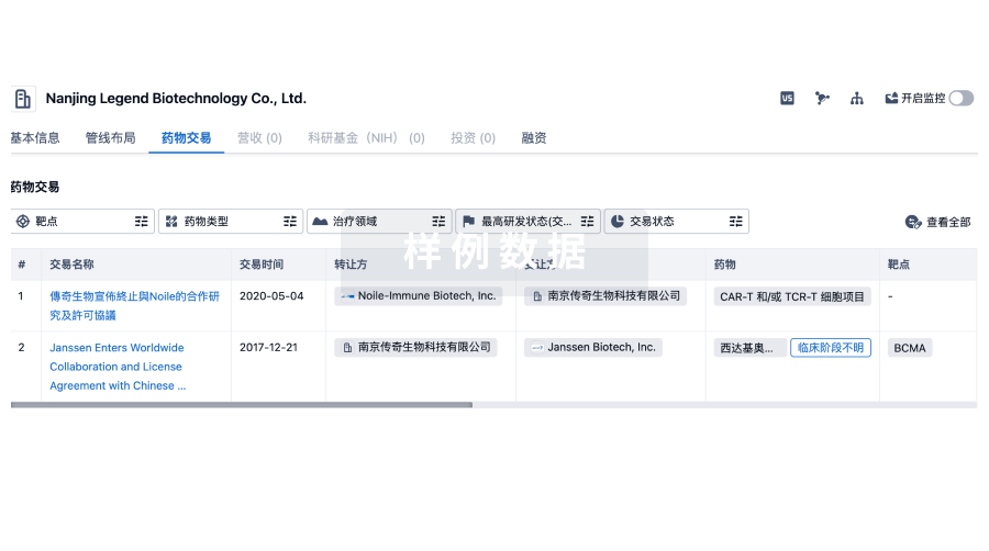
100 项与 Singapore Health Services Pte Ltd. 相关的药物交易
登录后查看更多信息
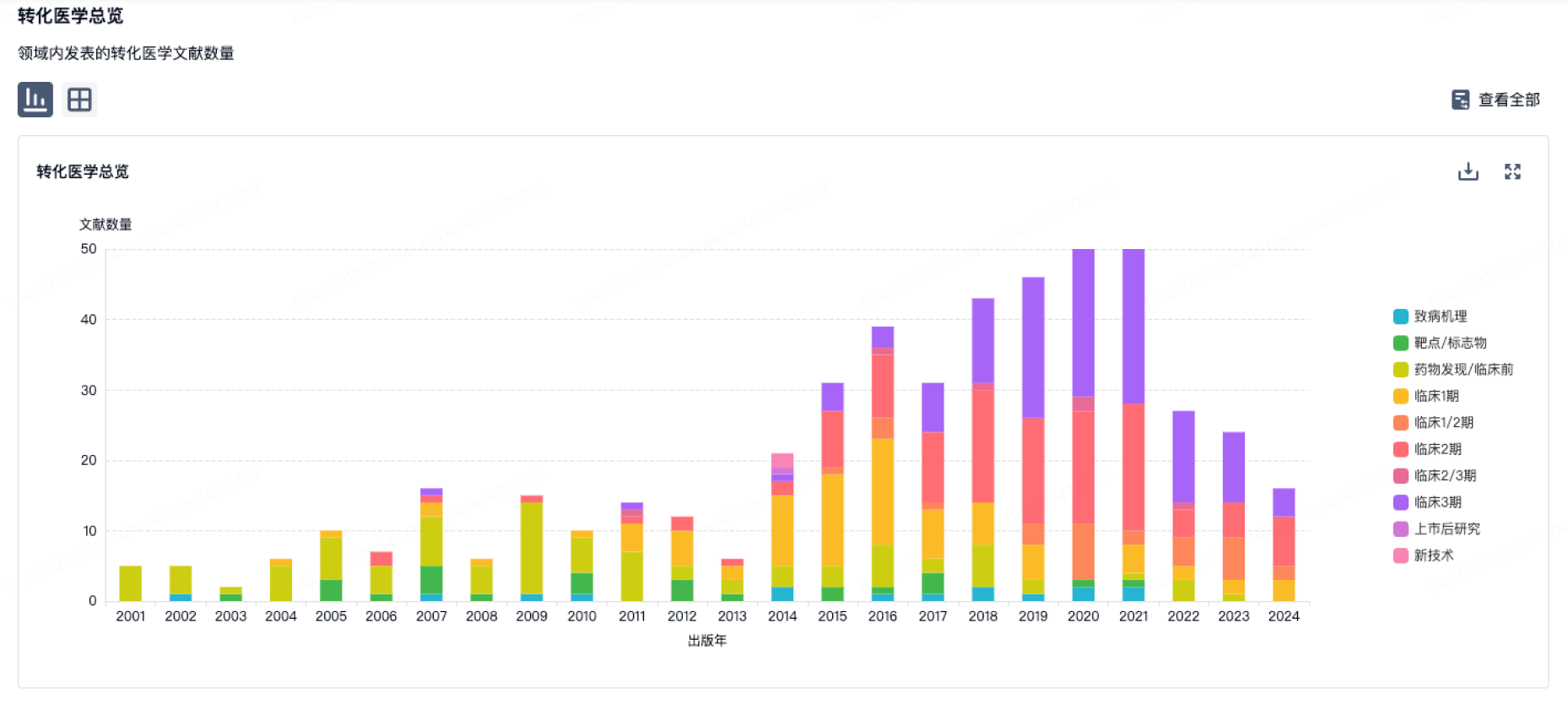
100 项与 Singapore Health Services Pte Ltd. 相关的转化医学
登录后查看更多信息
组织架构
使用我们的机构树数据加速您的研究。
登录
或
管线布局
2026年07月30日管线快照
管线布局中药物为当前组织机构及其子机构作为药物机构进行统计,早期临床1期并入临床1期,临床1/2期并入临床2期,临床2/3期并入临床3期
药物发现
4
5
临床前
临床2期
3
1
批准上市
其他
11
登录后查看更多信息
当前项目
登录后查看更多信息
药物交易
使用我们的药物交易数据加速您的研究。
登录
或
转化医学
使用我们的转化医学数据加速您的研究。
登录
或
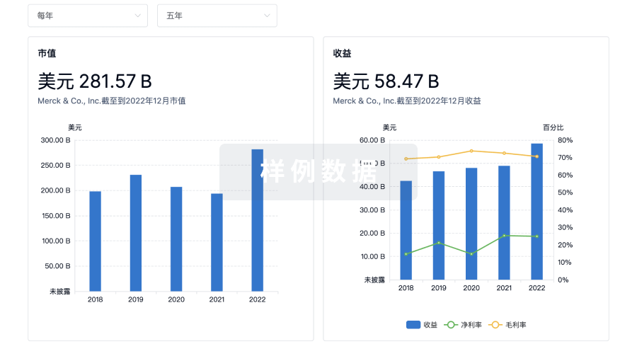
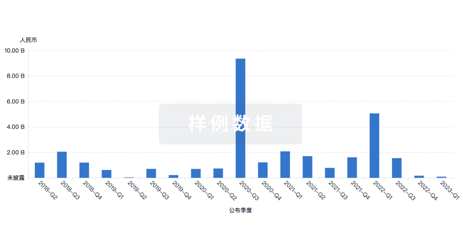
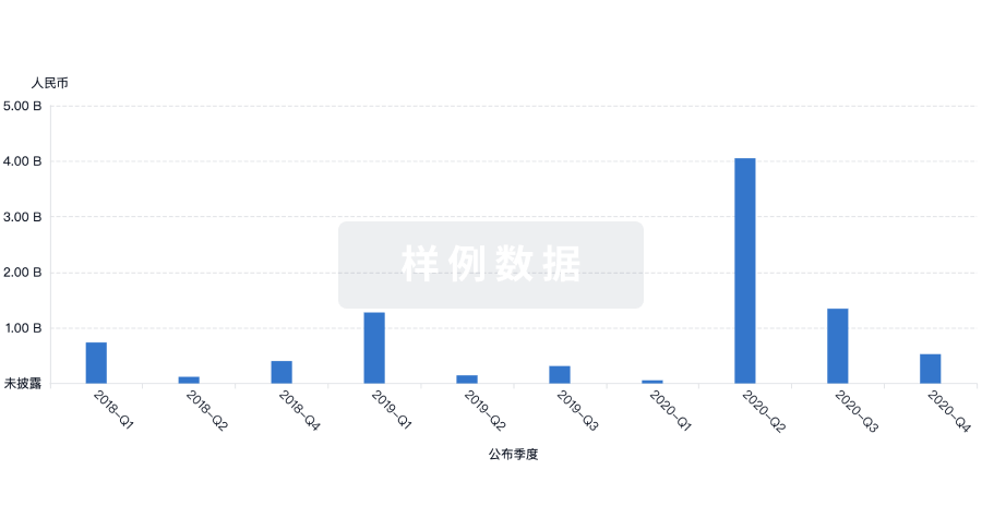
营收
使用 Synapse 探索超过 36 万个组织的财务状况。
登录
或
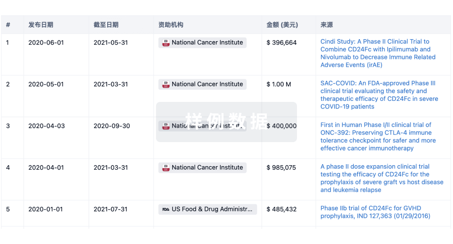
科研基金(NIH)
访问超过 200 万项资助和基金信息,以提升您的研究之旅。
登录
或
投资
深入了解从初创企业到成熟企业的最新公司投资动态。
登录
或
融资
发掘融资趋势以验证和推进您的投资机会。
登录
或
芽仔
全新生物医药AI Agent 覆盖科研全链路,让突破性发现快人一步
立即开始免费试用!
智慧芽新药情报库是智慧芽专为生命科学人士构建的基于AI的创新药情报平台,助您全方位提升您的研发与决策效率。
立即开始数据试用!
智慧芽新药库数据也通过智慧芽数据服务平台,以API或者数据包形式对外开放,助您更加充分利用智慧芽新药情报信息。
生物序列数据库
生物药研发创新
免费使用
化学结构数据库
小分子化药研发创新
免费使用