预约演示
更新于:2026-05-10

McMaster University
更新于:2026-05-10
概览
标签
感染
肿瘤
其他疾病
小分子化药
单克隆抗体
合成多肽
疾病领域得分
一眼洞穿机构专注的疾病领域

技术平台
公司药物应用最多的技术
靶点
公司最常开发的靶点
关联
靶点 |
作用机制 |
在研机构 |
原研机构 |
在研适应症 |
非在研适应症 |
最高研发阶段 |
首次获批国家/地区 |
首次获批日期 |
靶点 |
作用机制 |
在研机构 |
原研机构 |
在研适应症 |
非在研适应症 |
最高研发阶段 |
首次获批国家/地区 |
首次获批日期 |
靶点 |
作用机制 |
在研机构 |
原研机构 |
在研适应症 |
非在研适应症 |
最高研发阶段 |
首次获批国家/地区 |
首次获批日期 |
NCT06231069
Effects of Mixed Exercise Training and a Novel Multi-Nutrient Supplement on Muscle Fitness in Young Adults
NCT07300579
Vascular Events in Noncardiac Surgery Patients Cohort Evaluation Study 2

ISRCTN16668828
Intramedullary screw versus Kirschner wire fixation of extraarticular proximal and middle phalanx fractures: a multicenter randomized controlled trial
100 项与 McMaster University 相关的临床结果
登录后查看更多信息
登录后查看更多信息
2026-12-31Human Vaccines & Immunotherapeutics
Hematologic adverse events following COVID-19 vaccination and revaccination in persons aged 12 and above: A Canadian Immunization Research Network study
Article
作者: Lam, Godfrey ; McHenry, Mary ; Barber, Colin ; Castellucci, Lana A. ; Rosenfield, Lana ; Belga, Sara ; Deeks, Shelley ; Rex, Greg ; Sadarangani, Manish ; Constantinescu, Cora ; Roches, Anne Des ; Cameron, Scott B. ; Zaborniak, Karver ; Bosonea, Ana ; Viel-Theriault, Isabelle ; Cowan, Juthaporn ; Pham-Huy, Anne ; Finlay, Jane ; McConnell, Athena ; Chew, Jo Lin ; Vaudry, Wendy ; Lacuesta, Gina ; Abdurrahman, Zainab ; Quach, Caroline ; McNeil, Shelly ; Halperin, Scott ; Morris, Shaun K. ; De Serres, Gaston ; Mah, Allison ; Wilson, Sarah ; Burton, Catherine ; Pernica, Jeffrey M. ; Naus, Monika ; Chapdelaine, Hugo ; Grant, Ian ; Hildebrand, Kyla ; Pourshahnazari, Persia ; Gantt, Soren ; Salvadori, Marina ; Falcone, Emilia ; Buchan, C. Arianne ; Piche-Renaud, Pierre-Philippe ; Rubin, Tamar ; Kalicinsky, Chrystyna ; Suresh, Sneha ; Kanani, Amin ; Top, Karina A. ; Morris, Shaun ; Shivakumar, Sundeep ; Drolet, Jean-Philippe ; Blaquiere, Martin ; Upton, Julia ; Mak, Raymond ; Betschel, Stephen ; Comeau, Jeannette ; Salvo, Grazia ; Sturtevant, Doris ; Simons, Elinor ; Cook, Victoria ; Wright, Alissa ; Vostretsova, Kateryna ; Gagnon, Remi ; Song, Christine ; Carignan, Alex ; Smolinski, Michael ; Fitzpatrick, Tiffany
Data on safety of revaccination after a hematologic adverse event following immunization (AEFI) with COVID-19 vaccines are limited. This study characterized hematologic AEFIs in patients assessed by Canadian Special Immunization Clinic (SIC) network physicians from January 2021 to February 2023 and estimated recurrence rates of hematologic events after revaccination. Following clinical assessment of 475 individuals, revaccinated participants were followed for recurrence of AEFI. Thirty-eight cases (21 [55.3%] males and 17 [44.7%] females) with hematologic AEFIs and aged ≥12 were included in the analysis; 31/38 participants (81.6%) were 18 to 64 y old. Immune thrombocytopenia, deep vein thrombosis and pulmonary embolism were the most common diagnoses, accounting for 11 (28.9%), 9 (23.7%) and 9 (23.7%), cases. Two cases of thrombosis with thrombocytopenia syndrome (TTS) were reported after ChAdOx1. The vaccines associated with hematologic AEFIs were BNT162b2 (17, 44.7%), ChAdOx1 (11, 28.9%), and mRNA-1273 (10, 26.3%). Twenty-seven (71.1%) participants were revaccinated; 14 (51.9%) received the same vaccine product as their initial vaccine while 13 (48.1%) received a different product. There were no recurrences of the same AEFI reported after revaccination with an mRNA vaccine, including the two patients with TTS, suggesting that hematologic AEFI recurrences following COVID-19 vaccines are likely uncommon.
2026-12-31Human Vaccines & Immunotherapeutics
Meta-analysis of the efficacy of palivizumab versus nirsevimab at preventing medically attended respiratory syncytial virus infections in non-hospitalized preterm infants
Review
作者: Waghorne, Nicola ; Carbonell-Estrany, Xavier ; Keary, Ian ; Paes, Bosco ; Fullarton, John ; Rodgers-Gray, Barry
Palivizumab has >27 y of proven effectiveness in the prevention of severe respiratory syncytial virus (RSV) infection in high-risk infants. However, there remains limited data on its efficacy at preventing medically attended, non-hospitalized RSV infections (MARI). A systematic literature review was undertaken to identify randomized, placebo-controlled studies of prophylactic interventions against MARI and RSV-related hospitalization in healthy infants born ≤35 weeks' gestational age (wGA). The findings informed a meta-analysis of three studies (N = 2,464) that found palivizumab significantly reduced MARI by 70.5% compared with placebo in 29-35 wGA infants, with broadly similar efficacy to nirsevimab.
2026-12-31Gut Microbes
The putative role of the microbiota in the development of neuropsychiatric disorders following early childhood malnutrition
Review
作者: Khan, Waliul ; Collins, Stephen M. ; Jama, Yahya
Early childhood malnutrition (ECM) is robustly associated with increased risk of cognitive impairment and neuropsychiatric disorders across the lifespan, yet the biological mechanisms underlying this vulnerability remain incompletely defined. Accumulating clinical evidence indicates that ECM is associated with delayed maturation and reduced diversity of the intestinal microbiota, including depletion of taxa involved in short-chain fatty acid production and complex carbohydrate fermentation. These microbial alterations coincide with broader metabolic, immune, and barrier dysfunctions - such as reduced availability of neuroactive metabolites, low-grade inflammation, and impaired intestinal and vascular integrity - that plausibly intersect with critical processes in brain development. Experimental studies in animal models demonstrate that perturbation of microbiota-derived signaling during sensitive early periods is sufficient to induce lasting neurodevelopmental and behavioral changes, providing proof of concept for a causal role. However, in human populations, the microbiota remains best viewed as a biologically plausible intermediary rather than a proven determinant of outcome. Future progress will require integrative longitudinal studies and developmentally timed interventions to test whether restoration of microbiota function can modify neurodevelopmental trajectories. Clarifying these relationships has important implications for understanding the long-term consequences of early nutritional adversity and for identifying preventive strategies in settings where ECM remains prevalent.
2026-04-30
高管变更放射疗法
100 项与 McMaster University 相关的药物交易
登录后查看更多信息
100 项与 McMaster University 相关的转化医学
登录后查看更多信息
组织架构
使用我们的机构树数据加速您的研究。
登录
或
管线布局
2026年07月21日管线快照
管线布局中药物为当前组织机构及其子机构作为药物机构进行统计,早期临床1期并入临床1期,临床1/2期并入临床2期,临床2/3期并入临床3期
药物发现
6
16
临床前
临床1期
1
2
临床2期
其他
22
登录后查看更多信息
当前项目
登录后查看更多信息
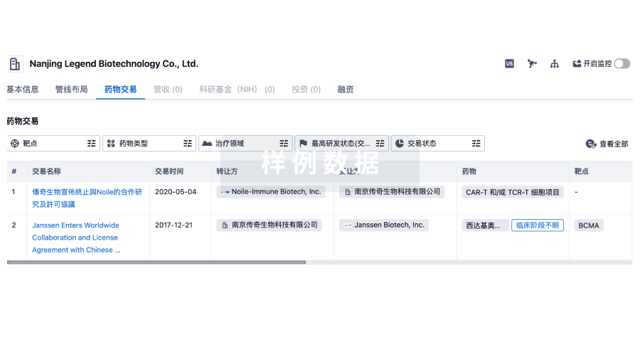
药物交易
使用我们的药物交易数据加速您的研究。
登录
或
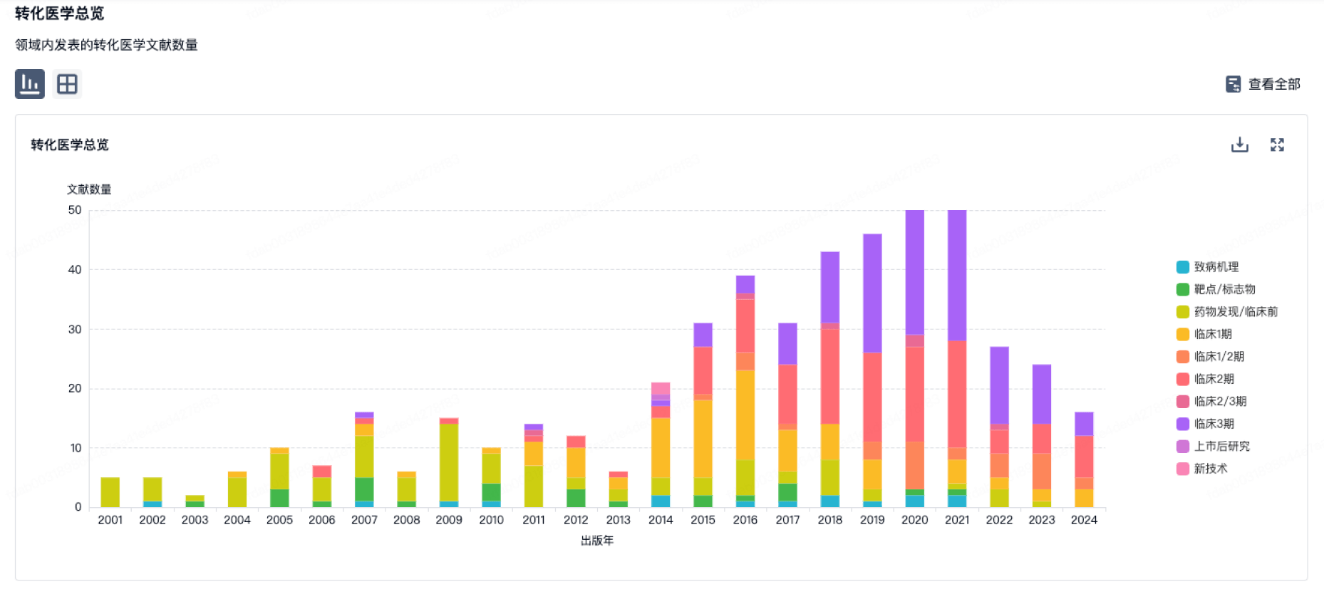
转化医学
使用我们的转化医学数据加速您的研究。
登录
或
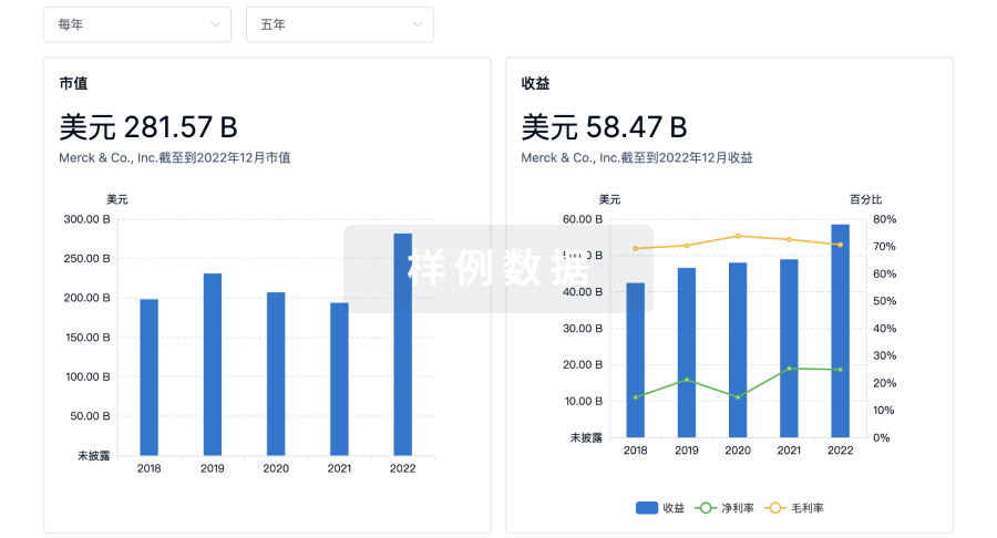
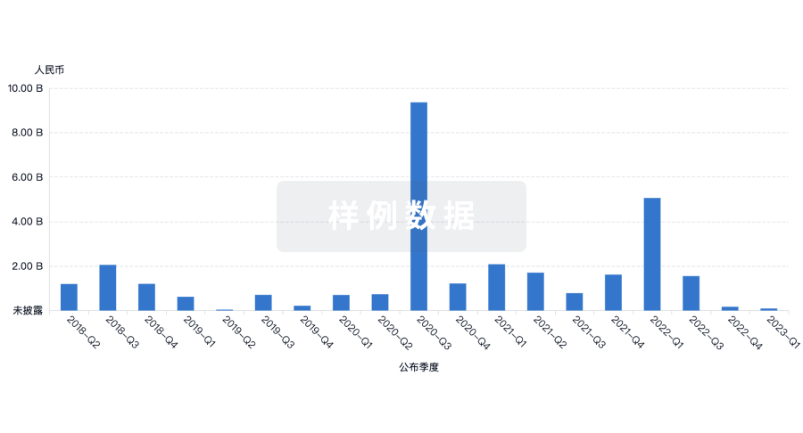
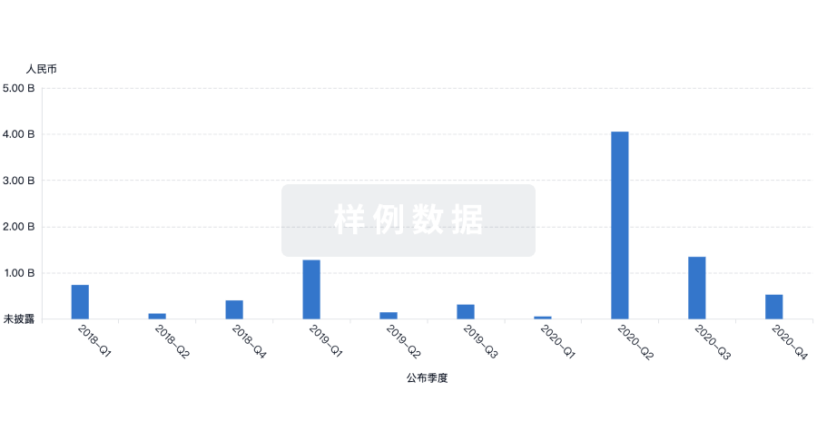
营收
使用 Synapse 探索超过 36 万个组织的财务状况。
登录
或
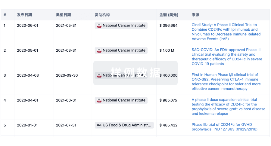
科研基金(NIH)
访问超过 200 万项资助和基金信息,以提升您的研究之旅。
登录
或
投资
深入了解从初创企业到成熟企业的最新公司投资动态。
登录
或
融资
发掘融资趋势以验证和推进您的投资机会。
登录
或
芽仔
全新生物医药AI Agent 覆盖科研全链路,让突破性发现快人一步
立即开始免费试用!
智慧芽新药情报库是智慧芽专为生命科学人士构建的基于AI的创新药情报平台,助您全方位提升您的研发与决策效率。
立即开始数据试用!
智慧芽新药库数据也通过智慧芽数据服务平台,以API或者数据包形式对外开放,助您更加充分利用智慧芽新药情报信息。
生物序列数据库
生物药研发创新
免费使用
化学结构数据库
小分子化药研发创新
免费使用