预约演示
更新于:2026-06-15
Gansu College of Traditional Chinese Medicine
更新于:2026-06-15
概览
标签
神经系统疾病
其他疾病
消化系统疾病
小分子化药
化学药
多糖药物
疾病领域得分
一眼洞穿机构专注的疾病领域

暂无数据
技术平台
公司药物应用最多的技术
暂无数据
靶点
公司最常开发的靶点
暂无数据
| 排名前五的药物类型 | 数量 |
|---|---|
| 小分子化药 | 6 |
| 化学药 | 1 |
| 多糖药物 | 1 |
关联
7
项与 甘肃中医药大学 相关的药物作用机制 BK channel激动剂 [+2] |
非在研适应症- |
最高研发阶段批准上市 |
首次获批国家/地区 中国 |
首次获批日期1993-01-01 |
作用机制 BAX调节剂 [+1] |
在研机构 |
非在研适应症 |
最高研发阶段批准上市 |
首次获批国家/地区- |
首次获批日期- |

27
项与 甘肃中医药大学 相关的临床试验ChiCTR2600118363
Development and validation of an individualized efficacy prediction model for lumbar disc herniation based on machine learning and multimodal data
开始日期2026-01-01 |
申办/合作机构 |
ChiCTR2400085974
Clinical efficacy and timeliness of filiform fire needling on herpes zoster patients and the influence on Th17/Treg cellular immune balance
开始日期2024-01-01 |
申办/合作机构 |
ChiCTR2300074554
Study on the effect of mask use on physiological indexes and subjective feelings of COPD patients
开始日期2023-10-07 |
申办/合作机构 |
100 项与 甘肃中医药大学 相关的临床结果
登录后查看更多信息
0 项与 甘肃中医药大学 相关的专利(医药)
登录后查看更多信息
493
项与 甘肃中医药大学 相关的文献(医药)2026-07-01·JOURNAL OF NEUROSCIENCE METHODS
2.5D HAU-Net with gated spatial attention for automatic hippocampus segmentation in MRI
Article
作者: Li, Xue ; Gong, Piqiang ; Chen, Fuming ; Ding, Xiaohan ; Lin, Dongmei ; Liu, Zhao
BACKGROUND:
The hippocampus is a key brain region and biomarker for Alzheimer's disease (AD). Accurate automated hippocampal segmentation is essential for anatomical and pathological analysis. However, the gap between model complexity and available computational resources hampers the clinical deployment of computer-aided diagnosis (CAD) systems, often resulting in limited accuracy and generalization.
NEW METHOD:
This study introduces a 2.5D U-Net-based framework that integrates an attention mechanism for efficient and accurate MRI hippocampal segmentation. Three consecutive slices (anterior, middle, posterior) are stacked to form 2.5D input representations, enhancing spatial context. The proposed HAU-Net incorporates a gated spatial attention module to improve feature selectivity and robustness. A hybrid Dice-BCE loss is used to address class imbalance and accelerate convergence.
RESULTS:
Experiments on the MSD Task04 Hippocampus and HarP datasets demonstrate strong performance, achieving Dice scores of 91.05% and 90.62%, respectively, with stable results across datasets.
COMPARISON WITH EXISTING METHODS:
Compared with the baseline U-Net and other widely used segmentation models, the proposed 2.5D attention-enhanced network achieves higher Dice similarity coefficients and better generalization while maintaining computational efficiency suitable for practical use.
CONCLUSIONS:
The attention-guided 2.5D HAU-Net provides an effective, robust, and resource-efficient solution for automated hippocampal segmentation. Its strong performance and low computational demand support its potential for real-world clinical application and broader use in neuroscience and medical imaging.
2026-06-01·DIABETES RESEARCH AND CLINICAL PRACTICE
Alterations in gray matter structure associated with cognitive decline in T1DM patients and mice: A voxel-based morphometry study
Article
作者: Yang, Xueni ; Min, Hang ; Liu, Xingqi ; Zhao, Lianping ; Tian, Limin
BACKGROUND:
Gray matter (GM) structural alterations and its correlation with cognitive decline in type 1 diabetes mellitus (T1DM) remain unclear.
METHODS:
We investigated GM structural alterations associated with cognitive decline in T1DM, utilizing a Voxel-based Morphometry (VBM) method to analyze structural magnetic resonance imaging (MRI) data from T1DM patients and mice. In clinical study, we recruited 73 adults, including 31 with T1DM, and 42 healthy controls (HCs). The GM volumes of different brain regions were quantified by utilizing VBM method. Correlation and mediation analysis were conducted to explore relationships between aberrant MRI indices and clinical variables and neuropsychological scores. In animal experiment, we conducted Morris water maze test and cranial MRI examinations on normal control mice (NC, n = 12) and T1DM mice (T1DM, n = 24) to detect GM structural alterations in T1DM mice with cognitive decline.
FINDINGS:
In clinical research, compared to HCs, T1DM patients exhibited a significant decrease in GM volume of the right cerebellar Crus I. The GM volumes of cerebellar lobules were closely related to cognitive scores and serum lipid levels in T1DM patients. In animal experiments, compared to the NC group, the T1DM mice exhibited the GM atrophy of 23 brain regions, including cerebellum, prefrontal cortex.
INTERPRETATION:
The GM volumes of cerebellar lobules were associated with cognitive scores in T1DM patients, suggesting that GM structural alterations in these lobules may be involved in the neuropathological mechanisms of brain damage in T1DM. And the GM atrophy in T1DM mice confirmed those changes in T1DM patients and provided valuable information for identifying potential vulnerable brain areas associated with cognitive decline in T1DM.
2026-05-01·Journal of Minimally Invasive Gynecology
Regarding “Complications Following Laparoscopic Hysterectomy With Concomitant Appendectomy: A National Analysis”
Letter
作者: Wang, Xiali ; Sun, Xiaotong
28
项与 甘肃中医药大学 相关的新闻(医药)2026-06-05
本期内容聚焦于2026年5月26日至6月2日的中药方剂研究动态,精心筛选并汇总了多项具有代表性的前沿研究进展。为了方便读者更好地把握研究方向,我们将这些文献进行分类整理,力求呈现一个系统、全面的研究概览。 本文汇总了中药方剂领域的最新高影响力研究,涵盖药物开发、治疗策略、诊断技术及基础机制。
发表在Phytomedicine上的文章阐述了二至丸通过激活IDO1-KYN-AhR信号通路,显著增强甲氨蝶呤治疗类风湿性关节炎骨稳态的疗效,促进骨形成并抑制骨吸收,为RA骨代谢紊乱的综合治疗提供新思路。
另一篇Phytomedicine综述系统总结了二仙汤的化学成分、药代动力学及临床应用,揭示其多成分多靶点机制在抗骨质疏松和抗衰老中的作用,推动了传统方剂的现代化研究。
Phytomedicine另一篇研究发现,清肺通络方通过双靶点抑制PTGS2和EPHX2,恢复花生四烯酸代谢平衡,有效减轻COPD相关的肺细胞铁死亡,展现了其疾病修饰潜力。
发表于Free Radical Biology and Medicine的研究揭示补肾调经汤通过抑制铁超载及调控p53/CDK1/ESPL1通路,改善高龄卵母细胞减数分裂障碍,提升卵母细胞质量,为晚育女性提供新的治疗策略。
另一篇Phytomedicine报道活血解毒方通过调节LAPTM4B/mTORC1/TFEB通路恢复自噬流,显著缓解心肌缺血再灌注损伤,揭示了中药治疗心脏疾病的新分子机制。
整体研究为中药方剂的个性化治疗和精准管理提供了重要理论基础和临床指导。
药物类:
1.Phytomedicine(IF:8.3):二至丸通过激活IDO1-KYN-AhR通路增强甲氨蝶呤对类风湿性关节炎骨稳态的疗效;
2.Phytomedicine(IF:8.3):二仙汤:化学成分、质量控制、药代动力学、药理作用及临床应用的综合综述;
3.Phytomedicine(IF:8.3):清肺通络方通过双靶点抑制COX-2与可溶性环氧化酶2恢复花生四烯酸代谢平衡,减轻COPD相关的铁死亡;
4.Free Radic Biol Med(IF:8.2):补肾调经汤通过阻断铁超载诱导的p53/CDK1/ESPL1通路改善衰老期卵母细胞减数分裂障碍,提高卵母细胞质量;
5.Antioxidants (Basel)(IF:6.6):菲丽克及其成分光果甘草素B通过调控突变型p53-钙/内质网应激-ROS-MAPK轴,诱导三阴性乳腺癌细胞Caspase-3/GSDME介导的焦亡;
6.J Ethnopharmacol(IF:5.4):犀角地黄汤通过调节巨噬细胞极化,激活伊他康酸介导的Nrf2/HO-1通路改善脓毒症;
7.J Ethnopharmacol(IF:5.4):基于网络药理学与代谢组学及体内吸收成分揭示雄参益肾方治疗迟发性性腺功能减退的潜在活性成分及作用机制;
8.J Ethnopharmacol(IF:5.4):三乌黄芩汤通过诱导NCOA4/FTH1介导的铁蛋白自噬触发结直肠癌细胞铁死亡;
9.Front Pharmacol(IF:4.8):荆防颗粒预防酒精性宿醉的随机、双盲、交叉试验;
10.Pharmaceuticals (Basel)(IF:4.8):整合网络药理学与蛋白质组学揭示二仙汤通过GSTA1介导的抗氧化应激作用对抗绝经后骨质疏松;
11.Pharmaceuticals (Basel)(IF:4.8):新舒宝片舒张冠脉的活性成分及机制;
12.Molecules(IF:4.6):从御肺宁到山奈酚:靶向STAT3-TP53-IL1B信号网络的多靶点抗炎、抗氧化与抗凋亡机制在COPD治疗中的作用;
13.J Pharm Biomed Anal(IF:3.1):基于UPLC-TWIMS-QTOF-MS与分子网络的多维数据采集与处理策略全面表征清热散结胶囊化学成分;
14.Front Med (Lausanne)(IF:3.0):金黄解毒方通过阻断RBD-ACE2结合与HIF-1α依赖性炎症减轻SARS-CoV-2感染;
15.Curr Pharm Des(IF:2.8):基于非靶向代谢组学与分子动力学模拟揭示桃红四物汤抗肺动脉高压的多靶点机制;
治疗类
1.Phytomedicine(IF:8.3):活血解毒方通过调节LAPTM4B/mTORC1/TFEB通路介导的自噬流改善心肌缺血再灌注损伤;
2.Int J Mol Sci(IF:4.9):调控糖代谢酶用于骨质疏松症治疗:现状与未来方法;
3.Front Pharmacol(IF:4.8):植物代谢物及中药复方在调控胃癌前病变及胃癌糖酵解-OXPHOS可塑性中的潜在作用:循证批判性评述;
4.Pharmaceuticals (Basel)(IF:4.8):参附汤在海水浸泡诱导意外低体温大鼠模型中的活性成分及治疗机制;
5.Pharmaceuticals (Basel)(IF:4.8):六味地黄作为认知障碍辅助治疗的有效性与安全性:系统评价、荟萃分析及网络药理学分析;
6.Curr Drug Deliv(IF:3.0):经典中药复方治疗阿尔茨海默病的研究进展;
手术类:
近期在该领域未有新的文章,敬请期待
诊断类:
近期在该领域未有新的文章,敬请期待
其他类:
近期在该领域未有新的文章,敬请期待
药物类:
1. 二至丸通过激活IDO1-KYN-AhR通路增强甲氨蝶呤对类风湿性关节炎骨稳态的疗效
期刊名称:Phytomedicine
影响因子:8.3
JCR分区:Q1
作者:Xiaoya Li(一作),Wen Jin(通讯)
单位:中国医学科学院中国协和医科大学药用植物研究所;教育部中草药生物活性物质与资源利用重点实验室;国家中医药管理局糖脂代谢疾病中医药防治效果评价重点实验室,北京 100193,中国
DOI:https://doi.org/10.1016/j.phymed.2026.158290
摘要:类风湿关节炎(RA)的进行性关节破坏及系统性骨质流失导致残疾和继发性骨质疏松,现有临床药物如甲氨蝶呤(MTX)对骨保护作用有限。本文研究二至丸(EZP)这一补肾中药方是否能通过激活IDO1-KYN-AhR信号通路增强MTX调节RA骨稳态的疗效。通过体外骨细胞模型及肾虚型胶原诱导关节炎大鼠(KD-CIA)模型,结果显示MTX+EZP组合优于MTX单药,促进成骨细胞分化与功能,抑制破骨细胞生成,减轻骨破坏与骨质流失,且恢复了受抑的IDO1-KYN-AhR信号活性。IDO1或AhR的siRNA或药理抑制逆转了该组合疗法的骨保护效应。最终鉴定出二至丸中的红景天苷和阿魏酸为潜在活性成分。研究表明,EZP通过激活IDO1-KYN-AhR通路提升MTX调节RA骨代谢的疗效,为RA骨代谢紊乱的综合治疗提供新策略。
总结:本研究发现传统中药方剂二至丸能显著增强甲氨蝶呤治疗类风湿关节炎骨损伤的效果,机制与激活IDO1-KYN-AhR信号通路密切相关。联合用药不仅促进骨形成、抑制骨吸收,还能缓解骨破坏和骨质流失,且关键成分为红景天苷和阿魏酸,提示该中西医结合方案有望成为RA骨代谢紊乱的有效治疗策略。
2. 二仙汤:关于化学成分、质量控制、药代动力学、药理特性及临床应用的综合综述
期刊名称:Phytomedicine
影响因子:8.3
JCR分区:Q1
作者:Gao-Ce Chen(一作),Qiao-Yan Zhang(通讯)
单位:浙江中医药大学药学院,杭州市310053,中国;海军军医大学药学院,上海市200433,中国
DOI:https://doi.org/10.1016/j.phymed.2026.158298
摘要:[背景] 二仙汤(Er-xian Decoction, EXD)是由张伯讷教授于20世纪50年代创制的著名中药方剂,含六味药材,具有类雌激素作用、抗骨质疏松、抗衰老及抗抑郁等多种药理活性,适用于更年期综合征、骨质疏松、早衰及高血压等疾病。
[目的] 本文旨在系统总结EXD的化学成分、质量控制、药代动力学、药理作用及临床应用,阐明其多组分多靶点的作用机制,为质量标准制定提供依据,并探讨未来在老龄化及代谢疾病中的应用前景。
[方法] 通过PubMed、中国知网、Web of Science等数据库检索1965年至2025年相关文献,采用叙述与表格形式整理EXD的成分结构及生物活性。
[结果] 识别出128种化合物,包括生物碱、黄酮、酚酸和萜类,质量控制以淫羊藿苷和小檗碱为标志物,提取工艺影响成分含量。药代动力学研究显示成分能被机体吸收并分布至肾脏和卵巢,黄酮类成分滞留时间较长。药理机制涵盖调节下丘脑-垂体-性腺轴、抗氧化及激活PI3K/Akt和Nrf2信号通路,临床试验证明EXD联合激素治疗能改善更年期综合征、骨质疏松及早衰症状。
[结论] 二仙汤作为整体性中药复方,具有协同多靶点作用,未来研究应聚焦活性成分鉴定、综合质量控制、药代动力学及联合用药机制,开展循证临床试验以推动精准医疗发展。
总结:二仙汤是一种含六种中药成分的传统方剂,具有类雌激素、抗骨质疏松和抗衰老等多种药理作用。它通过多成分、多靶点机制调节内分泌和抗氧化信号通路,有效治疗更年期综合征、骨质疏松和早衰等疾病。质量控制主要基于关键成分含量,药代动力学显示成分能有效吸收并分布于相关器官。临床应用显示其安全有效,未来研究需聚焦活性成分明确、质量标准完善和临床证据积累,以实现个体化精准治疗。
3. 清肺通络方通过双重抑制前列腺素内过氧化物合酶2和可溶性表环氧化酶2恢复花生四烯酸代谢平衡,减轻COPD相关的铁死亡
期刊名称:Phytomedicine
影响因子:8.3
JCR分区:Q1
作者:Yanru Wang(一作),Yongqi Liu(通讯)
单位:甘肃中医药大学敦煌医学教育部重点实验室;甘肃中医药大学重大疾病分子医学与中医药防治甘肃省重点实验室;甘肃敦煌医学基础学科研究中心
DOI:https://doi.org/10.1016/j.phymed.2026.158302
摘要:慢性阻塞性肺疾病(COPD)作为全球重大健康挑战,缺乏能改变其病程的治疗方法。经典中药方剂清肺通络方(QFTLF)对COPD具有疗效,但其分子机制和活性成分尚不明确。本研究通过COPD大鼠模型的药效评价和UPLC-MS/MS鉴定QFTLF血清原型成分,结合转录组、蛋白组、复杂网络及单细胞RNA测序等多模态分析,确定核心靶点和信号通路,并在体内外验证。结果显示,QFTLF显著改善肺功能、肺组织病理和系统性炎症,调控PTGS2、EPHX2及CYP2J2表达,恢复花生四烯酸代谢平衡,增加抗炎抗氧化代谢物11,12-EET,减少促炎促氧化的11,12-DHET,进而抑制炎症因子、脂质过氧化及铁死亡指标。计算机辅助设计和表面等离子共振验证7种关键血清活性成分与PTGS2、EPHX2高亲和结合。研究首次揭示QFTLF作为PTGS2/EPHX2双靶点天然抑制剂,通过调节代谢通路减轻COPD肺上皮细胞铁死亡,具有潜在疾病修饰价值。
总结:本研究揭示了清肺通络方通过双靶点抑制PTGS2和EPHX2,恢复花生四烯酸代谢平衡,从而减轻COPD肺上皮细胞的铁死亡和炎症反应,改善肺功能和病理状态。7种关键血清成分被鉴定为主要活性物质,支持其作为疾病修饰药物的潜力,填补了COPD治疗领域的临床需求。
4. 补肾调经汤通过阻断铁超载诱导的p53/CDK1/ESPL1通路改善老化过程中卵母细胞减数分裂障碍以提升卵母细胞质量
期刊名称:Free Radical Biology and Medicine
影响因子:8.2
JCR分区:Q1
作者:Li Li(一作),Huilan Du(通讯)
单位:河北中医药大学肝肾辨证整合医学河北省重点实验室、再生医学中西医结合协同创新中心、整合医学研究所
DOI:https://doi.org/10.1016/j.freeradbiomed.2026.05.333
摘要:卵母细胞质量下降是女性年龄相关生育能力下降的关键因素,高龄女性卵母细胞发育障碍主要表现为减数分裂异常。铁超载相关的线粒体功能障碍是其重要病理机制。传统中药方剂补肾调经汤(BSTJD)被认为能改善卵母细胞质量,但其在高龄女性中具体保护作用及机制尚不清楚。本研究通过自然衰老及FeSO4诱导铁超载小鼠模型,结合BSTJD干预及体外实验,发现BSTJD通过抑制铁超载及增强线粒体功能,减少ROS积累,下调p53和p21表达,促进CDK1表达并降低ESPL1表达,从而防止ESPL1引起的染色体粘连蛋白过早裂解,支持正常减数分裂完成,减少纺锤体异常和非整倍体现象。该研究为BSTJD临床应用提供实验依据,并为高龄女性卵母细胞质量改善提供潜在干预靶点。
总结:该研究证实了补肾调经汤(BSTJD)通过抑制年龄相关的铁超载,改善线粒体功能,从而降低活性氧水平,调控p53/CDK1/ESPL1信号通路,有效防止卵母细胞减数分裂异常,提升高龄小鼠卵母细胞质量,为改善女性晚育卵子质量提供了新的中医药治疗思路和潜在分子机制依据。
5. Feilike及其成分甘草铵B通过调控突变p53-钙/内质网应激-ROS-MAPK轴触发三阴性乳腺癌中Caspase-3/GSDME介导的焦亡
期刊名称:Antioxidants (Basel)
影响因子:6.6
JCR分区:Q1
作者:Jue Yang(一作),Jue Yang(通讯)
单位:贵州医科大学中药功能成分发现与利用国家重点实验室,贵州省天然产物研究中心,中国贵州贵阳550014
DOI:[https://doi.org/10.3390/antiox15050649](https://doi.org/10.3390/antiox15050649)
摘要:三阴性乳腺癌(TNBC)是一种侵袭性强且缺乏有效靶向治疗的乳腺癌亚型,亟需新的治疗策略。传统中药方剂Feilike(FLK)具有清热解毒功效,且其多种成分表现出抗肿瘤活性,可能适用于TNBC治疗。本研究结合网络药理学、转录组分析与实验验证,发现FLK显著抑制TNBC细胞系及患者来源类器官增殖,并诱导典型焦亡特征。机制上,FLK激活突变p53信号通路,导致钙离子失衡、内质网应激激活、线粒体功能障碍及ROS积累,进而激活P38/JNK-Caspase-3/GSDME通路诱导焦亡。体内4T1小鼠模型中,FLK显著抑制肿瘤生长并增强环磷酰胺抗肿瘤效果。甘草铵B(LCB)为关键活性成分,复制了FLK的焦亡诱导作用。研究揭示了FLK通过突变p53-ERS-ROS-MAPK轴诱导焦亡的新机制,为TNBC治疗提供潜在策略。
总结:该研究系统阐明了传统中药方剂Feilike及其主要成分甘草铵B对三阴性乳腺癌的抗肿瘤作用,主要通过诱导细胞焦亡实现。其机制涉及突变p53调控的钙离子失衡、内质网应激、ROS积累及MAPK信号通路激活,最终启动Caspase-3/GSDME介导的焦亡途径。体内外实验均证实FLK具有显著的抗肿瘤活性,且能增强化疗药物环磷酰胺的效果,提示其作为一种潜在的治疗策略具有重要价值。
6. 玳瑁地黄汤通过激活伊塔康酸介导的Nrf2/HO-1通路调节巨噬细胞极化从而改善脓毒症
期刊名称:J Ethnopharmacol
影响因子:5.4
JCR分区:Q1
作者:Fan Ge(一作),Jun Lu(通讯)
单位:南京中医药大学附属医院,江苏省中医院,南京,210029,中国
DOI:https://doi.org/10.1016/j.jep.2026.121884
摘要:脓毒症是危重病患者死亡的主要原因,通常由感染引发。经典中药方剂玳瑁地黄汤(XJDH)在脓毒症早期干预中显示出临床疗效,但其机制尚不明确。本研究采用脂多糖(LPS)建立小鼠脓毒症模型,评估系统性炎症及多器官损伤,并通过免疫荧光检测巨噬细胞极化状态。体外采用LPS刺激的THP-1细胞模型验证XJDH对巨噬细胞极化及炎症反应的调控作用。结果显示,XJDH显著改善脓毒症小鼠的系统症状和器官损伤,促进巨噬细胞向抗炎M2表型极化。阻断伊塔康酸合成酶Irg1后,炎症未见改善。体外实验证实XJDH剂量依赖地促进M1向M2转变,降低促炎细胞因子,提升抗炎因子,机制涉及上调Irg1表达及激活Nrf2/HO-1信号通路。该研究首次揭示XJDH通过伊塔康酸介导的Nrf2/HO-1通路调节巨噬细胞极化,减轻脓毒症炎症及器官损伤,为其临床应用提供理论依据。
总结:本研究表明,传统中药方剂玳瑁地黄汤能有效缓解脓毒症引起的全身炎症和多器官损伤,其机制是通过上调伊塔康酸合成酶Irg1,激活Nrf2/HO-1信号通路,促进巨噬细胞极化向抗炎的M2表型转变,从而抑制炎症反应。该发现为玳瑁地黄汤治疗脓毒症提供了新的分子机制支持,并增强了其临床应用的科学依据。
7. 雄蚕益肾方治疗晚发型性腺功能减退的潜在活性成分及机制研究:基于网络药理学结合代谢组学与血药成分分析
期刊名称:J Ethnopharmacol
影响因子:5.4
JCR分区:Q1
作者:Haoyu Wang(一作),Xing Zhou(通讯)
单位:湖南中医药大学附属第一医院
DOI:https://doi.org/10.1016/j.jep.2026.121897
摘要:[民族药理学相关性] 雄蚕益肾方(XCYSF)是一种用于治疗男性晚发型性腺功能减退(LOH)的传统中药方剂,但其活性成分及作用机制尚不明确。[研究目的] 本研究结合血清药物化学、网络药理学和代谢组学,探讨XCYSF治疗LOH的潜在药效物质基础及机制。[方法] 采用UPLC-Q-TOF-MS鉴定大鼠血清中XCYSF的血入组分,利用网络药理学预测其作用靶点及LOH相关靶点,应用代谢组学分析环磷酰胺诱导LOH大鼠血清差异代谢物,整合分析核心靶点和通路并通过分子对接验证。体内采用LOH大鼠模型,体外采用H2O2诱导的TM3细胞进行验证。[结果] 鉴定出31种血入组分,对应255个LOH相关靶点。代谢组学发现48种差异代谢物,主要涉及甘油磷脂代谢、胆汁酸代谢和自噬等通路。整合分析聚焦PI3K/Akt/mTOR信号通路及AR、mTOR、GSK3β、APP四个核心靶点。体内实验显示XCYSF恢复LOH大鼠睾酮水平并通过抑制PI3K/Akt/mTOR通路发挥治疗作用;体外实验表明含XCYSF血清及其关键成分瓜蒌苷促进睾酮分泌,机制相同。[结论] 雄蚕益肾方及其活性成分瓜蒌苷通过抑制PI3K/Akt/mTOR信号通路促进睾酮合成,缓解LOH。
总结:本研究利用现代分析技术系统性揭示了传统中药方剂雄蚕益肾方治疗男性晚发型性腺功能减退的活性成分及分子机制,确定其通过抑制PI3K/Akt/mTOR信号通路,调节关键靶点如AR和mTOR,促进睾酮合成,恢复性腺功能。研究不仅阐明了其多成分、多靶点的作用特点,还为临床应用和新药开发提供了理论依据。
8. 三吴黄芩汤通过激活NCOA4/FTH1介导的铁蛋白自噬诱导结直肠癌细胞铁死亡
期刊名称:J Ethnopharmacol
影响因子:5.4
JCR分区:Q1
作者:Xuanjing Tan(一作),Jinjun Wu(通讯)
单位:国家中医药辨证论治重点实验室,广东省中医药转化癌症研究重点实验室,国际转化中医药研究院,广州中医药大学,广州,广东,510006,中国
DOI:https://doi.org/10.1016/j.jep.2026.121932
摘要:传统中医认为结直肠癌与湿热积聚、热毒和阴虚有关。三吴黄芩汤(SWHQD)由黄芩、苦参和地黄组成,具有清热燥湿、滋阴作用,临床上作为辅助治疗结直肠癌的中药方剂。本研究在体外人结直肠癌细胞系及裸鼠异种移植模型中证实SWHQD抑制肿瘤增殖,诱导铁死亡特征如细胞内Fe2+升高、脂质活性氧和丙二醛增加、谷胱甘肽耗竭。机制上,SWHQD上调核受体辅激活因子4(NCOA4),促进其与铁蛋白重链(FTH1)结合,引发铁蛋白自噬导致铁释放,激活铁死亡。敲低NCOA4显著减弱以上效应。研究揭示SWHQD通过NCOA4/FTH1通路介导铁蛋白自噬诱导铁死亡,支持其作为结直肠癌辅助治疗的潜力。
总结:本研究探讨了传统中药三吴黄芩汤对结直肠癌的抗肿瘤作用,发现其通过激活NCOA4介导的铁蛋白自噬,释放铁离子并诱导癌细胞铁死亡,抑制肿瘤细胞增殖及生长。该机制为三吴黄芩汤的临床应用提供了分子基础,提示其作为结直肠癌的辅助治疗具有良好前景。
9. 井方颗粒预防酒精性宿醉的随机双盲交叉试验
期刊名称:Front Pharmacol
影响因子:4.8
JCR分区:Q1
作者:Feng Wu(一作),Laichun Lu(通讯)
单位:北京同仁医院药物临床试验国家研究所,首都医科大学,中国北京
DOI:https://doi.org/10.3389/fphar.2026.1717099
摘要:
【引言】井方颗粒(JFG)为含有11种植物药的传统中药方剂,传统用于祛寒除湿。本研究旨在临床评估JFG加速酒精代谢及减轻宿醉症状的疗效与安全性。
【方法】采用随机、双盲、两期交叉设计,纳入48名健康成人。受试者服用JFG(6袋×15g)或安慰剂,30分钟后饮用100mL 56%酒精度白酒。主要终点为饮酒后24小时内血浆酒精浓度;次要终点包括血浆酒精24小时曲线下面积(AUC0-24h)、酒精脱氢酶(ADH)和乙醛脱氢酶(ALDH)活性、急性宿醉量表(AHS)评分、尿量及安全性。
【结果】JFG显著降低饮酒后30分钟至8小时的血浆酒精浓度(P < 0.005)及24小时AUC(P < 0.001),AHS评分虽有下降趋势但无统计学意义(P > 0.05)。服用JFG伴随尿量增加(P < 0.001)和排尿次数减少(P = 0.032)。未报告严重不良事件。
【讨论】JFG显著降低饮酒后血浆酒精水平并有减轻宿醉症状趋势,具体作用机制尚待明确。
【临床试验注册】https://www.chictr.org.cn/, 标识号ChiCTR2400084155。
总结:本研究首次通过随机、双盲、交叉设计临床验证了井方颗粒在健康成年人中能显著加快酒精清除,降低血浆酒精浓度,并提高尿量,有助于缓解宿醉症状但未达显著统计学差异。该中药配方安全性良好,未见严重不良反应。结果支持井方颗粒作为预防酒精性宿醉的潜在辅助治疗方案,未来需进一步探明其具体代谢机制。
10. 二仙汤通过GSTA1介导的抗氧化应激作用对抗绝经后骨质疏松症的机制研究
期刊名称:Pharmaceuticals (Basel)
影响因子:4.8
JCR分区:Q1
作者:Jingdi Li(一作),Hui Yan(通讯)
单位:福建中医药大学,融合医学研究院
DOI:https://doi.org/10.3390/ph19050708
摘要:背景:绝经后骨质疏松症(PMOP)是一种因雌激素缺乏导致骨重塑失衡的常见代谢性骨病。二仙汤(EXD)作为传统中药方剂,已显示出治疗PMOP的临床效果,但其活性成分及分子机制尚不明确。方法:采用去卵巢小鼠模型评估EXD的治疗效果,通过UHPLC-Q鉴定其吸收成分,结合网络药理学和定量蛋白组学预测关键通路和靶点。通过Western blot、谷胱甘肽(GSH)水平、谷胱甘肽S转移酶(GSTs)活性及脂质过氧化物指标(MDA、4-HNE)体内验证候选靶点,分子对接评估活性成分与靶蛋白结合亲和力。结果:EXD显著改善骨微结构和骨重塑平衡,鉴定出137种核心吸收成分。综合分析显示EXD主要调控谷胱甘肽代谢通路以抵抗氧化应激,锁定谷胱甘肽S转移酶A1(GSTA1)为潜在靶点。体内试验证实EXD上调GSTA1表达,恢复GSTs活性,补充GSH储备,降低MDA和4-HNE水平。分子对接表明EXD活性成分与GSTA1结合稳定。结论:EXD通过激活GSTA1介导的谷胱甘肽代谢,抑制氧化应激,从而缓解PMOP,提供了其临床应用的机制依据。
总结:本研究阐明了传统中药方剂二仙汤对绝经后骨质疏松的治疗机制,发现其通过调节谷胱甘肽代谢途径,激活关键抗氧化酶GSTA1,显著减轻骨骼氧化应激和骨质退化。该研究结合动物模型、组学技术及分子对接,全面揭示了EXD的活性成分及分子作用靶点,为二仙汤临床应用和新药开发提供了科学依据。
11. 新舒宝片在冠状动脉血管舒张中的活性成分及机制研究
期刊名称:Pharmaceuticals (Basel)
影响因子:4.8
JCR分区:Q1
作者:Zhenkun Li(一作),Hongjun Yang(通讯)
单位:长春中医药大学药学院
DOI:https://doi.org/10.3390/ph19050704
摘要:
背景:新舒宝片(XSB)由五味中药组成,临床用于缓解心血管疾病。本研究旨在探究XSB及其单味药材对冠状动脉的血管舒张作用,挖掘其活性成分及作用机制。方法:通过体外冠状动脉血管环实验评估XSB及各单味药的肠道吸收液(IAS)的舒张效果,采用UPLC-Q-TOF-MS鉴定最活跃药材IAS的化学成分,结合分子对接及体外验证预测活性成分及其机制。结果:XSB-IAS及各单味药均表现不同程度的血管舒张,山楂(Crataegus pinnatifida Bge)IAS在6-18 mg/mL浓度下的舒张率最高,达26.45%-45.16%。UPLC-Q-TOF-MS鉴定出山楂IAS中50种成分。蛋白互作网络显示NOS3为核心靶点。分子对接发现异氯原酸B和白桦脂醇等成分与NOS3结合力强。异氯原酸B在0.05-2.5 mM范围内验证了其舒张血管作用,机制涉及抑制电压依赖及受体依赖的钙通道,激活钾通道,并依赖血管内皮功能。结论:本研究揭示了新舒宝片的物质基础及血管舒张机制,首次发现异氯原酸B具有冠状动脉舒张效应,为中药质量控制提供了依据。
总结:新舒宝片作为一种中药复方,在冠状动脉血管舒张方面具有显著作用,尤其是其中的山楂成分表现最佳。研究通过化学成分鉴定及分子对接确认异氯原酸B为主要活性成分,能够通过多种离子通道调控实现血管舒张,且依赖血管内皮功能。该发现不仅揭示了新舒宝片的作用机制,也为中药有效成分的质量控制提供了科学依据。
12. 从玉飞宁到槲皮素:靶向STAT3-TP53-IL1B信号网络的多靶点抗炎、抗氧化及抗凋亡机制在COPD治疗中的应用
期刊名称:Molecules
影响因子:4.6
JCR分区:Q2
作者:Xiubo Li(一作),Kan He(通讯)
单位:吉林大学基础医学院药理学系
DOI:https://doi.org/10.3390/molecules31101674
摘要:[背景] 慢性阻塞性肺疾病(COPD)是一种导致不可逆且进行性气流受限的异质综合征。玉飞宁(YFN)是一种用于治疗COPD的中药复方,但其关键成分及机制尚未完全明确。[方法] 采用网络药理学方法识别YFN的关键活性成分及其靶点。利用CIBERSORT算法分析COPD免疫浸润特征及其与主要靶点的关联。通过COPD小鼠模型评估槲皮素对炎症、氧化损伤及凋亡的影响。在烟草烟雾提取物(CSE)处理的BEAS-2B细胞中评估槲皮素的保护作用,并利用流式细胞术分析其抗凋亡效果。转录组分析探讨CSE诱导BEAS-2B细胞的转录变化。[结果] 槲皮素为YFN的关键成分,STAT3、TP53及IL1B为核心靶点。免疫浸润分析显示STAT3-TP53-IL1B信号网络与炎症细胞浸润显著相关。槲皮素缓解COPD小鼠模型中的炎症、氧化损伤及细胞凋亡。在CSE诱导的BEAS-2B细胞中,槲皮素抑制炎症反应、氧化损伤及凋亡。转录组测序发现223个差异表达基因,槲皮素处理后STAT3转录水平显著上升,TP53和IL1B显著下降。[结论] 槲皮素通过抑制炎症反应和氧化损伤及减少细胞凋亡发挥治疗COPD的作用,提示YFN治疗COPD的机制可能通过槲皮素靶向STAT3-TP53-IL1B信号网络实现。
总结:本研究通过网络药理学及免疫细胞浸润分析确定玉飞宁中槲皮素为治疗COPD的关键活性成分,主要靶点为STAT3、TP53和IL1B。实验验证显示槲皮素能显著缓解COPD模型的炎症、氧化损伤及细胞凋亡,并调控相关基因表达。该发现为玉飞宁治疗COPD的作用机制提供了分子基础,表明槲皮素通过抑制STAT3-TP53-IL1B信号网络发挥多靶点抗炎、抗氧化及抗凋亡的治疗效果。
13. 多维数据采集与处理策略整合用于清热散结胶囊化学成分的全面表征:基于UPLC-TWIMS-QTOF-MS与分子网络分析
期刊名称:J Pharm Biomed Anal
影响因子:3.1
JCR分区:Q2
作者:Shiyu Zhang(一作),Ji Ye(通讯)
单位:海军军医大学药学院
DOI:https://doi.org/10.1016/j.jpba.2026.117577
摘要:分子网络(MN)广泛应用于草药和复杂配方的成分分析,尽管其通过共享的MS2碎片模式和结构同源性有效关联化合物,但常受源内碎片和加合离子干扰,导致光谱相关性减弱和冗余节点。本文提出结合离子身份分子网络(IIMN)与基于特征的分子网络(FBMN)的新策略,分别减少共洗脱离子干扰和增强异构体辨识能力。结果显示IIMN显著提升网络连通性、降低数据冗余并增强注释可信度。以清热散结胶囊为案例,采用UPLC-TWIMS-QTOF-MS结合数据依赖采集(DDA)和高清DDA扫描模式,系统鉴定了143种化合物,包括3种首次报道的新成分。该策略有效降低冗余节点干扰,提升新化合物发现能力,为复杂体系系统分析提供了有力方法。
总结:本文针对传统分子网络在复杂中药组分分析中存在的碎片离子和加合离子干扰问题,创新整合IIMN和FBMN技术,提高了异构体鉴别和网络连通性。通过对清热散结胶囊进行超高效液相色谱-离子迁移-质谱联用分析,成功鉴定大量化学成分并发现新物质,展示该策略在复杂天然产物系统的应用潜力,推动中药成分研究的精细化和系统化。
14. 金黄解毒(JHJD)方通过阻断RBD-ACE2结合及HIF-1α依赖性炎症减轻SARS-CoV-2感染
期刊名称:Front Med (Lausanne)
影响因子:3.0
JCR分区:Q1
作者:Zhan-Qun Yang(一作),Long Chen(通讯)
单位:北京大学第三医院药学部
DOI:https://doi.org/10.3389/fmed.2026.1817137
摘要:
【背景】自2019冠状病毒疾病(COVID-19)爆发以来,中国广泛使用中药及其配方预防和治疗COVID-19,且临床效果显著。本研究旨在评估新设计的中药配方金黄解毒(JHJD)的治疗潜力及作用机制,该配方由金银花、黄芩、藿香、白芷、蒲公英和薄荷组成。
【方法】体外检测JHJD对SARS-CoV-2刺突蛋白RBD与ACE2结合及伪病毒感染的抑制作用。通过网络药理学分析预测JHJD的活性成分、靶基因及富集通路,并在体外验证关键靶点对病毒诱导炎症的缓解作用。
【结果】JHJD有效阻断野生型及奥密克戎变异株RBD与ACE2结合,抑制伪病毒感染。网络分析鉴定375种成分、338个靶点(主要分布于肺、胃和脾),及63条与病毒感染和炎症相关的重要通路。交叉分析提示CCND1和HIF1A为关键基因,体外验证显示JHJD通过调控HIF-1α减轻炎症。
【结论】金黄解毒方有效抑制SARS-CoV-2进入细胞,对奥密克戎亚型表现出优越的抑制效果。其双重作用机制包括阻断RBD-ACE2相互作用及通过调节HIF-1α缓解病毒诱导炎症,显示出作为COVID-19潜在治疗候选药物的前景。
总结:本研究开发的中药配方金黄解毒(JHJD)能够有效阻断新冠病毒刺突蛋白与细胞受体ACE2的结合,阻止病毒进入宿主细胞,同时通过调节关键基因HIF-1α,减轻病毒引发的炎症反应。JHJD对包括奥密克戎在内的多种病毒变异株均表现出良好抑制作用,显示其作为新冠病毒治疗药物的潜力。
15. 桃红四物汤抗肺动脉高压多靶点机制的代谢组学和分子动力学模拟研究
期刊名称:Curr Pharm Des
影响因子:2.8
JCR分区:Q2
作者:Xinyue Wang(一作),Yucai Chen(通讯)
单位:北京中医药大学中医学院,北京,100029,中国
DOI:https://doi.org/10.2174/0113816128446449260213204759
摘要:[引言] 肺动脉高压(PH)是一种病因复杂且治疗选择有限的疾病。桃红四物汤(THSWD)作为传统中药配方,显示出治疗PH的潜力,但其药理机制尚不明确。本研究旨在利用整合药理学策略系统解析THSWD抗PH的具体机制。
[方法] 采用体内实验结合多组学分析建立PH大鼠模型,评估THSWD的治疗效果,包括血流动力学参数、肺血管重构及右心室肥厚的变化,检测增殖及炎症标志物。利用网络药理学筛选活性成分及核心靶点,并通过分子对接及分子动力学模拟验证。开展非靶向代谢组学分析相关代谢物及通路。
[结果] THSWD显著改善PH病情,降低右心室收缩压,减轻肺血管重构和右心室肥厚,抑制PCNA表达和炎症反应。网络药理学识别出槲皮素、黄酮类、β-谷甾醇等关键成分,HIF-1α/VEGF信号通路为核心靶点,分子模拟显示成分与靶点结合稳定。代谢组学提示其调控花生四烯酸代谢相关代谢物。
[讨论] 研究揭示THSWD通过多靶点机制,主要抑制HIF-1α/VEGF介导的肺血管重构及右心室肥厚,同时调节花生四烯酸代谢恢复代谢平衡,解释了传统配方的现代药理基础。
[结论] THSWD有效延缓实验性PH进程,其治疗作用通过调控HIF-1α/VEGFA信号通路和矫正代谢失衡发挥,支持其作为PH多组分治疗策略的潜力。
总结:本研究通过体内模型和多组学技术,揭示了桃红四物汤治疗肺动脉高压的多靶点机制。该方剂能够显著降低肺动脉高压相关的心肺病理改变,抑制细胞增殖与炎症反应,关键成分与HIF-1α/VEGF信号通路紧密结合,并通过调节花生四烯酸代谢恢复代谢稳态,体现了传统中药复方多成分、多靶点治疗复杂疾病的优势,提供了其现代药理学证据基础。
治疗类
1. 活血解毒方通过调节LAPTM4B/mTORC1/TFEB通路介导的自噬流缓解心肌缺血再灌注损伤
期刊名称:Phytomedicine
影响因子:8.3
JCR分区:Q1
作者:Feifei Liao(一作),Hua Qu(通讯)
单位:中国中医科学院西苑医院
DOI:https://doi.org/10.1016/j.phymed.2026.158306
摘要:
[背景] 心肌缺血再灌注损伤(MIRI)严重限制了急性心肌梗死再血管化的疗效,其中心理机制之一是自噬流受损。活血解毒方(HXJDF)作为一种中药复方展现出心脏保护作用,但其作用机制尚不明确。
[目的] 本研究旨在探讨HXJDF是否通过恢复受损自噬流缓解MIRI及其分子机制。
[方法] 通过GEO转录组数据差异表达分析和加权基因共表达网络分析筛选MIRI相关基因,并与HXJDF预测靶点交集得到候选基因,结合机器学习算法优先确定核心基因。利用网络药理学、图神经网络虚拟筛选、分子对接及分子动力学模拟筛选关键活性成分和靶点。通过体内大鼠MIRI模型和体外缺氧复氧损伤H9c2细胞验证药效及机制,包括梗死面积测定、组织病理及酶学检测、自噬相关结构电镜观察、自噬流监测及蛋白和基因表达分析,构建LAPTM4B基因敲低细胞模型。
[结果] 综合分析确定LAPTM4B为核心靶点,活性成分白花苷与LAPTM4B稳定结合。体内实验显示HXJDF显著减小梗死面积,改善组织损伤,降低CK-MB和cTnI水平,减少异常自噬体积累,上调LAPTM4B和LAMP1,抑制mTOR磷酸化,降低LC3B和p62表达。体外实验证实HXJDF提高细胞存活率,促进TFEB核转位,恢复自噬流。机制上,HXJDF通过上调LAPTM4B,抑制mTORC1过度激活,降低mTOR和S6K1磷酸化,缓解自噬体积累,调节自噬相关蛋白表达,改善溶酶体功能。LAPTM4B敲低消除HXJDF保护作用及mTORC1/TFEB调控。
[结论] HXJDF通过调节LAPTM4B/mTORC1/TFEB通路恢复自噬流,从而保护心肌免受缺血再灌注损伤。
总结:
本研究创新性地揭示了活血解毒方缓解心肌缺血再灌注损伤的分子机制,首次通过整合转录组数据、机器学习和图神经网络虚拟筛选,精准定位LAPTM4B为核心靶点,并证明其与活性成分白花苷的稳定结合。该方剂通过调控LAPTM4B/mTORC1/TFEB通路,有效恢复受损的自噬流,改善心肌细胞自噬功能和溶酶体活性,显著减轻心肌损伤。这一发现为中药治疗心肌缺血再灌注损伤提供了新的科学依据和靶点,推动了中药现代化及精准治疗的发展。
2. 调控葡萄糖代谢酶治疗骨质疏松的现状与未来方法
期刊名称:Int J Mol Sci
影响因子:4.9
JCR分区:Q1
作者:Ziwen Zhang(一作),Yanqiu Liu(通讯)
单位:山东中医药大学中医健康产业技术研究院药学院
DOI:https://doi.org/10.3390/ijms27104536
摘要:骨质疏松症是一种系统性骨骼疾病,特征为骨量减少、骨微结构退化及骨折风险增加。其发病机制与能量代谢异常密切相关,尤其是骨细胞中的葡萄糖代谢重编程。在骨质疏松状态下,成骨细胞与破骨细胞的平衡被打破,伴随氧化磷酸化受损、糖酵解失调和三羧酸循环效率降低,最终导致线粒体功能障碍。这些代谢改变引发能量供应不足,加速骨丢失。因此,调节葡萄糖代谢关键酶成为有前景的治疗策略。当前策略包括利用天然化合物、中药复方及特异性抑制剂调控葡萄糖代谢相关途径,恢复细胞能量稳态及骨重塑平衡。文章提出一个分层治疗框架:首先抑制破骨细胞的病理性糖酵解(尤其是通过LDHA和PKM2),其次恢复成骨细胞的氧化磷酸化(如通过COX I-V或ATP合酶),最后采用多靶点中药复方作为辅助。该框架通过针对细胞类型和途径的分级,旨在为骨疾病的代谢干预提供理论基础和未来研究方向。
总结:本综述创新性地系统整合了骨质疏松症中葡萄糖代谢异常的机制及其治疗潜力,提出了基于细胞类型和代谢途径的分层治疗策略,强调精准抑制破骨细胞的糖酵解和促进成骨细胞的氧化磷酸化,结合中药复方多靶点调控,为骨质疏松代谢治疗提供了新的理论指导和临床应用思路。此策略有望推动骨疾病代谢干预领域的研究和药物开发。
3. 植物代谢物和中医药方在调控胃癌及胃癌前病变糖酵解-氧化磷酸化可塑性中的潜在作用:证据的批判性评估
期刊名称:Front Pharmacol
影响因子:4.8
JCR分区:Q1
作者:Shenghao Li(一作),Bingjie Huo(通讯)
单位:河北医科大学第四医院中西医结合肿瘤科,河北石家庄
DOI:https://doi.org/10.3389/fphar.2026.1791005
摘要:
肿瘤细胞中的Warburg效应表现为葡萄糖摄取增加、糖酵解通量增强,以及即使在有氧条件下丙酮酸转化为乳酸而非进入氧化磷酸化(OXPHOS)用于能量生成。这种代谢重编程及OXPHOS功能障碍与胃癌前病变(GPL)和胃癌(GC)的发生、侵袭、转移、化疗耐药及预后不良密切相关。调控与糖酵解和OXPHOS相关的信号通路及其与微生物的交互,成为GPL和GC治疗的新策略。植物代谢物和中医药方在调节GPL和GC代谢方面显示出优势,能够多靶点调控糖酵解各步骤及纠正OXPHOS异常,从而恢复线粒体功能稳态。研究发现,这些天然药物通过PI3K/AKT、Hippo、HIF-1α、c-Myc及非编码RNA等信号网络调节GPL和GC代谢,影响相关酶活性,抑制肿瘤细胞增殖、侵袭、转移并增强化疗敏感性。然而,目前多为相关性证据,缺乏直接靶点验证。综上,利用植物代谢物和中医药方调节GPL和GC的糖酵解-氧化磷酸化代谢重编程,具有重要的治疗潜力和发展前景。
总结:
本文系统综述了植物代谢物和传统中医药方在调控胃癌及其前病变代谢重编程中的作用机制,特别聚焦于糖酵解与氧化磷酸化(OXPHOS)途径的动态调节。创新点在于强调这些天然药物通过多个信号通路如PI3K/AKT、Hippo、HIF-1α、c-Myc及非编码RNA网络多靶点调节代谢,纠正线粒体能量代谢异常,从而抑制肿瘤进展和提升化疗敏感性。文章指出,尽管现有研究多为相关性发现,缺乏直接靶点证据,但植物代谢物和中药复方的多靶点代谢调控特性为胃癌治疗提供了新的策略和理论基础。未来研究需进一步明确分子机制和临床转化潜力。
4. 海水浸泡引起意外低体温大鼠模型中参附汤的活性成分及治疗机制
期刊名称:Pharmaceuticals (Basel)
影响因子:4.8
JCR分区:Q1
作者:Yanrong Gong(一作),Wei Gu(通讯)
单位:上海海军军医大学中医学院
DOI:https://doi.org/10.3390/ph19050793
摘要:背景/目的:参附汤(SFD)是一种由人参和附子组成的传统中药方剂,具有恢复生命体征和对抗休克的功效。但其如何缓解海水浸泡引起的低体温尚不清楚。方法:制备不同人参与附子比例(1:1,1:2,2:1)的SFD,并用UPLC-Q-TOF-MS分析其化学成分。采用15℃海水浸泡低体温大鼠模型,评估不同剂量及比例SFD单次灌胃预处理对大鼠存活时间的保护作用,并筛选最佳干预条件。基于SFD吸收成分进行网络药理学分析预测作用机制,随后通过RT-PCR、Western印迹、ELISA及组织切片验证。结果:SFD含54种化合物,包括人参皂苷和乌头碱类,且不同配比中各成分相对浓度不同。动物实验显示,比例为1:1且剂量为1.35 g/kg的SFD预处理显著延长存活时间并减缓体温下降。网络药理学基于28种血浆吸收成分预测503个靶点,富集于cAMP及MAPK信号通路。该配方增强褐色脂肪组织(BAT)内脂滴形成及呼吸代谢率,其产热作用可能通过激活p38 MAPK/PGC1α/PPARγ和NE-(β3-AR)-cAMP-PKA通路上调解偶联蛋白1 (UCP1)实现。结论:单次预防性给予SFD可显著延长低温海水浸泡大鼠的存活时间,机制与UCP1上调及BAT产热增强相关,表明SFD在低体温管理中具备潜在应用价值。
总结:本研究系统揭示了参附汤在海水浸泡诱发意外低体温中的保护作用及其机制,首次明确了其活性成分及最佳配比,证明1:1比例和1.35 g/kg剂量的单次给药能显著延长大鼠存活时间。通过结合网络药理学和分子生物学验证,发现参附汤通过激活p38 MAPK/PGC1α/PPARγ和β3-肾上腺素受体相关cAMP-PKA信号通路,促进褐色脂肪组织的UCP1表达及产热功能,缓解低体温。这为传统中药干预低温损伤提供了科学依据和创新思路,具有重要的临床转化潜力。
5. 六味地黄汤作为认知障碍辅助治疗的有效性与安全性:系统综述、荟萃分析及网络药理学研究
期刊名称:Pharmaceuticals (Basel)
影响因子:4.8
JCR分区:Q1
作者:Jihyun Hwang(一作),Jungtae Leem(通讯)
单位:Research Center of Traditional Korean Medicine, College of Korean Medicine, Wonkwang University, 460, Iksan-daero, Iksan-si 54538, Jeollabuk-do, Republic of Korea
DOI:https://doi.org/10.3390/ph19050776
摘要:
背景/目的:六味地黄汤(LWDH)是一种广泛用于认知功能下降的经典植物性草药配方。本研究旨在评估其在认知障碍中的疗效和安全性,并利用网络药理学探讨其潜在药理机制。方法:我们检索了11个数据库至2024年11月,纳入比较LWDH联合常规治疗与单纯常规治疗的随机对照试验,进行临床结局的荟萃分析,并整合草药-成分-靶点和疾病靶点数据集以识别核心分子模块。结果:共纳入12项随机对照试验,涉及1137名参与者。辅助使用LWDH与认知功能(MMSE评分MD=2.34,95%CI 0.88-3.79)、日常生活能力及生活质量的改善相关,但研究存在较大异质性及随机化和盲法不明确等方法学局限,存在潜在偏倚风险。联合用药组不良事件较少,但报告质量有限,整体偏倚风险评估为“有一定顾虑”。网络药理学分析发现LWDH靶点与认知障碍相关基因存在广泛重叠,涉及神经退行性病变、细胞凋亡及中枢神经系统功能相关通路,然而这些机制基于计算预测,仍需进一步验证。结论:六味地黄汤可能对认知障碍具有辅助益处,但鉴于纳入研究方法及临床异质性限制,结果应谨慎解读,相关药理机制需通过后续实验验证,未来需设计严谨的随机对照试验以确立更稳固的证据。
总结:
本研究系统评估了六味地黄汤作为认知障碍辅助治疗的疗效与安全性,结合临床荟萃分析和网络药理学方法,首次整合了临床和分子层面的证据,揭示其可能通过多条神经退行性、凋亡及中枢神经系统相关通路发挥作用。尽管现有临床研究存在异质性及方法学不足,提示六味地黄汤具有改善认知功能和生活质量的潜力,同时安全性较好。其创新之处在于采用网络药理学解析复杂草药的多靶点机制,为后续机制研究和临床验证提供方向。未来需要高质量的随机对照试验及实验验证以进一步确认这些初步发现。
6. 阿尔茨海默病的经典中药方剂研究进展
期刊名称:Curr Drug Deliv
影响因子:3.0
JCR分区:Q2
作者:Jiarui Zhao(一作),Fengmao An(通讯)
单位:内蒙古通辽蒙医防治痴呆研究所
DOI:https://doi.org/10.2174/0115672018401009251117113526
摘要:阿尔茨海默病(AD)由复杂的病理变化引起,是全球公共卫生系统无法忽视的问题。其主要临床表现包括进行性认知障碍、精神心理变化,晚期则出现瘫痪和生活自理能力丧失。AD患者及其照护者面临的经济和心理负担日益加重,全球各国及联合国在AD治疗上的年度支出持续上升。AD的治疗依然具有挑战性,目前可用的治疗手段有限。中医药因其多靶点、多成分、安全低毒的特点,受到关注,为AD的预防和治疗及新药开发提供思路。本研究汇总了27个在基础或临床研究中报道对AD具有预防和治疗作用的中药方剂。中医药可通过清除Aβ沉积、抑制Tau蛋白磷酸化、降低神经炎症、缓解线粒体功能障碍和氧化应激、调节肠道菌群来干预AD进程。本文旨在为进一步探索中医药在AD预防及治疗中的作用及潜在机制提供证据,并为新型抗AD药物开发提供参考。
总结:该综述系统总结了27种经典中药方剂对阿尔茨海默病的防治潜力,突出中医药多靶点、多成分和低毒性的优势,涵盖从减少β-淀粉样蛋白沉积到调控肠道微生态等多重机制,显示出中医药在AD治疗中的广泛干预路径和应用前景。此研究不仅丰富了AD病理机制的理解,也为未来中药新药研发提供了理论依据和实践指导,具有创新性地推动了中医药现代化及其在神经退行性疾病中的应用研究。
手术类:
近期在该领域未有新的文章,敬请期待
诊断类:
近期在该领域未有新的文章,敬请期待
其他类:
近期在该领域未有新的文章,敬请期待
免责声明
本文旨在为医疗卫生专业人士传递更多医学资讯前沿,不能以任何方式取代专业的医疗指导,也不应被视为诊疗建议,本平台不推荐任何未获批的药品/适应证使用。部分内容由AI辅助生成,请注意甄别。
本内容系编译、摘录自公开出版或发表的学术研究论文。我们的目的是推动前沿临床知识的传播与普及,助力临床医疗质量发展。原始论文的全部著作权归原作者及/或出版方所有,本内容仅为学术报道与信息分享。如文章作者或版权持有者不希望被报道,请联系本媒体编辑(留言/添加微信 Jackzhao361),我们将立即处理并删除相关内容。感谢您的理解与支持!
2026-06-02
·大眼鱼
音频速读
朋友,在你动笔填志愿之前,我先跟你说句实话:这篇文章,不负责替你的人生做决定。它既不兜售“二十一世纪是生物的世纪”那种过期鸡汤,也不会抛出“生化环材四大天坑”的粗糙恐吓。它的任务只有一个——像一个跟你家有点交情、又恰好在行业里泡了二十年的老大哥,大过年的把你拉到阳台,把中药学这个专业的里里外外、沟沟坎坎,掰开揉碎了摊在你面前。
这篇文章提供的一切信息,不管说得多么斩钉截铁,本质上都只是“情报”和“参考”。看完之后,你是觉得“这就是我命中注定的专业”,还是觉得“幸好没选,赶紧绕着走”,都是对的。因为你的路,只能你自己选;后果,也只能你自己背。咱们谁也别劝谁。
好了,丑话说在前头,接下来说点正事。
板块1:三个故事,看穿中药学的“里子”
先别急着翻那些干巴巴的专业介绍。我给你讲三个真事,比任何官方定义都管用。
故事一:深山里的GPS失灵了
北京中医药大学有个传统,大一暑假要上山采药实习,一般去河北雾灵山或者北京妙峰山一带的野外实习基地。半个月时间,认两百种药用植物,做标本,考试的时候老师随手一指,你得在一分钟之内报出这棵草的科、属、种、入药部位、功效。
有一年,一个小组跟着老师往深山里走。本来计划下午三点下山,结果山里起了大雾,能见度不到五米,带路的当地向导也迷了方向。更要命的是,所有人的手机都没信号,GPS也失灵了。
队伍里开始有人慌。这时候,一个平时不怎么吭声的男生站出来了。他说,咱们顺着这片卷柏走。大家问为什么。他说,卷柏又叫“还魂草”,在咱们刚才上山的地方,它都是长在南坡石缝里的,南坡干燥,阳光足。咱们现在要找的是北坡,北坡湿冷,肯定能找到溪流,顺着溪流就能下山。
带队老师当时就乐了,说,课本上的“药用植物生态学”你没白学。四十分钟后,他们真的顺着溪流找到了护林站。这个男生后来保研到了中科院,做药用植物资源保护。
这个故事告诉你什么?中药学不是你想象中那种摇头晃脑背汤头歌的玄学。它从根子上是一门基于观察、归纳和实践的学科。山上的每一棵草,长在什么地方,什么时候开花,和周围什么植物伴生,这背后全是规律。你学的东西,关键时候能救命。
故事二:一张图谱,价值三百万
我认识一个师姐,现在在某省药检所工作。她读研的时候,导师接了一个企业的委托,检测一批从东南亚进口的“血竭”。
血竭你听着名字可能觉得吓人,它是棕榈科植物麒麟竭果实渗出的树脂,外号“活血圣药”,治跌打损伤、心脑血管淤阻效果很好。正品价格不菲,一公斤能卖到几千块。那批货有八吨,合同金额接近三百万。
企业采购那边本来已经准备付款了,例行送检的时候,师姐接手了这个活。她一看样品就觉得不对劲。颜色比正品偏暗,断面光泽也不一样。但她没有只凭经验下结论,而是按照《中国药典》的标准,跑了一趟高效液相色谱。
所谓高效液相色谱,你可以理解成一把能把复杂中药里的化学成分拆开来、一个个看清楚的高科技标尺。不同产地、不同品种的中药,它的化学成分构成就像人的指纹一样,有独一无二的图谱特征。
结果出来了,图谱跟正品血竭完全对不上。师姐的导师又让追加了DNA条形码鉴定,直接测这批样品的基因序列。最终确认,这根本不是血竭,是用松香加色素做出来的假货,成本不到真品的十分之一。
企业那边吓出一身冷汗,三百万差点打水漂不说,这要是做成了药投放到市场,就是重大质量安全事故。
这个故事又告诉你什么?中药学早就不是李时珍时代“眼看、手摸、口尝”的初级阶段了。现代中药学,核心是两个字:标准。怎么确定真伪?用化学。怎么控制质量?用仪器。怎么保证安全?用数据和流程。这是一个高度依赖现代分析技术的学科,谁再跟你说中药学“土”,你可以把这个故事甩在他脸上。
故事三:第191号样品的背面
这个故事你大概听说过,但我保证你听到的版本不够细。
屠呦呦当年翻古籍,在东晋葛洪的《肘后备急方》里看到一句话,治疟疾,“青蒿一握,以水二升渍,绞取汁,尽服之”。她注意到一个字:渍。古人没有说“煮”,而是用冷水浸泡后绞汁。她由此推测,高温可能会破坏青蒿里的有效成分。
于是改用乙醚低温萃取。后面的事情,教科书上都写了:第191号样品,对鼠疟的抑制率达到了100%。青蒿素由此问世。2015年,屠呦呦拿了诺贝尔生理学或医学奖,这是中国科学家在中国本土做出的科研成果第一次拿诺贝尔科学奖。
但很多人不知道的是,当年那个“523”抗疟项目,全国有几十家单位参与,筛选了四万多种化合物和中草药,都没有取得突破。为什么偏偏是屠呦呦的团队找到了门?
因为她同时懂两套语言。一套是古籍里那些“治疟”“截疟”的经验描述,那是几千年人体试验积累下来的模糊智慧;另一套是植物化学、药理学、有机化学的现代科学方法论。模糊智慧告诉她“这里可能有宝”,科学方法论给了她精确挖宝的工具。
这两个东西缺一不可。只懂古籍,她就是旧社会的郎中,说不出为什么有效,拿不出让世界信服的证据。只懂化学,她就是对着海量化合物大海捞针,没有方向。而中药学这个专业,培养的恰恰就是这种能把两套语言打通的人。
青蒿素的发现不是终点,它是一个路标。它告诉后来人,中药这个巨大的天然化合物库里,一定还藏着治疗癌症、阿尔茨海默病、糖尿病的新分子。问题是,你能不能找到它,论证它,让世界接受它。这是挑战,也是这个专业最激动人心的地方。
三个故事讲完了。你现在应该对中药学有了一个模糊但比刚才接近真相的印象。接下来,咱们从历史的脉络开始,把这件事的来龙去脉理清楚。
板块2:从神农尝百草到“健康中国”——这个专业到底在学什么
先说一个冷知识。
中药学的专业代码是100801,属于医学门类下的中药学类,本科毕业授予的是理学学士学位。
注意,是理学学士,不是医学学士。这意味着什么?意味着从国家学位制度设计的层面,就把中药学和能当医生的中医学给彻底分开了。中医学专业代码是100501,授予的是医学学士学位。这个区别,我后面会反复强调,因为它直接关系到一个致命问题:你能不能开处方。现在先记住就好。
“药食同源”的古老基因
中药的起源,说到底是人类在找食物的过程中不断“试错”试出来的。
你想,远古时期,祖先们满山遍野找吃的。吃了大黄的根茎,拉肚子了;吃了乌梅,便秘缓解了;某次打猎受伤,随手抓了一把草嚼碎敷在伤口上,血止住了。这些经验口口相传,一代代积累,慢慢就形成了早期的药物知识。
“神农尝百草,一日而遇七十毒”,这个神话讲的就是这个集体试错的过程。神农不是一个人,是所有参与过这个过程的先民的集合体。
到了文字出现,这些经验被记录下来,就有了药物学著作。《诗经》里记载了五十多种药用植物,《山海经》里有一百二十多种。而公认现存最早的中药学经典,是成书于东汉的《神农本草经》。这本书收了365种药物,对应一年365天,分上中下三品。上品无毒,可久服,属于滋补类;中品有小毒,治病用,斟酌其宜;下品多毒,不可久服,是治重病用来攻邪的。
这个“三品分类法”,是全世界最早的药物分类系统之一。你想想,同时期的欧洲人在干什么?罗马帝国的医生还在用放血疗法。
随后一千多年,中药学著作不断涌现。南朝陶弘景的《本草经集注》,把药物数量扩充到730种;唐朝官修的《新修本草》,是世界上第一部由国家颁布的药典,比欧洲最早的《纽伦堡药典》早了将近九百年;到了明朝李时珍的《本草纲目》,1892种药物,附图1100多幅,附方11000多个,集十六世纪以前中国药学之大成。达尔文把它称为“中国古代的百科全书”。
这个历史脉络说明什么?说明中药学从来不是一个封闭僵化的体系。从365种到1892种,历代医家一直在做“加法”,不断把新的药物、新的用法、新的理论整合进来。这种开放和积累,才是中医药能活到今天的内在生命力。
现代转型:从经验到科学的惊险一跳
然而古代本草著作再伟大,它也解决不了现代人关心的问题。
你说这味药能治什么什么病,古人用了上千年。好,现代医学问你:起作用的具体是哪个化学成分?分子结构是什么?它通过什么机制作用于人体的哪个靶点?有效剂量是多少?致死剂量是多少?和别的药一起吃会不会出事?对肝肾功能有没有影响?孕妇能不能用?
这一连串问题,古籍回答不了。过去的中药靠的是一个“信”字,祖祖辈辈用过来没出大事,就继续用。但现代社会需要的是“证”字,你需要拿出让全世界都能理解和接受的科学证据。
这个转型,大概从二十世纪初开始。一批留学归国的科学家,开始用化学的方法分析中药。比如1920年代,协和医学院的陈克恢先生从麻黄里分离出了麻黄碱,证实了它的药理作用跟肾上腺素类似。麻黄碱后来成为全世界广泛使用的平喘药和鼻减充血剂。这是中药现代化最早的成功案例之一。
新中国成立后,这个进程明显加快。1950年代开始,全国中医院校和研究所陆续建立,中药学从中医里面分出来,成为一个独立的学科体系。过去中药是中医的附庸,郎中自己开方自己抓药。现在,它有了自己的学术框架:中药化学、中药鉴定学、中药炮制学、中药药剂学、中药药理学……每一门分支学科,背后都对应着一整套现代科学的方法和标准。
2015年屠呦呦拿诺贝尔奖,2020年中医药在新冠疫情防治中表现突出,张伯礼院士他们搞的那个“三药三方”,都是循着“从经验到证据”这条路走下来的。你说它完全转型成功了吗?还没有。很多中药的作用机制仍然说不清楚,质量标准也还在不断完善中。但方向是明确的,也是唯一的:用现代科学的语言,把古老智慧翻译出来,验证清楚,让全世界看得懂、用得上。
时代意义:大健康时代的“原料供应商”
你现在翻开国家层面的文件,《“健康中国2030”规划纲要》《中医药发展战略规划纲要》《关于加快中医药特色发展的若干政策措施》,这些都是在给中医药行业打政策强心针。
背后是什么逻辑?人口老龄化。中国六十岁以上人口已经超过了两亿九千万,再过十年会更多。这个庞大的人群需要什么?需要慢性病管理,需要康复治疗,需要养生保健。而中药在治未病、慢病调理、功能康复方面的优势,恰好是契合这个需求的。
另一个逻辑,是全球天然药物市场的扩张。全世界范围内,从植物、动物、矿物中寻找新药,始终是新药研发的一条重要路径。紫杉醇是从太平洋紫杉的树皮里发现的,现在是治疗卵巢癌、乳腺癌的一线化疗药;阿司匹林的源头是柳树皮。中药这个经过几千年人体筛选的天然化合物库,对全球药物研发来说就是一座超级富矿。
所以你看,中药学这个专业,往小了说,学的是认药、制药、管药的本事;往大了说,它卡在的位置,是连接中国传统医学智慧与现代生命科学的那个桥梁,是大健康产业链最前端的“内容供应商”。不管你是想搞研发,做生产,管质量,卖产品,起点都在这里。
这个定位,比任何好听的空话都实在。你先把它记住了,后面选方向、做规划的时候,能帮你少走很多弯路。
板块3:一个带刺儿的自测——你到底是不是学中药的料
好了,铺垫得差不多了。我知道你现在最关心的问题不是“这个专业好不好”,而是“这个专业适不适合我”。
说句得罪人的话,我在咨询现场见过太多家长,拽着一个看见化学公式就哆嗦的孩子往中药学这边塞。理由是“中医好啊,铁饭碗,越老越吃香”。
铁不铁的先不说,如果你孩子进去之后门门挂科,大二结束哭着喊着要退学,你那时候再说铁饭碗,就真成了打自己的脸。
所以这个部分,请你认真对待。下面有十个问题,每一个都直击要害。别自欺欺人,诚实回答。答完之后,你自己心里就有数了。
问题一:你对“背东西”这件事,是怕还是恨?
中药学对记忆力的要求,丝毫不亚于法学和医学。你需要背的东西,至少包括:几百种常用中药的性味归经、功效主治、用法用量、配伍禁忌;方剂学里上百个经典方子的组成、功用、主治;药用植物学里几百个科属特征……这还是打底的。
如果你属于“理解型”选手,任何事情必须搞懂为什么才能记住,光靠死记硬背就犯困,那你学这个专业的前两年会很痛苦。因为它有大量的基础知识,在初学阶段就是需要你先背下来,后面才能慢慢理解。
我见过最惨的一个案例,一个挺聪明的男生,理科学霸,高考数学将近满分,因为喜欢中国传统文化选了中药学。大一上学期,《中药学》期末考试,要求写出80味药的完整信息,他挂了。补考,又挂了。不是他不努力,是他的记忆方式从来都是推导型的,突然给他一堆需要纯粹记忆的内容,他的大脑处理不过来。
当然,如果你的记忆力不错,或者虽然记忆一般但你特别有耐心、肯下苦功,这一关是能过的。背东西这件事,说白了也是可以训练的,就跟练肌肉一样。关键是你得有一个基本预期,知道前面有这个坎在等着。
问题二:你对化学的感情,是爱过、恨过,还是压根没碰过?
这个问题和第一个问题并列,是筛掉最多人的两大门槛。
中药学是一个浸泡在化学里的专业。四大化学——无机化学、有机化学、分析化学、物理化学,你是逃不掉的。后续的专业核心课,中药化学、中药鉴定学、中药药剂学、中药药理学,每一门都需要你熟练运用化学的原理和方法。
这么说吧,如果你高中化学就学得痛不欲生,看见结构式就头晕,闻到实验室的味道就想吐,那我真诚地劝你,不要报中药学。这不是你努不努力的问题,是这个专业的学习内容本身就会成为对你日常的折磨。大学四年,与其在一堆化学课里挣扎,不如选一个真正适合你的方向,放过自己也放过化学老师。
反过来,如果你化学成绩不错,甚至觉得有机合成、物质结构分析这类东西挺有意思,那中药学会给你打开一个特别的应用场景。同样的苯环、羟基、羧基,别人在化工原理里算管道,你在中药化学里提取分离黄酮、生物碱,后者显然要有趣得多。
问题三:你是个细致、坐得住的人吗?
中药鉴定学实验,给你一包未知的药材粉末,可能是黄芪,也可能是红芪,甚至可能是某种掺了淀粉的假货。你需要把它放在显微镜下,观察纤维、导管、石细胞的形态特征。这跟你刷短视频的体验完全不同。它需要你把眼睛贴在目镜上,一个视野一个视野地找,找到那个决定性的细胞结构,把它的形状画下来,标注清楚。这个过程快则二十分钟,慢则一个小时。整个过程你的身体基本不动,注意力高度集中,稍一走神就前功尽弃。
如果你是个连课间十分钟都坐不住,做作业五分钟就要摸一次手机的人,这样的场景你能扛得住吗?
中药学不管是做鉴定、做化学实验还是做药理学动物实验,大量的时间都是在和精密的仪器、繁琐的操作、漫长的等待打交道。耐心和细致,在这个领域不是锦上添花,是基本配置。
问题四:你对中医的基础理论和思维方式,是发自内心的认可,还是抱着“学技术混饭吃”的心态?
这个问题不讲对错,只讲现实。现实就是,中药学的整个理论根基是建立在中医理论基础之上的。四气五味、升降浮沉、归经、君臣佐使……这些概念如果你从内心深处就觉得是“迷信”“不科学”“玄学”,那你学起来会非常拧巴。每一堂课你都在和自己的价值观较劲,这太累了。
你不一定非得是个中医的铁杆粉丝,但你至少得有一个开放的心态,先承认这套理论在解释和指导用药方面有它的有效性和逻辑自洽性,然后再去学习、研究、批判。全盘接受和全盘否定,都不利于学好这个专业。
问题五:你选择这个专业,是不是因为想“逃避物理和数学”?
如果是,那你可能要失望了。
中药学属于理学,基础课里有高等数学、医药数理统计。有些方向,比如中药药剂学里的制剂动力学、中药药理学里的定量构效关系研究,数学和统计还用得挺深。
虽然整体上数学的深度和频率比不上工科,但这绝不意味着你可以完全抛开数理逻辑。如果你以为选了中药学就可以告别计算和公式,一心只读圣贤书,那你对现代高等教育的课程设置有误解。
问题六:你的嗅觉和视觉,有没有什么特殊的问题?
这听起来像废话,但其实是真有人踩过的坑。中药鉴定、中药炮制这些课程和实践,高度依赖感官。你需要用眼睛辨别药材细微的颜色和纹理差异,用鼻子闻香气,甚至用舌头尝味道。部分方向还需要一定的辨色能力,比如化学实验中的滴定终点颜色变化。
如果你有红绿色盲,或者嗅觉失灵,选这个专业之前一定要慎重。它不是完全不能学,但在很多实操环节会面临实际的障碍,可能影响成绩和未来的职业选择。
问题七:你想象中的大学中药学课堂,是不是仙风道骨的老师带你在林间采药、在药圃种花?
如果有这个幻想,我现在就给你泼冷水。
上山采药、标本制作这些,确实有,而且很有趣。但是,它在你的本科四年课表里占的比重,可能连百分之五都不到。你绝大部分的时间,和所有理工科学生一样,在教室上理论课,在实验室摆弄瓶瓶罐罐,在自习室刷题备考。生化环材的学生过的什么日子,你过的就是什么日子,甚至可能因为要背的东西更多而更累。
把那一抹浪漫的想象收一收。它只是艰苦学习中间的一点调剂,不是常态。
问题八:如果毕业后的第一份工作,月薪四千到六千,你能接受吗?
我不是在危言耸听。这是全国范围内中药学专业本科应届生的一个大致起薪区间。不排除有拿得更高的,比如去做医药代表的,或者进了头部企业研发岗(但这个岗位现在本科越来越难进)。大部分进药厂做质量检验、进医药公司做销售内勤、进小城市医院药房的基本都落在这个范围里。
对于一个读了四年本科的大学生来说,这个数字确实不算高。如果你家庭条件比较困难,有迫切的改善经济状况的需要,或者你自己对于“体面收入”的定义远高于此,那你需要认真权衡。这个专业不是一个能让你快速致富的专业,它是一个需要长期积累、厚积薄发的领域。五年十年之后,有经验、有职称、有资源的资深药师和研发人员,收入会有一个明显的跃升,但那是熬出来的。
问题九:你的家人,对你选择这个专业是什么态度?
我知道,十八九岁的年纪,最烦父母指手画脚。但是选专业这件事,在绝大多数情况下,你还需要家里的经济支持和精神支持。如果你父母强烈反对,甚至到了要断绝生活费的程度,你硬选,大学四年也会很难过。
可以试试拿我前面讲的这些客观信息去和他们沟通,看能否达成共识。同时你也听听他们的反对理由是什么,是“找不到工作”,还是“太辛苦”,还是“不如学临床有面子”。看看这些理由是否站得住脚,也看看自己能不能拿出有力的回应。
问题十:你能否面对亲友聚会时,那句灵魂拷问——“学中药啊,以后就是抓药的吧?”
我不是在开玩笑。这句话,你未来的很多年里会反复听到。从亲戚、邻居、父母同事,甚至是从你正在学临床医学的高中同学嘴里冒出来,带着一种微妙的优越感。
你能不能每次都心平气和地解释?能不能不被这种外行的偏见影响心情?能不能在自己内心深处,对这个专业有一份笃定的认同感,而不需要通过别人的认可来获得自信?
这十个问题,你可以自己拿张纸,打勾打叉。如果大部分答案都是积极的、正面的,那你和中药学的匹配度已经算相当高了。如果有一半让你心里打鼓,那就要仔细掂量。如果有七八个都让你摇头,那或许,你需要重新考虑一下填报方案。
板块4:专业连连看——中药学到底不是什么
有句老话叫“名不正则言不顺”。高考选专业也一样,很多学生和家长之所以选错,不是因为信息不够,而是因为想当然。看到“中药”两个字,脑子里自动补出一整套画面,结果进去才发现根本不是那么回事。
这一节,我的任务就是帮你把中药学从一堆容易混淆的专业里摘出来,告诉你它到底不是啥,免得你张冠李戴。
第一对冤家:中药学 VS 中医学
这是每年填报志愿时,误解最深、后果最严重的一对。我必须把字放大加粗地说:
中药学专业毕业生,不能考执业医师资格证,没有处方权,不能当医生。
中医学专业才是培养中医临床医生的,人家学五年,毕业拿医学学士学位,可以考执业医师资格证,未来可以在医院独立出门诊开方子。
中药学培养的是什么样的角色呢?是那个在医生开出方子之后,确保这张方子上的每一味药都来源正确、质量合格、炮制规范、剂量安全的人。是那个把药材变成汤剂、丸剂、颗粒剂的人。是那个研发新中成药、制定质量控制标准的人。是那个在药检所里用仪器和数据判断药材真伪优劣的人。
你可以把医生和药师的关系理解成指挥官和军火专家。指挥官说,我需要火力支援打掉前方那个碉堡。军火专家说,好,我给你选这种炮弹,射程够,威力足,稳定性好。指挥官决定“打哪儿”和“用什么打”,军火专家确保“打得准”和“弹药不炸膛”。
如果你的人生理想就是穿着白大褂坐在诊室里给病人望闻问切,那请你出门右转,去中医学。别来中药学,来了你拿不到处方权,到时候后悔都来不及。
第二对冤家:中药学 VS 药学
这俩专业,在课程体系上的重合度高达百分之六七十。有机化学、分析化学、生物化学、药理学、药剂学这些核心课,两边都要学。
区别在于“底色”不同。药学是建立在现代医学和化学生物学基础上的,它研究的是化学药物——分子结构明确、作用靶点清楚、剂量反应关系精准。从阿司匹林到青霉素到靶向抗癌药,都是它的势力范围。
中药学则以中医理论为底色,研究的对象是中药——成分复杂、多靶点作用、讲究配伍协同。它也要用现代化学和药理学的工具来分析中药,但它的出发点、解释体系和质量控制思路,都多了一层中医理论的维度。
就业面来说,药学比中药学更广。因为化学药的市场规模和岗位数量目前还是绝对大头。药学毕业生去外资药企、跨国CRO公司、大型化药厂的机会要明显多于中药学。薪资方面,整体上药学也略高一些,尤其是在一线城市的研发岗和外企销售岗。
但中药学有一条隐形的政策护城河。国家对中药的保护和支持力度一直在加码,很多跟中医药相关的检测、监管、评审岗位,明确要求中药学背景。药学的人想跨过来,反而会遇到专业不对口的壁垒。而且中药大健康产业这些年增速很快,中药饮片、配方颗粒、中药保健品、中药日化产品这些赛道,对中药学人才的需求在持续增长。
简单概括一下:药学的路更宽,但竞争也更拥挤;中药学的路稍窄,但赛道内的人更少,政策加持下有自己的专属机会。
第三对冤家:中药学 VS 中药资源与开发
这俩专业同属中药学类,课程重合度很高,很多学校大一大二的课程安排基本一样,所以容易搞混。
中药学是“从药材到成药”的全流程覆盖,重心在鉴定、炮制、制剂、质量标准这些环节,未来大部分进了药厂、药检、医院。
中药资源与开发,重点在“资源”两个字。这个专业更偏重药用植物的种植、培育、资源普查、野生抚育和可持续利用。它需要你上山下乡去调查某个地区的药用植物资源分布,研究怎么让濒危的野生药材实现人工种植,怎么通过育种技术提高药材的产量和有效成分含量。
就业方向上,资源专业的毕业生除了药企,还有相当一部分去了种苗公司、药材种植基地、自然保护区、农业农村相关单位。工作环境相对艰苦,跟土地打交道多。但好处是,随着国家对中药材源头质量控制的重视,这个方向的需求在上升。老百姓都讲究“道地药材”,谁来保证道地?就是这群人。
如果你喜欢植物、喜欢野外工作、对现代农业和生态保护有热情,中药资源与开发可能比中药学更适合你。如果你只想在实验室或者医院药房里工作,那中药学更对路。
第四对:中药学 VS 蒙药学、藏药学等民族药学
中国除了汉族的中医药,还有藏族、蒙古族、维吾尔族、傣族等多个民族的传统医药体系。在本科专业目录里,蒙药学、藏药学这些是单独设立的专业,主要分布在内蒙古、西藏、青海等地的高校。
它们和中药学的关系,不是包含和被包含,而是并列。民族药学学的是本民族的理论体系、药物资源、炮制方法和临床用法。比如藏药学,它要学藏医理论、藏药经典著作比如《晶珠本草》,认的是青藏高原特有的药用植物。
这类专业的就业范围更窄,更定向。一般来说,毕业生主要回本民族地区的医院、药厂、科研机构工作,和当地的医疗体系绑定较深。如果你不是该民族的学生,或者对那里的文化体系缺乏深厚的兴趣和了解,报考时需要谨慎考虑未来的就业地域问题。
择专业小结
说了这么多“是”和“不是”,我帮你总结一个简单粗暴的判断流程。
如果你的目标是当临床医生,不管是中医还是西医,都不要报中药学。那是中医学、临床医学的范畴。
如果你喜欢中医药的思维体系,同时又对化学、生物、实验感兴趣,能接受在实验室或者生产车间工作,中药学是值得认真考虑的选择。
如果你化学是短板,也不喜欢做实验,但对中国传统医学文化感兴趣,不如去看看中医学、针灸推拿学,或者干脆把中医药当课外爱好,本科选一个更适合自己的专业。
专业没有绝对的好坏,只有适合不适合。把位置坐对,你的大学四年才会是增值的过程,而不是相互折磨的过程。
板块5:核心课程全拆解——舌尖上的化学与生物学交响曲
这一节,是给那些已经决定或大概率要报中药学,想提前知道“前方有什么妖怪等着我”的同学准备的。
中药学的本科课程体系,按功能可以分成三大块。我把它掰开来,逐个讲透。
第一部分:地基三兄弟
这三门课,一般安排在大一、大二,是所有后续课程的理论基础。
《中医学基础》 这是你进入中医药世界的语言入门课。阴阳五行、藏象经络、气血津液、病因病机、四诊八纲……中医那套独特的话语体系,就是在这门课里教给你的。
很多刚接触这门课的理科生会感到一阵强烈的“不适”。因为它的思维方式和你过去十几年接受的数理化训练完全不同。数理化讲究精准的定义、严密的因果链。而中医基础理论在很多地方用的是类比、归纳、取象比类的方法。比如“肺与大肠相表里”,为什么肺和大肠会成为一对?因为从功能系统上看,肺主肃降,大肠主传导,两者在人体气机运行上相互配合,而不是因为它们解剖位置离得近。
这门课学不好的后果是,后面学中药学、方剂学的时候,你完全听不懂老师在说什么。为什么要讲“四气五味”?因为“气”和“味”是药物纠正人体阴阳偏颇的手段。如果你连阴阳偏颇是什么都理解不了,那四气五味对你来说就只是一堆需要死记的标签。
《中药学》 这是整个专业的灵魂课程,也是你们平时说的“背功地狱”。各论部分按解表药、清热药、泻下药、祛风湿药、芳香化湿药等等章节排列,每一章讲十几二十味药。每一味药,你要掌握它的药性(四气、五味、归经、有毒无毒)、功效、临床应用、用法用量、使用注意。
这门课的学习有一个窍门,就是一定要把理解和分类结合起来。比如清热药,分清热泻火、清热燥湿、清热解毒、清热凉血、清虚热五个小类。你先要把这五个类的概念吃透,知道它们在病机和适应证上的区别,然后再把具体药物往这个框架里面填。石膏是清热泻火,黄连是清热燥湿,金银花是清热解毒,生地是清热凉血,青蒿是清虚热。理解了大框架,记忆量至少能降低三分之一。
《方剂学》 方剂学讲的是药物的组方规律。“君臣佐使”这四个字,你在这门课里会听到耳朵起茧。一个方子里,哪味药是君药,针对主病起主要治疗作用;哪味是臣药,协助君药或者治疗兼证;哪味是佐药,制约毒性或者反佐以防拒药;哪味是使药,调和诸药或者引药归经。
考试的时候,给你一个病例,让你分析某个经典方剂的配伍意义,或者根据证候来设计一个组方,是高阶玩法。这门课学通了之后,你对中药的理解会上一个台阶,从单味药的功效上升到药物协同作战的层面。
地基打完,后面就是重头戏了。
第二部分:四大金刚
《中药化学》 这是整个专业课程里,挂科率最高、让最多人头疼的硬骨头,没有之一。
它研究的是中药里的化学成分:黄酮、生物碱、萜类、挥发油、皂苷、多糖……这些名词你现在听起来可能还有点陌生,但很快它们就会像你高中同学的名字一样熟悉。你需要掌握这些成分的结构类型、理化性质、提取分离方法、鉴定检识技术。
这门课的本质就是天然产物化学。你想象一下,一味中药,比如黄连,它里面含有几十上百种化合物。你的任务就是,从这一堆混合物里,用溶剂提取、柱色谱分离、重结晶等各种手段,把其中的有效成分,比如小檗碱,给单独拎出来,纯度要够高,然后鉴定它的结构,测它的含量。整个过程每一步都充满了细节和坑,提取溶剂选错了,产率极低;色谱条件没摸好,分离度不够;结晶溶剂没选对,出来的不是晶体是油状物。
学中药化学需要你有扎实的有机化学和分析化学底子。如果有机化学的官能团反应你都没搞明白,到这一步你会寸步难行。
《中药鉴定学》 这就是我之前故事二里提到的那个师姐学的核心本领。它的任务是用各种方法——来源鉴定(基原)、性状鉴定(眼看手摸鼻闻)、显微鉴定(看粉末看切片)、理化鉴定(化学试剂反应)——来判断一味药材的真伪优劣。
这门课有个特别大的优势,就是“看得见摸得着”。你会在大三上学期集中接触三四百种常用中药饮片标本,看它们的外形、断面、气味,记住每一个的鉴别特征。何首乌有云锦状花纹,大黄有星点,蛤蚧的尾巴有银灰色的环纹,羚羊角有一个个隆起的环脊……这些特征记住了,你在药材市场里就能分辨李逵和李鬼。
考试的实操部分,老师会把药材标本用白纸盖住,只露出一个很小的部分,让你根据这个线索说出药材名和鉴别要点。或者给你一张显微粉末图,让你根据特征细胞判断是什么药材。很考验观察力和记忆力。
《中药炮制学》 炮制,可以说是中药最有特色的一门技术,也是中药和天然药物化学最大的分水岭。同样一味药,生用和制用,功效可能完全不同。
地黄,生的叫生地黄,清热凉血养阴生津;蒸熟了叫熟地黄,滋阴补血益精填髓。化学成分上,生地黄含梓醇,熟地黄因为加热这个苷键断裂了,变成了另一类物质。炮制工艺的变化,直接导致化学成分的变化,进而导致药效的变化。这就是炮制学的精髓所在:通过水火共制、净制切制、加辅料制等手段,对药材进行“驯化”,以达到减毒增效、改变药性、便于调剂的目的。
这门课的实验课会比较“有烟火气”。你会亲手操作炒王不留行,看种子在锅里受热膨胀爆开白花;炒阿胶珠,用蛤粉烫出来的阿胶珠外表酥脆内里空心;还有复制法做半夏,水火共制做熟地。别的专业同学经过你们实验室,常常会闻到各种炒货的香味。
《中药药剂学》 前面学的都是怎么认药、怎么制药性,到了这门课,讲的是怎么把药材变成患者最终吃到嘴里的药品形态。汤剂、丸剂、散剂、膏剂、片剂、胶囊、注射液、气雾剂……每一种剂型都有它的制备工艺和质量要求。
这门课和工业生产的衔接是最紧密的。粉碎、过筛、混合、制粒、压片、包衣、灭菌、包装……你会在实验课上亲手操作压片机,把中药粉末压成片剂,然后测它的硬度、崩解时限、含量均匀度。你也会自己熬膏滋,掌握火候和收膏的标准。无菌制剂车间里那些洁净区管理规范,你也要学。
想进药厂做生产和质量的,这门课是安身立命的根本。
第三部分:隐形大Boss
最后必须单独提一下两门化学课:《有机化学》和《分析化学》。
我已经反复强调了化学的重要性,这里再总结一次。在中药学本科四年的课程体系中,有机化学和分析化学是一切专业课程的技术基础。中药化学需要有机化学的结构知识和分析化学的分离分析原理,中药鉴定学依赖分析化学的仪器分析手段,中药炮制学涉及有机化学反应,中药药剂学需要分析化学做制剂质量评价。
如果你高中化学不够扎实,这个暑假建议你重点补一下有机化学的入门知识,什么官能团、同分异构体、基础的有机反应类型。也不用太紧张,大学老师会从头讲起,但你早做一点准备,开学后就不容易掉队。
第四部分:那抹独特的亮色
最后说点轻松的。前面提到过,中药学有几门特色实践课,是你向高中同学炫耀的资本。
药用植物学野外实习:通常安排在大一或者大二暑假,一到两周时间,去学校选定的实习基地,一般是山区或者自然保护区。每天上午跟着老师爬山采药,老师边走边指,这是什么科什么属什么植物,有什么药用价值。你挎着标本夹,拿着枝剪,边走边采边记。下午回来压制标本,换吸水纸,整理笔记。晚上老师会做一天的总结和答疑。半个月下来,你能认识两百来种植物,顺便治好你的手机依赖症——山里没信号。
标本制作:药用植物腊叶标本的制作是每个中药学学生必须掌握的手艺。怎么修剪、怎么压制、怎么换纸、怎么上台纸、怎么缝制、怎么贴标签,每个步骤都有讲究。一份制作精美的标本,可以保存几十年甚至上百年,看到自己亲手做的标本被收入学校标本馆,那种成就感是很实在的。
炮制实训:大三的炮制课实验,你会拥有一个自己专属的炮制工位,煤气灶、铁锅、铲子、筛子一应俱全。穿白大褂戴帽子和口罩,在一排排煤气灶前挥铲翻炒的画面,常被我们戏称为“新东方中药分校”。这个实训是真能学到手艺的,毕业之后进饮片厂,上手比别人快得多。
好了,课程就说这么多。总结一下:中药学是一个理科属性极强的专业,化学是它的第一语言,实验是它的基本工作方式。那种“传统文化熏陶、远离理科折磨”的想象可以收起来了。但如果你恰好对化学不反感,甚至还有一点兴趣,那这些课程会把你的大学生活填得非常充实,每一门课都有实实在在看得见的技能增长。
板块6:AI+中药学——当李时珍遇上GPU
这一节,我估计是很多考生和家长最想看的。AI这两年的发展速度大家有目共睹,选专业的时候不把这个变量考虑进去,四年后毕业可能措手不及。
先说结论:中药学不会被AI取代,但会用AI的中药师,会淘汰不会用的。
下面我详细拆解,AI到底能在中药学里干什么,以及你在大学四年可以做哪些准备,让自己成为那个“会用AI”的人。
AI能干什么:七个已经落地的方向
智能鉴定:传统的药材鉴定靠眼看手摸鼻闻和理化实验,对鉴定师的经验要求极高,培养周期长。现在已经有团队在做了,用手机拍一张饮片的照片,AI通过深度学习模型,比对数据库里几万张不同批次、不同产地、不同等级的正品和伪品图像,几秒钟给出真伪优劣的判断。准确率目前还比不上顶尖的老专家,但已经超过大部分初级药师。而且AI不会累,二十四小时都能干活,这在药材集散地、药企采购质检环节有巨大的应用空间。
网络药理学和AI辅助解析复方机制:这是目前最热的研究方向之一。中药复方,动不动十几味药,每味药里几十上百个化合物,这几个化合物同时作用于人体的几十个靶点。这个多成分-多靶点的复杂网络,用传统实验方法一个一个去验证,成本高到无法想象,周期长到让人绝望。现在用AI,可以先在计算机上做虚拟筛选和网络分析。用公共数据库把已知的中药成分、蛋白靶点、信号通路都关联起来,然后用算法预测哪个成分最可能作用于哪个靶点,哪些成分之间有协同效应。这相当于在新药研发最早期、最烧钱的试错阶段,先用AI画一张可能的“作战地图”,再用实验去验证,极大提高效率。
智慧煎药和智能调剂:这个在医院和大型中药房已经在铺开了。过去抓中药,药师拿着戥子对着处方一味一味地抓,效率低,容易出错。现在很多智慧中药房,电脑接收到医生开的电子处方,自动调剂机根据系统指令,从几百个药斗里自动称取所需药材,误差比人工小得多。煎药环节也用上自动化设备,煎药的时间、火候、加水量都由系统精确控制,标准化程度大大提高。你拿到手里的,不再是塑料袋里淌着水的汤剂,而是密封的、条码可追溯的标准化煎药成品。
高通量筛选天然产物:青蒿素之后,全世界的科学家都在中药这座天然化合物库里挖宝。但中药里的化合物数量是海量的,谁也不知道哪个分子对哪种疾病有效。以前靠人海战术,筛一个算一个。现在AI可以把这个过程加速成千上万倍。用机器学习模型,根据已知有效分子的结构和靶点信息,去预测数据库中其他分子的潜在活性,把最有希望的那一批挑出来优先做实验验证。这个思路,大大缩短了从“一锅汤”到“一颗药”的距离。
中药质量标准的智能化研究:中药质量控制的难点在于,药材是活的,不同产地、不同年份、不同加工方式的同一味药,化学成分构成都会有波动。用一个固定单一的标准去卡,往往不够合理。现在有研究者尝试用AI对大量样本的指纹图谱数据进行深度学习,建立能自适应判断质量优劣的模型。这个方向如果落地,对中药国际化是巨大的推动,因为它解决了“一致性评价”这个老问题。
中医古籍的数字化与知识挖掘:从《神农本草经》到《本草纲目》,再到散落在民间的各种手抄本,中药的古籍文献浩如烟海,很多有价值的信息没有被充分发掘。自然语言处理技术,可以让AI去“读”这些古籍,把里面提到的药物、病症、用法等信息自动抽取出来,建立知识图谱。比如AI可以自动整理出,古籍里所有治疗“头痛”的方子涉及到哪些药物,哪些出现的频率最高,为现代研究提供方向和依据。
药物警戒与安全性评价:中药的安全性问题这些年越来越受重视。马兜铃酸肾毒性事件、何首乌肝毒性争议等等,都在倒逼行业加强对中药不良反应的监测和预测。AI可以通过对已有不良反应报告的大数据分析,建立药物-不良反应之间的关联预测模型,帮助药监部门和企业在药物上市后持续监测安全信号,及时发现潜在风险。
你在大学四年能做些什么
听我说了这么多,你大概能感受到,AI对中药学的赋能不是虚无缥缈的概念,而是正在发生的产业变革。
但问题是,现在大部分中医药院校的本科课程设置,在这些方面还很滞后。你可能到毕业都没系统地学过编程和数据分析,怎么应对?
答案是,靠自己。
大一:把Python学了。这不是为了让你当程序员,而是为了让你掌握一门能和计算机对话的基础语言。不用学得多深,会基础语法,会用pandas做数据处理,会用matplotlib画图,就够你本科阶段用了。现在网上免费的Python入门课铺天盖地,B站上找一套播放量高的,跟着敲代码就行。不要觉得难,现在的Python入门门槛已经很低了,关键是你得动起来。
大二:在课程压力最大的时候,也要保持对AI+医药领域资讯的关注。可以关注几个相关的公众号和学术期刊,了解一下行业里都在用AI做什么。如果有余力,开始接触简单的机器学习概念,什么是监督学习,什么是分类和回归,神经网络是个什么玩意。不用深究数学推导,先建立感性认识。
大三:主动去找本院系的老师,打听谁在做中药信息学、网络药理学、计算化学相关的研究。如果能进实验室帮忙做做数据整理和分析,哪怕是给师兄师姐打下手,也是非常宝贵的经历。将来考研保研的时候,你有这段经历在手,导师会高看你一眼。如果你能在毕业设计里融入一点AI的元素,比如建一个简单的药材分类模型,哪怕很粗糙,在本科生里就已经很出挑了。
大四:做考研方向选择的时候,可以重点考虑中药信息学、中药系统生物学等交叉方向。这些方向目前国内开设的还不多,但趋势很明显,属于新兴的蓝海领域,早入场者优势明显。
AI是工具,和你手里的枝剪、显微镜、色谱仪没什么本质区别。不要恐惧它,也无需神话它。把它纳入自己的技能包里,你会比其他只背药的同学多一条腿走路。
板块7:大学四年通关地图
这一节是一份拿来就能用的行动指南。按时间线走,每一年该做什么,节点在哪儿,坑在哪儿,我都标出来。
大一:启蒙与筑基
关键词:适应、英语四级、基础课、职业感知。
大一上学期你会经历一个适应期。大学的上课方式、考试方式和高中完全不同。没有人盯着你交作业,没有周考月考排名,自由时间一大把,很容易就滑进“上课玩手机下课打游戏”的模式。所以大一最重要的任务不是学多少东西,而是尽快建立一个自律的节奏。
具体有几件事:
不管你们学校大一下学期才允许考四级,还是大一上就可以考,你在大一这一年,必须、一定、绝对要把大学英语四级拿下。这个证早拿早省心,越拖越难考。
高等数学和基础化学课千万别挂。挂科意味着失去当年所有的评奖评优资格,保研也基本没戏。大一课业相对最轻,是刷绩点的黄金窗口。后面专业课难起来,绩点再往上拉就费劲了。
可以有意识地去了解本专业的职业方向。放假回家,去本地的中医院中药房看看,去中药店转转,去药材市场逛逛。不用深入,就是感受一下,这个行业的人在干什么,工作环境是什么样的。这种零星的职业感知,对后续做选择很有帮助。
大一暑假,如果有条件,在回家之前,去自家厨房认识十种药食同源的东西,山药、枸杞、薏苡仁、生姜、大枣、桂圆、莲子、花椒、八角、薄荷……看着实物,查一下它们的性味归经和功效。就当预习大二要学的《中药学》。
大二:核心硬课的攻坚战
关键词:化学、中药学、方剂学、进实验室。
大二是本科四年最累的一年。有机化学、中药学、方剂学集中在这一年,门门都是硬骨头。
这一年没有别的巧,就是拼勤奋。有机化学课前预习,课后马上复习做题,不懂的及时问。每一类反应,用一张白纸把它的反应式、机理、条件、适用范围自己总结一遍,把书读薄。中药学按前面说的,先建框架再填细节,每天早起半小时,雷打不动背二十味药,一学期下来刚好差不多两遍。
方剂学要背方歌,别嫌土,方歌是千锤百炼提炼出来的记忆口诀,比自己编的顺口多了。找个伴一起背,互相抽背,效果比一个人闷头背好得多。
还有一件重要的事:开始接触实验室。打听一下本院系各个老师的课题组都在做什么方向,哪个方向你听着有兴趣。找那些老师手下的研究生师兄师姐聊聊,问问实验室接不接受本科生。一般老师都愿意接收积极主动的本科生来帮忙,虽然一开始可能只是洗瓶子、配试剂、养动物,但你在实验室里待着,就能耳濡目染学到真东西。这种经历,后续参加大学生创新创业项目、毕业论文选题、考研联系导师的时候,是你最硬的筹码。
大三:专业高峰和方向分岔
关键词:四大金刚、技能大赛、考研or就业的抉择。
大三全面进入专业核心课,中药化学、中药鉴定、中药炮制、中药药剂四座大山压过来。到这个阶段,你对这个专业是爱是恨,已经非常清楚了。有的人越学越带劲,实验课做出来的结果好看能高兴一整天;有的人越来越痛苦,每天都想转行。不管怎样,坚持到这一步了,至少把学分修满,学位拿到。
大三上学期,学校通常会组织参加全国中药学类专业学生知识技能大赛的校内选拔。这个比赛含金量很高,初赛考理论,复赛决赛考实操,包括药材鉴定、炮制操作、化学实验操作等。如果能拿到国家级奖项,对于保研是一块极有分量的敲门砖。哪怕不为了比赛,赛前集训本身也是一次系统梳理专业知识的好机会。
大三下学期,必须做出一个决定:考研还是直接就业。这个问题我不替你选,但我帮你列出决策依据。
如果你将来想进三甲医院药房、省级药检所、知名药企研发岗,或者想留高校,研究生是门槛,必须考,不考没路走。如果你目前学习成绩还不错,有希望拿到保研资格,那就全力确保最后一个学年的绩点,同时积极参加目标院校的暑期夏令营。
如果你对做实验已经厌倦,或者家里经济条件需要你尽快工作,那就业也可以选。中药学本科就业不是找不到工作,但要做好从基层做起的心理准备,薪资和岗位不会太惊艳。
做出选择之后,大三暑假就是分水岭。考研的,暑假就是第一轮复习冲刺期,政治英语专业课都要提上日程。就业的,暑假必须找一份实习,药企的质检、生产、销售,或者医院的药房见习都行。暑假回来就是秋招,到时候简历上空空如也,你拿什么和同学竞争?
大四:收获与最后冲刺
关键词:秋招、考研初试复试、毕业论文。
进入大四,时间会过得飞快。
考研党,九月份预报名,十月份正式报名,十二月底初试。这段时间是心理压力最大的时期,保持每天的学习节奏就好,别跟周围人攀比进度。考完之后短暂休息,马上开始准备复试,不要等成绩出来再动手。
就业党,秋招是机会最多的时段,好的企业岗位大部分在秋招就放出来了。不要因为自己在考研就完全不去秋招,哪怕拿一个offer保底,压力都会小很多。春招机会少,岗位也多是补录,选择面窄。
大四下学期,所有人共同的挑战是毕业论文。本科毕业论文不要求你有什么惊天动地的创新,核心是完整走一遍科研流程。选题别好高骛远,和导师充分沟通,确保实验方案可行性,早点动手做实验,数据处理完就开始写,别拖到最后。答辩的准备要重视PPT,逻辑清晰、图表规范、表达流畅,就成功了一大半。
临近毕业的五月六月,不管你即将步入职场还是继续读书,都值得对自己说一声辛苦了。中药学本科能顺利毕业,说明你既扛住了化学的毒打,又熬过了背书的磨砺,还完整地体验了中国传统学问与现代科学之间的冲撞和融合。这些经历,比那一纸学位证本身更让你不一样。
板块8:就业与升学——能者熬出天地,平者稳以立身
终于说到这个话题了。我知道,这是绝大多数家长最关心的问题。毕业了能干啥?收入怎么样?能不能考公务员?咱们一个一个说。
就业全景:六大流向
医院药房:这是最传统、最稳定、也是很多家长心目中最理想的去向。各级中医院的药剂科、中药房,负责处方调配、药品采购、库存管理、制剂生产等。合同制的招聘比较多,想要编制通常需要参加卫健系统的统一招考。三甲医院药房的岗位竞争非常激烈,一线城市已经普遍要求硕士起步。二三线城市本科还有机会。优点是稳定,社保公积金齐全,环境相对简单。缺点是起薪不高,晋升通道狭窄,在一个岗位可能一干就是很多年。如果你图安稳,这个方向可以考虑,但要有耐心。
药品检验与监管机构:各级药品检验所、食品药品检验检测中心、市场监督管理局等。主要工作是药品质量监督抽检、质量标准复核、药品安全事件应急处置等。很多岗位属于事业编或公务员,逢进必考。中药学的岗位数量明显少于药学,每年国考省考放出来的职位就那么几个,一个岗位常常几十上百人竞争。所以,考公这条路可以走,但不要把所有希望都押在上面。
中药生产企业:这是吸纳中药学毕业生的绝对主力。
研发岗:做新药发现、药效物质基础研究、工艺优化、质量标准提升的,一般要求硕士以上,知名企业核心研发岗博士更受青睐。工作环境在实验室,薪资天花板比较高。
生产质量岗:QA(质量保证)和QC(质量控制)。QA管体系,确保生产过程符合GMP规范,跟文件打交道多;QC管检测,用仪器和实验把关原辅料和成品的质量,跟瓶瓶罐罐打交道多。这两个岗位是药企的刚需,本科生的主要就业方向之一。工作稳定,但很多时候岗位比较基层,在车间或者实验室一待一整天,需要有耐心。
销售和市场岗:包括医药代表、学术推广专员、产品经理等。这是本科毕业薪资弹性最大的方向,做得好的一年拿十几二十万不稀奇,做得不好只能拿底薪。对人际沟通和抗压能力要求高,出差频繁,适合性格外向、想通过自己努力快速攒钱的年轻人。
中药流通企业:药材公司、饮片厂、连锁药店等。饮片厂需要鉴定和炮制人才,连锁药店需要驻店药师。驻店药师需要通过执业药师资格考试,拿到证之后在药店注册执业。工作环境相对简单,薪资和业绩挂钩。
大健康与快消行业:中药保健品公司、中药化妆品公司、药膳餐饮企业等。做产品研发、质量管理、技术推广。这是一个正在快速膨胀的新赛道,对既懂中药又懂产品开发的人才需求在增长。相比传统药企,工作氛围可能更活跃一些,但企业规模和稳定性参差不齐。
新兴赛道:智慧药房运维、医药数据分析、中药科普自媒体、医药知识产权服务等。这些领域还在发展初期,岗位数量不多,但代表着未来的方向。如果你在大学期间就有意识地积累了一些跨界技能,可以关注这些机会。
薪资真相:直面现实
这个问题,我不能给你画大饼,也不能故意制造焦虑。根据行业招聘数据和毕业生的反馈,中药学本科应届生在一线城市的起薪,大概在四千五到六千五之间;二三线城市在三千五到五千之间。这是平均水平,具体因城市、企业性质、岗位差异而浮动。
这个数字,和同时期的计算机、金融、通信等热门专业的本科起薪相比,确实有明显差距。中药学不是一个能让你一毕业就“逆天改命”的专业。选择它,你就要接受这个行业的薪酬规律:起薪不惊艳,增长靠积累。
五年之后,这个差距会开始分化。一个干了五年的QC组长,月薪可能在八千到一万二;一个有了稳定客户和业绩的医药代表,年收入可能到十五到二十万;一个拿到主管药师职称的三甲医院药房骨干,薪资福利加起来在当地可以过得很体面。十年之后,技术精湛的研发总监、经验老到的主任药师、手握资源的销售总监,他们的收入是能和同龄人中的佼佼者比肩的。
关键在于,这五到十年里,你有没有在技术上或资源上建立起别人难以替代的优势。如果有,这个行业能给你稳定且越来越好的回报。如果只是在低水平岗位上重复劳动,那十年后你的薪资大概率还是原地踏步。
升学指南:考研怎么选
中药学本科毕业后选择考研的学生比例很高,很多学校能达到一半以上。如果你决定考研,方向怎么选?
常见的考研方向主要有这几个:中药化学方向,做天然产物提取分离和结构鉴定;中药药理学方向,做药效和安全性评价;中药资源学方向,做药材的资源调查和质量评价;中药药剂学方向,做新剂型和新制剂研发;中药分析学方向,做质量标准研究和成分分析;还有相对小众的中药炮制学、临床中药学等。
选方向有两个原则。第一,看自己的兴趣和特长。你是有机化学特别强,还是动物实验做得顺手,还是对野外资源调查感兴趣。跟自己的长项匹配,读研三年会好过很多。第二,看导师。选导师比选方向有时候更重要。一个好的导师,给你提供方向、资源、人脉,带你发表高水平论文,毕业时给你推荐好工作。一个不好的导师,把你当廉价劳动力,三年下来学术上颗粒无收,身心俱疲。报考之前,一定要想办法联系到目标导师课题组在读的研究生,从侧面了解一下组里的氛围和导师的风格。
还有一个重要提醒:中药学本科可以跨考药学相关专业,比如药物化学、药理学、药剂学。考上了,你就拿到了“药学”的学位,就业面会更宽。但这需要你在考研专业课上下更多功夫,毕竟药学和中药学的课程侧重点有差异,需要自己补课。如果你考研目标院校的药学专业实力很强,这个跨考值得一试。
板块9:众生问答——直捣黄龙,回答你最痛的那20个问题
这个部分是我认为整篇文章最解渴的地方。以下问题全部来自历年高考咨询中考生和家长最真实的发问,有些很扎心,有些很现实。我尽量每个都一针见血。
毕业就是“抓药”的吗? 是,也不是。你抓的可能是技术,可能是标准,可能是数据,而不仅仅是戥子上的那几克药。本科毕业进医院药房,初期确实要轮转抓药,这是基本功。但你的职业天花板显然不在这里。如果你毕业十年还在抓药,那问题不在专业,在你。
是不是要背很多书?记忆力不好能学吗? 要背,量还很大。但中药学的背诵不是背圆周率那种毫无逻辑的死记硬背。大量的药物有性味归经功效的内在关联,化学结构相似的成分常常有类似的活性。理解其中的关联逻辑,能在很大程度上减轻记忆负担。纯记忆力差一些但逻辑思维好的学生,靠理解加反复也能学下来。
物理/历史组合能报吗? 要看你所在省份的高考选科要求和目标院校的具体规定。在新高考省份,多数中医药院校的中药学专业要求选考物理加化学,或者物理加生物,有些要求化学加生物。历史类考生能报的空间极小,很多学校干脆不在历史组投放中药学计划。填报前务必查清楚。
女生多还是男生多?女生就业受歧视吗? 中医药院校普遍女生占多数,中药学专业女生比例常在七成以上。就业时,实验室、药房、质量控制这类岗位对女性没有明显偏好,甚至因为细心耐心的特质,女生更受欢迎。但在生产车间一线、长期出差的销售岗,有些单位可能更倾向于招男生,这不叫歧视,是用工需求的差异。女生完全可以在自己的优势领域找到好工作。
这专业算“生化环材”天坑吗? 这个问题我真诚地回答你:可以算半个。它和生化环材一样,都是实验学科,本科课程硬核,就业起薪不算高,深造能明显提升竞争力。但它的独特之处在于,有中医药政策和传统文化两重加持,细分赛道内竞争不如纯化学纯生物那么拥挤。如果你在这半个坑里挖对了方向,是能挖出金子的。
学中药能当医生吗? 重要的事情说三遍:不能。不能。不能。处方权的问题我前面已经详细解释过了,这里不重复。希望这是你在读完这篇文章之后记得最牢的一句话。
考公务员有优势吗? 不明显,甚至可以说没有优势。专业对口的岗位少,主要集中在药监系统和少数基层卫生健康部门。以“中药学”专业可报的岗位数量,远少于法学、财会、中文、计算机这些传统考公大户。如果你以考公为第一目标,中药学不是一个高性价比的选择。
想进三甲医院,硕士够吗? 一线城市三甲,博士起步已成常态。强二线城市三甲,硕士是门槛,但竞争依然惨烈,还要看当年的招聘计划和你的学校出身。普通地级市三甲或县市级二甲,硕士进去还比较稳。本科进三甲,除了极少数紧缺岗位或者边远地区,基本不用想。
本科学校一般,考研能翻身吗? 能。中药学考研的公平性相对较高。你考的是专业课、英语、政治,分数硬碰硬。每年都有不少来自普通二本的学生,通过考研考进北中医、上中医、南中医这些顶级学府。关键是你在本科期间,要把目标专业的考研专业课学扎实,初试分数尽可能高。
这专业对化学要求有多高? 极高。我在这篇文章里至少提了四五次了,不再展开。化学不行,慎选。
文科生思维方式是不是不适合学? 我不能绝对地说不适合。但中药学本科的学习内容,带有极强的理工科属性——讲逻辑、讲实证、讲定量分析。如果对一个问题的思考习惯是模糊的、感悟式的、拒绝量化分析的,那在大量化学和实验课程面前会非常痛苦。反之,如果你虽然是文科生,但逻辑清晰、不排斥数理和实验,也有学好的可能,只是过程会更辛苦。
去外地读书,回老家好就业吗? 中医药行业的就业有较强的地域性。一个学校的附属医院、合作药企、校友人脉常常集中在学校所在省及周边。如果你打算毕业后回老家所在省份工作,优先考虑该省份的中医药大学或者在该区域有影响力的院校。名气很大的外省名校,回来之后校友资源可能不如本省强校。
未来会被AI取代吗? 只懂照着方子抓药、对着药典做固定检验的人,被取代的风险中等偏高。因为重复性、标准化的操作,机器和算法完成得更快更好。但能理解临床需求、设计新药研发策略、解读复杂分析数据、在模糊条件下做专业判断的中药师,AI是取代不了的,只会变成他的得力工具。
收入真的像网上说的那么低吗? 起薪阶段,是的,不算高。这个行业需要长期积累。那种“干到三十岁就能年薪百万”的故事,在中草药领域几乎没有。但“干到四十岁,凭手艺和经验过上体面稳定的中产生活”,是比较现实的图景。你能接受这种节奏,它就适合你。
读这个专业,上大学要买很多药材吗? 个人不需要。上课和实验用的药材、饮片、试剂,学校实验室都会提供。你真正需要自备的,可能是一个好一点的放大镜,用于药材性状观察;几件合身的白大褂;一本趁手的笔记本,用来画显微鉴定图。
中药学和蒙药学藏药学是什么关系? 前面讲过了,并列关系。中药学是汉族医药体系,蒙药藏药是各自民族的医药体系,理论、药物、炮制方法都不一样。毕业证上写的专业也不同,就业方向也各有侧重。
做中药研发,一定要读博吗? 想进头部药企做核心研发、想留在高校做科研,博士学位现在是入场券。如果只是在企业研发部门做一些辅助性的实验工作,硕士也够。但想做课题负责人、带团队,博士是绕不过去的。
中药学专业可以出国留学吗? 可以,但需要转方向。直接对口“中药学”的海外硕士博士项目极少。绝大多数出国深造的学生,会选择天然药物化学、药理学、植物化学、生物技术等相关方向。这些方向在欧美日都有不错的学校和导师。
本科毕业不想干本行,能转行做什么? 医药代表、医药媒体编辑、医药行业分析师、医药专利代理、大健康产品经理、科普自媒体……这些岗位接受药学中药学背景,但又不等同于传统的制药岗位,属于跨界。你需要在校期间就积累相关技能和实习经验。
说了这么多,到底推不推荐报中药学? 我没有标准答案给你。我只能说:如果你对化学不排斥,对中医药文化有认同感,有耐心和细致,不介意起薪低但看重职业的长期积累性和稳定性,那中药学是值得认真考虑的选择。如果你化学是死穴,或者迫切需要在三十岁之前实现较高收入,或者将来一定要考公务员,那你要三思。
这个决定,只能你自己做。我的任务是给你做决定所需的全部关键信息,而不是替你做决定。
板块10:择校定终身——中国中药学高校格局概览
高校的选择,直接关系你未来几年的学习资源、城市生活、人际圈层和就业去向。中药学在全国开设院校不少,从顶级的“双一流”到各省属地方院校都有,层次分明。我按梯队给你梳理一下。
第一梯队:顶峰的皇冠
北京中医药大学:业内公认的中医药最高学府,211工程、“双一流”建设高校。中药学是其王牌专业之一,在教育部学科评估中长年位居前列。拥有中药学国家重点学科、国家中药学实验教学示范中心。地处北京,学术资源、政策信息、国际交流机会都是顶级的。当然,分数线也是顶级的,高考没考进全省前多少名基本不用想。考研竞争同样激烈。
上海中医药大学:坐落上海的区位优势无可比拟,同样是国家“双一流”建设高校。中药学学科实力非常强,尤其在中药标准化、中药药理学、中药国际化方面走在前列,与上海大量的生物医药企业联系紧密,学生实习就业质量高。分数线同样不低。
这两所是第一梯队,谁都不服谁的那种。北中医胜在历史积淀和首都平台,上中医强在国际化和产业对接。能进这两所里的任何一所,都意味着你拿到了这个领域能拿到的最好的起点之一。
第二梯队:行业的脊梁
这批学校分布在全国各大区域,是支撑中医药行业的中坚力量。
南京中医药大学:中药炮制学的大本营,在这个细分领域有深厚传承。地处长三角,就业资源丰富。
广州中医药大学:南药研究的重镇,对岭南道地药材和民间用药经验的研究独树一帜。华南地区中医药行业的人脉半壁江山。
成都中医药大学:西南地区中医药的老大哥,中药资源学研究实力突出,背靠四川这个中药资源大省,有得天独厚的资源优势。
天津中医药大学:张伯礼院士坐镇,在中药现代化、中药安全性评价方面有很强的平台和团队,近年发展势头很好。
浙江中医药大学、黑龙江中医药大学、山东中医药大学、湖南中医药大学等也都各有特色,在各自省内及周边区域有很强的影响力。
第二梯队的学校,专业实力很强,但分数线比北中医上中医略低,对于分数不那么顶尖但又想学出点名堂的考生来说,性价比很高。选择的时候,把地域因素考虑进去,你未来想在哪个片区发展,优先考虑该片区的龙头中医药大学。
第三梯队:地方豪强
全国各省份基本都有一所省属的中医药大学或学院,比如河南中医药大学、湖北中医药大学、广西中医药大学、贵州中医药大学、甘肃中医药大学等等。它们的分数线通常在省属本科院校的中等水平,是本地及周边考生的主要选择。
这些学校在省内就业市场的认可度很高,当地的中医院、药企里大量都是这些学校的校友。如果你的目标是在本省就业,而这些学校的分数线对你来说很匹配,那选它们比硬够一个外省同层次的学校更明智。
一个特殊的第三选择:药科大学的中药学
这里要单独提一下中国药科大学和沈阳药科大学。这两所是药学界的名校,但它们也开设有中药学专业。课程设置上,化学和药学的权重会更大,中医药理论课时相对少一些。毕业生出来,药学味更浓,就业去药企研发岗的比例高。分数一般不低于中医药大学的中药学专业。如果你化学特别强,更偏向做现代药物研发,那么这类学校的培养风格可能更适合你。
择校观:名气还是手艺?
这个问题没有标准答案。北中医的文凭拿出去全国通用,牌子硬;地域性强校在当地有扎实的人脉网络,找工作可能比拿一个外省名校的文凭还顺畅。你怎么选,取决于你对未来就业地域的规划。别盲目追最高,也别随便选一个就近的,把这个决定放在你自己的未来规划里去衡量。
板块11:宝藏资源库——从入门到入迷的书籍与工具
学习任何专业,只靠课内那点东西远远不够。这里推荐一些值得你在不同阶段接触的资源。
启蒙读本,建立感性认识
《本草纲目》原文你可能读不下去,但我建议你至少翻一翻《本草纲目》的图鉴类选本,认认图,感受一下古代药学家是怎么观察和记录自然界的。走进中药的世界,从认识一棵草开始,总是没错的。
纪录片《本草中华》《本草中国》拍得很美,画面精致,讲了采药人、炮制师傅、中医师的故事。它不是严格的学术资料,但对于培养对这个领域的感情和兴趣,非常有帮助。你在B站上就能搜到。
硬核工具,专业路上离不开
当你开始学专业课,国家药品标准查询数据库和药智网是你绕不开的利器。查一个药材的标准、查一个制剂的批文、查行业的最新政策,都能在这些平台上完成。有些高校图书馆购买了机构账号,你在校期间可以免费用。
B站上有一大批高质量的中药学课程视频,很多是名校老师录制的公开课。中药化学听不懂?搜一下,换一个老师听一遍,可能就通了。中药鉴定学记不住?找个视频,看老师拿着实物标本讲,比看课本上的静态图片直观得多。
AI时代的科研利器
前面讲了AI加中药的广阔前景。你如果对这个交叉领域感兴趣,可以开始了解一些基础工具。Python是基础语言。Scikit-learn是入门机器学习的经典工具包。网络药理学常用的数据库和工具,比如TCMSP中药系统药理学数据库、Cytoscape网络可视化软件,都可以在本科高年级开始摸索。哪怕只是跑一遍别人教程里的流程,对这套方法的理解也会比光看论文深刻很多。
假期翻阅,培养兴趣
推荐你买一本带图鉴的《常见药用植物识别手册》,开本不用大,方便携带。假期去公园、去爬山、去乡下的路边,随手翻翻,看看能不能认出几种。认错了没关系,重要的是养成观察植物的习惯。对一个学中药的人来说,自然界的每一株植物都可以是一味药,这个视角会让你看到的世界比别人更丰富。
板块12:给准大学生的一封“情书”——愿你此生,做一味独当一面的“药”
同学,如果你看到了这里,我默认你已经对中药学有了足够的了解,并且愿意继续认真考虑它,甚至已经开始在志愿表上寻找它的位置。
那么在最后,我想换一个调子,用一种不那么“硬核”的方式,跟你聊聊你即将开始的这四年,以及更远的未来。
我想把你在大学的四年,比喻成中药的炮制过程。
中药炮制,说白了就是一句话:把植物动物矿物的原药材,通过一系列手段,变成安全、有效、便于使用的药物。这个过程,和你从一个高中生变成一个有专业知识、有独立人格、对社会有用的成年人的过程,何其相似。
大一,是净制。
药材采收回来,第一步是除去泥沙和非药用部位。你的大一,就是“净制”的过程。洗掉高中时代遗留下来的被动学习习惯,除去“考上大学就轻松了”的错误念头,挑出那些干扰你成长的“泥沙”——游戏成瘾、无效社交、浑浑噩噩。这一步做不干净,后面炮制出来的品质好不了。
大二大三,是水火共制。
这是最痛苦,也是最关键的阶段。你要在化学、药理、鉴定、炮制一锅又一锅的“火”上反复翻炒,在一次次实验失败、考试焦虑、课题挫折的“水”里浸泡淬炼。这个过程可能会让你怀疑自己,可能会让你崩溃大哭。但正是这些“水火”的反复锤炼,让你的药性开始发生质的变化。你身上的青涩和浮躁被去除了,知识结构变得清晰有力,看待问题开始有了专业的视角。你正在脱胎换骨,只是过程太疼了,你暂时感受不到。
大四,是干燥与成型。
经过水火共制之后,药材需要干燥、分装,才能成为可以进入临床的成品饮片。大四的你,结束实习,写完论文,拿到学位,准备离开学校的“炮制车间”,被抛入社会的洪流之中。你即将去验证,你这味“药”,到患者(社会)手里,到底管不管用。
此后经年,你或许会成为救人于危难之际的强效“附子”,是ICU里临床药师手中那个精准用药方案的关键一环;你或许会成为调和诸药的温厚“甘草”,在医院药房、社区药店,日复一日为普通人解除微小的病痛,平凡而不可或缺;你或许会成为不走寻常路、能穿山破石通络的“穿山甲”(当然现在已不用动物药,这里只是比喻),在科研的最前线,死磕一个困扰人类多年的疾病靶点,十年磨一剑;你也可能,成为看似平淡无奇却能在关键时刻起死回生的“大黄”,在药品监管、质量把关的岗位上,拦截住一批可能危害患者生命的假药劣药,用专业守住底线。
不管你最终成为哪一味药,都请记住,你首先是自己的“药王”。能疗愈自己在成长过程中受过的伤,接纳自己的不完美,保持对世界的好奇和善意,才能有足够的力量,去普济他人。
这四年,值得你全力以赴。
板块13:额外加餐——避坑指南与跨界者说
在正文的最后,我想补充一些零散但非常实用的内容,可能会在某个不经意的时刻帮到你。
十条后悔没早知道的事
这些是很多毕业多年的中药学人的肺腑之言:
英语和计算机是两条隐形的腿。不管你将来是做科研还是做贸易,英语文献阅读能力和数据分析能力,是拉开差距的关键。
跟对一个好导师,比读一所名校更重要。在研究生阶段,导师的人品、学术视野和带教能力,对你未来发展的影响是决定性的。
趁早把执业药师资格证考下来。本科毕业后有几年工作年限要求,满足条件尽快考。这个证是你职业生涯的基本保障。
大学期间,至少把一种实验技能练到精熟。HPLC操作也好,显微鉴定也好,细胞培养也好。有一手绝活,找工作的时候底气足很多。
不要忽视对医患沟通、药品管理法规的学习。技术上你是专家,但药品是给人用的,相关的软技能和法律知识,能保护你走得更远。
保持阅读本领域英文核心期刊的习惯。Journal of Ethnopharmacology,Phytomedicine,Journal of Natural Products,这些期刊定期翻翻,知道前沿在做什么。
本科期间争取发表一篇论文,哪怕是中文的普刊。这对保研和考研复试是极大的加分项。
人际关系很重要。你同学日后散布于各大药企、药监、医院,珍惜这份人脉。
别和学临床的同学比收入。不在一个赛道,硬比只会徒增焦虑,专注走好自己的路。
身体健康是革命的本钱。实验室久站久坐,注意腰椎颈椎;接触化学试剂注意防护。
跨界者说:中药学的N种打开方式
中药学背景不等于一辈子困在中药圈。一些走出来的跨界校友提供了这样的思路:
医药投资:本科中药学,硕士读了金融或MBA,去券商或投资公司做医药行业研究员。懂技术又懂财务的复合型人才在金融圈很吃香。
药品知识产权:通过了专利代理人资格考试,在律所或药企知识产权部,专门负责中药的专利撰写、申请和诉讼。这个细分领域人才极度稀缺。
健康产品经理:在一家互联网大健康公司,负责中药相关的知识图谱构建、在线问诊辅助系统设计。医学专业背景加互联网思维,价值加倍。
中药学给你的,不仅是一套具体的技能,更是一个接触和理解生命、健康、疾病、自然的独特视角。这个视角,在很多你意想不到的领域,都能发光。
粉碎几个流传最广的谣言
“中药无毒副作用。” 错。任何物质都有毒性,取决于剂量和用法。中药里有大量有毒药材,炮制和配伍就是为了减毒。盲目滥用中药导致肝肾损伤的案例并不少见。
“马兜铃酸事件毁了中药。” 马兜铃酸的肾毒性确凿无疑,含马兜铃酸的药材已被严格管控或禁用。这恰恰是科学研究的胜利,而不是中药的失败。
“古方就是最好的方子。” 古方是宝贵遗产,但必须经受现代科学的检验和筛选,不能不加甄别全盘照搬。
板块14:结语——百草有径,此路漫长但值得
文章写到这里,已经接近尾声。
我在这两万多字里,试图把这个专业的美好与艰辛、希望与落寞、传统与现代,都摊开来给你看。我没有试图把它包装成一个金光闪闪的完美选项,因为它本来就不是。任何选择都有代价,区别只在于,你是否在充分知情的情况下,心甘情愿地承担那个代价。
中药学这条路,不是一条可以快速致富的捷径,也不是一个能让你站在聚光灯下的舞台。它是一条需要耐得住寂寞、沉得下心去慢慢走的路。沿途有翻不完的化学书,有做不完的色谱实验,有背不完的药性歌诀。但这条路也通向青蒿素那样的发现,通向一个个被中医药治愈的鲜活生命,通向一个将古老智慧用现代语言重新讲述给世界的机会。
如果你在看清了这一切之后,仍然愿意选择它,那我祝福你。
百草有径,此路漫长,但走通的人都会告诉你,值得。
欢迎关注我们的公众号,更多专业介绍资料恭候您!
注:本文提及的课程设置、考研方向、就业数据等信息,基于截至2025年各高校普遍情况、行业调研数据和招聘平台统计信息撰写,具体请以目标院校最新官方培养方案和当年实际招聘市场情况为准。
2026-04-17
欢 迎 关 注
引用本文:李斌,熊帮发,伊琳. 大气颗粒物加重缺血性卒中炎症损伤的潜在分子机制研究[J]. 中华神经医学杂志, 2026, 25(3): 251-267. DOI: 10.3760/cma.j.cn115354-20251005-00600.
作者:李斌1 熊帮发2 伊琳1
作者单位:1甘肃中医药大学中西医结合学院;2甘肃中医药大学第一临床医学院
摘 要
目的 探讨大气颗粒物加重缺血性卒中炎症损伤的潜在分子机制。
方法 (1)网络毒理学研究:利用PubChem、STITCH、OMIM、STRING、DAVID等数据库,验证城市大气颗粒物标准参考物质SRM 1648a成分脑毒性、收集潜在靶基因、筛选与缺血性卒中的共同靶点,并构建蛋白-蛋白互作网络及进行基因本体(GO)分析、京都基因与基因组百科全书(KEGG)通路富集分析。(2)动物实验:将72只大鼠按随机数字表法分为假手术组、模型组、低浓度染毒组、中浓度染毒组、高浓度染毒组、Toll样受体4(TLR4)抑制剂组,每组12只。后5组制备大脑中动脉闭塞1 h再灌注24 h损伤模型,后4组于模型制备前采用滴鼻方式,每隔1天,分别连续予1.25、12.50、125.00及12.50 mg/mL SRM 1648a染毒液染毒4周,TLR4抑制剂组每次染毒前1 h腹腔注射1% TAK-242溶液(1.5 mg/kg),假手术组、模型组同步予灭菌生理盐水滴鼻。再灌注24 h后,采用Bederson分级法评估各组大鼠神经行为缺陷,TTC染色评估梗死体积,Nissl染色观察神经元损伤,TUNEL染色检测细胞凋亡,免疫组化染色检测TLR4表达,免疫荧光染色检测胶质纤维酸性蛋白(GFAP)⁺、离子钙结合衔接分子1(Iba1)⁺细胞数及TLR4⁺/Iba1⁺、TLR4⁺/GFAP⁺、C3d⁺/GFAP⁺、S100钙结合蛋白A10(S100A10)⁺/GFAP⁺细胞数,干湿重法检测脑组织含水量,ELISA法检测白细胞介素(IL)-1β、IL-6、肿瘤坏死因子-α(TNF-α)含量,蛋白质印迹法检测TLR4、髓样分化因子88(MyD88)、血管内皮钙黏蛋白、AQP4、紧密连接蛋白-1、核因子-κB(NF-κB)蛋白表达,实时荧光定量PCR检测TLR4、MyD88、NF-κB mRNA表达。
结果 (1)网络毒理学:SRM 1648a中3类53种成分中51种通过毒性测试,对应374个潜在靶基因,筛选出145个与缺血性卒中的共同靶点,最终确定33个核心靶点。GO/KEGG富集分析提示,SRM 1648a可能通过调控脑血管内皮细胞一氧化氮生物合成及神经元凋亡过程,激活晚期糖基化终末产物受体、Toll样受体信号通路,加剧缺血性卒中后炎症损伤。(2)动物实验:与模型组相比,低、中、高浓度染毒组大鼠Bederson评分均更高,梗死体积均更大,脑组织含水量均更高,血脑屏障相关蛋白(血管内皮钙黏蛋白、AQP4、紧密连接蛋白-1)表达异常,细胞凋亡率均更高,促炎因子(IL-1β、IL-6、TNF-α)水平升高,胶质细胞活化及促炎表型增强(GFAP⁺、Iba1⁺、C3d⁺/GFAP⁺、TLR4⁺/GFAP⁺、TLR4⁺/Iba1⁺细胞数均更高,S100A10⁺/GFAP⁺细胞数均更低),TLR4/MyD88/NF‑κB mRNA及蛋白表达均上调(模型组及低、中、高浓度染毒组TLR4蛋白表达分别为1.189±0.002、1.944±0.013、2.251±0.001、3.282±0.011,MyD88蛋白表达分别为1.170±0.066、1.843±0.057、2.046±0.033、2.941±0.129,NF-κB蛋白表达分别为1.122±0.040、1.657±0.070、1.705±0.016、2.515±0.048)(均P<0.05)。与中、高浓度染毒组相比,TLR4抑制剂组上述指标均显著改善(均P<0.05)。
结论 大气颗粒物可加重缺血性卒中大鼠脑内炎症损伤,机制与激活TLR4/MyD88/NF-κB通路有关;TAK-242可阻断该通路并减轻脑损伤。
前 言
卒中是全球范围内导致死亡和残疾的主要原因之一,其中缺血性卒中约占87%[1]。该病以颅内供血动脉阻塞为核心病理基础,可引起供血区脑组织缺血;若血管再通,也易诱发缺血再灌注损伤,进而激活炎症级联反应,加重脑组织继发性损害,最终导致关键脑区结构与功能受损,引发严重神经功能缺损[2]。在此过程中,炎症反应是介导继发性脑损伤的关键环节,多种炎症介质可通过破坏血脑屏障完整性、诱导细胞凋亡及氧化应激等途径扩大梗死范围,显著影响神经功能修复与预后[3]。
2015年第68届世界卫生大会第68.8号决议明确指出,空气污染是卒中、缺血性心脏病等非传染性疾病的重要危险因素[4]。空气污染物中以大气颗粒物危害最为突出,其为悬浮于大气中的固、液态颗粒混合物,可携带金属、病毒、细菌等多种致病成分[5-8]。研究证实,大气颗粒物可诱发炎症反应与细胞损伤,参与多种疾病发生发展,但其具体致病机制仍有待深入阐明[9]。
现有研究表明,Toll样受体4(TLR4)/髓样分化因子88(MyD88)/核因子-κB(NF-κB)信号通路异常激活是驱动继发性神经炎症的核心机制,该通路活化可显著上调肿瘤坏死因子-α(TNF-α)、白细胞介素(IL)-1β等促炎因子释放,引发并维持炎症级联反应,加剧血脑屏障损伤与神经元死亡[10]。同时也有研究显示,大气颗粒物可经嗅觉通路进入中枢并激活TLR4/NF-κB通路,破坏血脑屏障结构与功能,进而加重中枢神经系统炎症损伤[11]。
此外,空气污染物可通过破坏多种生物分子网络产生复杂毒性效应,而传统毒理学检测方法难以快速、全面地评估其生物学效应,无法从系统层面解析污染物的整体作用及相关毒性通路[12-14]。近年来,网络毒理学融合多组学数据整合与网络分析技术,已成为解析疾病发病机制中关键分子靶点及通路的重要研究方法[15-16]。
基于以上背景,本研究以城市大气颗粒物标准参考物质SRM 1648a为研究对象,首先运用网络毒理学方法筛选其毒性成分与靶基因,结合缺血性卒中疾病靶点鉴定共同作用靶点,并通过蛋白-蛋白互作(PPI)网络及功能富集分析揭示核心靶点与相关通路;随后建立大鼠短暂性脑缺血再灌注损伤模型,探讨SRM 1648a暴露对大鼠神经功能、脑组织结构及关键分子表达的影响,以期阐明大气颗粒物加重大鼠缺血性卒中炎症损伤的具体机制。
材料与方法
一、网络毒理学研究
1. SRM 1648a毒性鉴定:参考文献[8],从PubChem数据库(https://pubchem.ncbi.nlm.nih.gov/)检索SRM 1648a所含成分的化学结构及分子信息,利用ADMETlab3.0平台(https://admetlab3.scbdd.com/)和ProTox3数据库(https://tox.charite.de/protox3/)验证其神经毒性,将任一平台预测为神经毒性的成分定义为潜在神经毒性目标物质。
2. SRM 1648a靶基因收集:从STITCH(http://stitch.embl.de/)和Swiss Target Prediction数据库(http://www.swisstargetprediction.ch/)检索SRM 1648a所含成分的人类潜在靶基因,对数据进行合并、去重,构建其核心靶基因集。
3. 缺血性卒中相关靶基因收集:以“ischemic stroke”为关键词,从GeneCards(https://www.genecards.org/)和OMIM数据库(https://omim.org/)检索缺血性卒中相关靶基因。GeneCards数据库以“Relevance score”中位数为阈值,纳入评分高于中位数的基因,循环筛选将基因数量控制在1 000~2 000个;OMIM数据库纳入所有检索到的基因。对两数据库结果合并、去重,获得高质量缺血性卒中相关靶基因集。
4. SRM 1648a致缺血性卒中潜在靶点确定:采用Venny 2.1在线工具(https://bioinfogp.cnb.csic.es/tools/venny/index.html),筛选SRM 1648a核心靶基因集与缺血性卒中相关靶基因集的共同靶点,作为SRM 1648a致缺血性卒中的潜在靶点。
5. 潜在靶点的PPI网络构建及核心靶点筛选:将潜在靶点导入STRING数据库(https://cn.string-db.org/),限定物种为人类(Homo sapiens),设置最小相互作用评分为中等置信度(0.400),构建PPI网络;将网络数据导出并导入Cytoscape 3.10.3软件,通过CentiScaPe 2.2 Menu插件计算Betweenness unDir、Closeness unDir、Degree unDir拓扑参数,筛选同时高于各参数阈值的靶点作为核心靶点。再次利用STRING数据库对核心靶点进行PPI网络分析,并通过Cytoscape软件构建“SRM 1648a-成分分类-具体成分-核心靶点”可视化网络。
6. 核心靶点的功能富集分析:参考文献[12-13,17],利用DAVID数据库进行核心靶点的基因本体(GO)、京都基因与基因组百科全书(KEGG)通路富集分析,富集结果用微生信在线平台(https://www.bioinformatics.com.cn/)进行可视化处理。
二、动物实验
1. 实验动物:成年雄性SD大鼠73只,体质量250~300 g,购自兰州大学实验动物中心[生产许可证号:SCXK(甘)2023-0003,使用许可证号:SYXK(甘)2023-0004],标准化条件下饲养于甘肃中医药大学实验动物中心。本研究中实验操作均严格遵守《实验动物福利伦理审查指南》(GB/T 35892-2018),实验方案经甘肃中医药大学医学伦理委员会审批(批号:2023-566)。
2. 实验试剂:SRM 1648a购自美国NIST公司;TAK-242购自上海皓元生物医药科技有限公司;TTC染液购自北京索莱宝科技有限公司;山羊抗鼠C3d多克隆抗体购自美国R&D Systems公司;兔抗鼠胶质纤维酸性蛋白(GFAP)单克隆抗体、兔抗鼠离子钙结合衔接分子1(Iba1)单克隆抗体、兔抗鼠S100钙结合蛋白A10(S100A10)多克隆抗体、兔抗鼠TLR4多克隆抗体、兔抗鼠MyD88多克隆抗体、兔抗鼠血管内皮钙黏蛋白多克隆抗体、兔抗鼠水通道蛋白4(AQP4)多克隆抗体、兔抗鼠紧密连接蛋白-1多克隆抗体、鼠抗鼠NF‑κB p65单克隆抗体及鼠抗鼠β‑肌动蛋白(β‑actin)单克隆抗体均购自美国Immunoway公司。
3. 试剂配制:(1)SRM 1648a染毒液配制:依据《环境空气质量标准》(GB3095-2012)的限值,经体表面积等效换算为大鼠基础暴露量(20.09 μg),并以其10倍、100倍递增,设定低、中、高3个剂量组;为适配鼻腔滴注染毒,根据大鼠最大单次滴注体积,使用SRM 1648a配制并利用生理盐水倍比稀释获得低(1.25 mg/mL)、中(12.50 mg/mL)、高(125.00 mg/mL)浓度的染毒液[11]。(2)TAK-242配制:将TLR4抑制剂TAK-242溶解于二甲基亚砜中,然后在无菌无内毒素生理盐水中将浓度稀释为1%。
4. 动物分组及处理:采用随机数字表法,将72只大鼠分为假手术组、模型组、低浓度染毒组、中浓度染毒组、高浓度染毒组、TLR4抑制剂组,每组12只。后5组大鼠制备成大脑中动脉闭塞再灌注损伤模型,其中低、中、高浓度染毒组及TLR4抑制剂组于模型制备前4周起,采用滴鼻方式,每隔1天,分别连续予1.25、12.50、125.00及12.50 mg/mL染毒液染毒4周;TLR4抑制剂组于每次染毒前1 h,经腹腔注射1%TAK-242溶液,给药剂量参考文献[17]报道设为1.5 mg/kg体质量。假手术组、模型组大鼠按照染毒的频次、周期,同步予灭菌生理盐水鼻腔滴入,其中假手术组接受与模型组相同的手术操作过程,但不行线栓栓塞大脑中动脉操作。另外再单独取1只大鼠,同样制备成损伤模型,并在术前、缺血期、再灌注期使用激光散斑血流成像技术测量脑血流量变化,以验证模型是否制备成功。
5. 大脑中动脉闭塞再灌注损伤模型制备:大鼠经腹腔注射戊巴比妥钠(50 mg/kg)全身麻醉后,置于恒温毯上持续监测并维持直肠体温于36.5~37.5 ℃。采用线栓法制备左侧大脑中动脉闭塞再灌注损伤模型,即经颈外动脉插入线栓并向前推进至颈内动脉,以阻断大脑中动脉起始部,线栓闭塞1 h后拔除线栓恢复脑血流,行24 h再灌注。
6. 神经症状评分:再灌注24 h后,每组选取6只大鼠,采用Bederson分级法评估其神经行为缺陷,具体评分规则如下:0分,无明显缺陷;1分,前肢屈曲;2分,前肢屈曲,对侧向推动的阻力减小;3分,绕圈;4分,绕头颅-尾轴转圈;5分,无自发运动[18]。
7. 梗死体积评估:神经症状评分结束后,从中选取3只大鼠,予麻醉、脑标本取材、切片(2 mm等厚冠状切片)等操作,用TTC染色区分梗死组织(苍白色)与正常脑组织(红色),然后对脑片进行拍照,使用Image J软件进行轮廓勾画与定量分析,分别计算每张脑片的梗死面积及双侧大脑半球总面积,因脑片厚度均匀,故依据公式(总梗死面积/双侧大脑半球总面积)×100%计算梗死体积百分比(%)。
8. 脑组织切片染色:神经症状评分完成后,每组选取3只大鼠,经麻醉、脑标本取材、切片处理后,开展以下染色实验:(1)Nissl染色:取脑组织切片,严格参照试剂盒说明书完成Nissl染色。(2)TUNEL染色:取脑组织切片,按照试剂盒说明书操作,依据公式(TUNEL阳性细胞数/DAPI阳性细胞数)×100%计算细胞凋亡率(%)。(3)免疫组化染色:切片置于脱蜡修复液中煮沸30 min,自然冷却后用蒸馏水浸洗5~6次;加入过氧化物酶阻断剂室温封闭15 min,PBS冲洗3次、每次2 min;滴加TLR4一抗(稀释比1∶300),4 ℃孵育过夜,37 ℃复温30 min;PBS冲洗后加入HRP标记二抗,室温孵育30 min;DAB显色至阳性信号清晰,纯水终止反应;苏木素复染5 min,盐酸乙醇分化、碱水返蓝后,自来水冲洗5~10 min;依次经85%、95%、100%乙醇梯度脱水各3 min,透明处理后中性树胶封片;采用显微成像系统采集图像,利用Image J软件分析吸光度值。(4)免疫荧光染色:取脑组织冷冻冠状切片,经抗原修复、5%BSA封闭处理后,分别加入一抗(兔抗鼠GFAP单克隆抗体,稀释比1∶200;兔抗鼠Iba1单克隆抗体,稀释比1∶500;兔抗鼠TLR4多克隆抗体,稀释比1∶100;山羊抗鼠C3d多克隆抗体,稀释比1∶500;兔抗鼠S100A10多克隆抗体,稀释比1∶100),4 ℃孵育过夜;PBS漂洗后加入荧光二抗并滴加DAPI染核,甘油封片;荧光显微镜下采集典型视野,手动计数GFAP⁺、Iba1⁺及TLR4⁺/Iba1⁺、TLR4⁺/GFAP⁺、C3d⁺/GFAP⁺、S100A10⁺/GFAP⁺细胞。
9. 脑组织含水量检测:再灌注24 h后,每组选取3只大鼠,麻醉并迅速取出大脑。将取出的大脑半球沿矢状缝切开,分为缺血半球和非缺血半球。将缺血半球样本湿称重,然后在100 ℃真空烘箱中干燥48 h后重新称重,依据公式(湿重-干重)/湿重×100%计算脑组织含水量(%)。
10. 促炎因子含量检测:按照制造商的说明使用ELISA试剂盒检测脑组织匀浆中IL-1β、IL-6和TNF-α含量,通过酶标仪检测450 nm波长处吸光度值,并经标准曲线换算及结合样本蛋白浓度归一化后,最终得出IL-1β、IL-6和TNF-α含量,结果以pg/mg为单位表示。
11. 蛋白质印迹法检测TLR4、MyD88、NF-κB p65、血管内皮钙黏蛋白、AQP4及紧密连接蛋白-1表达:取冷冻组织剪碎,预冷生理盐水冲洗2遍,加入裂解液低温匀浆,4 ℃下12 000 r/min(离心半径10 cm)离心15 min,取上清;BCA法测定蛋白含量,取等量蛋白上样于100 g/L SDS-PAGE凝胶电泳分离,转至PVDF膜,室温下50 g/L脱脂牛奶封闭2 h,加入相应一抗(兔抗鼠TLR4多克隆抗体、兔抗鼠MyD88多克隆抗体、兔抗鼠血管内皮钙黏蛋白多克隆抗体、兔抗鼠AQP4多克隆抗体、兔抗鼠紧密连接蛋白-1多克隆抗体、鼠抗鼠NF-κB p65单克隆抗体、鼠抗鼠β-actin单克隆抗体,稀释比均为1∶2 000)于4 ℃下过夜孵育,TBST洗膜后,将膜与HRP标记的山羊抗兔IgG或山羊抗鼠IgG(稀释比均为1∶5 000)在室温下温育1 h,TBST再次洗膜。使用凝胶成像系统观察条带,并使用Image J软件分析条带的灰度值。目的蛋白相对表达量以目的蛋白条带灰度值与内参蛋白(β-actin)条带灰度值的比值表示。
12. 实时荧光定量PCR检测TLR4、MyD88、NF-κB mRNA表达:根据制造商的说明用Trizol试剂提取脑组织总RNA,并根据制造商的方案使用iScript cDNA Synthesis试剂盒将其逆转为cDNA。实时荧光定量PCR大致过程包括引物设计与验证、反应体系配制、PCR程序运行与荧光信号采集、数据分析。反应程序为:95 ℃预变性5 min;随后进行40个循环的95 ℃变性10 s、60 ℃退火20 s、72 ℃延伸20 s;最后进行熔解曲线分析以验证扩增特异性。引物序列为:TLR4正向:5’-CCACAAGAGCCGGAAAGTT-3’,反向:5’-AGAAGATGTGCCTCCCCAGA-3’;MyD88正向:5’-ACTTGGAGATCCGCGAGTTT-3’,反向:5’-ATTTCTGGCAGTCCTCCTCG-3’;NF-κB正向:5’-CGCATCCAGACCAACAATAACC-3’,反向:5’-GCCAGGTCCCGTGAAATACA-3’;β-actin正向:5’-ACCCGCGAGTACAACCTTCT-3’,反向:5’-GTCAGGATGCCTCTCTTGCT-3’。基因相对表达量采用2-ΔΔCt法进行计算。
三、统计学分析
所有统计分析均使用SPSS 26.0、GraphPad Prism 9.5共同完成。呈近似正态分布的计量资料以均数±标准差表示,多组间比较采用单因素方差分析,若方差齐性(Levene检验P>0.05),则进一步采用Tukey法进行多重比较;若方差不齐,则采用Games-Howell法进行多重比较。以P<0.05为差异有统计学意义。
结 果
一、网络毒理学
1. SRM 1648a成分与缺血性卒中靶基因筛选:SRM 1648a中3类53种成分有51种通过毒性测试(另2种因无SMILES号无法预测毒性);对其潜在人类靶基因筛选显示,25种无机元素对应248个靶基因,19种多环芳烃对应581个,7种多氯联苯对应163个,去重后共获得374个与SRM 1648a相关的靶基因。从GeneCards和OMIM数据库中分别获得7 548个和20个缺血性卒中相关靶基因,经筛选后纳入GeneCards数据库1 883个及OMIM数据库全部20个,合并去重最终得到1 898个缺血性卒中相关靶基因。通过Venny 2.1在线工具共筛获145个SRM 1648a致缺血性卒中的潜在靶点。
2. 靶点的PPI网络:SRM 1648a致缺血性卒中潜在靶点的PPI网络共有144个节点和1 557条边。利用Cytoscape软件CentiScaPe 2.2 Menu插件,共确定33个核心靶点(TTR、TXN、JUN、STAT1、NOS3、REN、SOD1、TP53、MAPK1、NR3C1、HIF1A、ESR1、MAOB、BACE1、MAOA、GPX3、IL6、SNCA、CASP3、TNF、PTGS2、AKT1、FN1、COMT、PPARG、APP、IL1B、GSR、HMOX1、ACHE、TFRC、SOD2、MYC),其PPI网络共有33个节点和358条边。
3. 核心靶点功能富集分析:GO分析显示,核心靶点共富集到261个差异有统计学意义的GO条目,包括185个生物过程、30个细胞组分及46个分子功能,主要涉及对氧化应激的反应、对脂多糖的反应和凋亡过程等生物学过程,神经元细胞体、线粒体基质和细胞表面等细胞组分以及DNA结合转录激活因子活性、蛋白结合和酶结合等分子功能。KEGG通路富集分析显示,核心靶点显著富集于112条信号通路,主要涉及Toll样受体信号传导通路、TNF信号传导通路和IL-17信号传导通路等。
二、动物实验
1. 脑血流量变化:术前大鼠左侧大脑中动脉管径正常,血流灌注良好,末梢血流存在;缺血期大鼠左侧大脑中动脉无血流灌注,动脉管径纤细;再灌注期大鼠左侧大脑中动脉重新获得血液充盈,血流部分灌注,末梢血流消失,动脉管径增粗、迂曲。具体内容见图1。
2. 抑制TLR4可逆转SRM 1648a暴露加重的脑缺血损伤:(1)Bederson评分:6组大鼠Bederson评分总体差异有统计学意义(F=156.211,P<0.001)。与假手术组相比,模型组大鼠Bederson评分更高(P<0.05);与模型组相比,低、中、高浓度染毒组大鼠Bederson评分均更高(均P<0.05);与中、高浓度染毒组相比,TLR4抑制剂组大鼠Bederson评分均更低(均P<0.05)。(2)梗死体积:6组大鼠梗死体积总体差异有统计学意义(F=1 158.466,P<0.001)。与假手术组相比,模型组大鼠梗死体积更大(P<0.05);与模型组相比,低、中、高浓度染毒组大鼠梗死体积均更大(均P<0.05);与中、高浓度染毒组相比,TLR4抑制剂组大鼠梗死体积均更小(均P<0.05)。具体内容见图2A、2B。
3. 抑制TLR4可逆转SRM 1648a暴露加重的脑水肿:6组大鼠脑组织含水量总体差异有统计学意义(F=64.900,P<0.001)。与假手术组相比,模型组大鼠脑组织含水量更高(P<0.05);与模型组相比,低、中、高浓度染毒组大鼠脑组织含水量均更高(均P<0.05);与中、高浓度染毒组相比,TLR4抑制剂组大鼠脑组织含水量均更低(均P<0.05)。6组大鼠血管内皮钙黏蛋白、AQP4和紧密连接蛋白-1表达总体差异均有统计学意义(F=270.573,P<0.001;F=301.526,P<0.001;F=234.597,P<0.001)。与假手术组相比,模型组大鼠血管内皮钙黏蛋白表达更低、AQP4和紧密连接蛋白-1表达均更高(均P<0.05);与模型组相比,低、中、高浓度染毒组大鼠血管内皮钙黏蛋白表达均更低、AQP4和紧密连接蛋白-1表达均更高(均P<0.05);与中、高浓度染毒组相比,TLR4抑制剂组大鼠血管内皮钙黏蛋白表达均更高、AQP4和紧密连接蛋白-1表达均更低(均P<0.05)。具体内容见图2C、3。
4. 抑制TLR4可逆转SRM 1648a暴露加重的神经元损伤:光镜下观察发现,假手术组大鼠神经元排列整齐,细胞紧邻,细胞大小正常,结构完整,Nissl小体丰富,染色均匀清晰。模型组大鼠神经元排列与假手术组相比不规整,细胞间距增宽,细胞稍肿胀,结构完整,Nissl小体开始崩解破坏,但染色较为均匀清晰。各染毒组大鼠神经元排列紊乱,细胞间距逐渐增宽,细胞肿胀,部分神经元结构破坏,随着浓度升高,神经元坏死增多,存活数量减少,Nissl小体着色浅甚至裂解消失。TLR4抑制剂组大鼠较中、高浓度染毒组,细胞结构完整,细胞肿胀减轻,染色较为清晰,但神经元排列仍较为紊乱。具体内容见图4。
5. 抑制TLR4可逆转SRM 1648a暴露促进的细胞凋亡:假手术组、模型组、低浓度染毒组、中浓度染毒组、高浓度染毒组、TLR4抑制剂组大鼠细胞凋亡率分别为(2.21±1.69)%、(7.62±2.83)%、(15.11±4.41)%、(19.10±2.13)%、(26.08±2.66)%、(14.03±2.86)%,总体差异有统计学意义(F=25.332,P<0.001)。与假手术组相比,模型组大鼠细胞凋亡率更高(P<0.05);与模型组相比,低、中、高浓度染毒组大鼠细胞凋亡率均更高(均P<0.05);与中、高浓度染毒组相比,TLR4抑制剂组大鼠细胞凋亡率均更低(均P<0.05)。具体内容见图5。
6. 抑制TLR4可逆转SRM 1648a暴露促进的促炎因子释放:6组大鼠IL-1β、IL-6、TNF-α含量总体差异均有统计学意义(F=9.386,P<0.001;F=9.698,P<0.001;F=10.272,P<0.001)。与假手术组相比,模型组大鼠IL-1β、IL-6、TNF-α含量均更高(均P<0.05);与模型组相比,低、中、高浓度染毒组大鼠IL-1β、IL-6、TNF-α含量均更高(均P<0.05);与中、高浓度染毒组相比,TLR4抑制剂组大鼠IL-1β、IL-6、TNF-α含量均更低(均P<0.05)。具体内容见图6。
7. 抑制TLR4可逆转SRM 1648a暴露促进的星型胶质细胞、小胶质细胞活化:荧光显微镜下观察发现,假手术组大鼠的星形胶质细胞、小胶质细胞均呈细长分枝状。模型组大鼠的星形胶质细胞胞体肥大,小胶质细胞向阿米巴样转化。各染毒组大鼠的星形胶质细胞、小胶质细胞活化程度随暴露水平升高呈增强趋势,星形胶质细胞出现极度肥厚、突起短粗的极端反应性形态,小胶质细胞则完全去分枝化,变为胞体圆润的巨噬细胞样,并密集聚集。TLR4抑制剂组大鼠中上述两种细胞的过度活化形态显著减轻。同时,部分活化的小胶质细胞与活化的星形胶质细胞在位置上接近,可能存在着协调活化。具体内容见图7。
8. 抑制TLR4可逆转SRM 1648a暴露促进的A1/A2型反应性星型胶质细胞比例失调:6组大鼠C3d⁺/GFAP⁺、S100A10⁺/GFAP⁺细胞数总体差异均有统计学意义(F=183.654,P<0.001;F=23.443,P<0.001)。与假手术组相比,模型组大鼠C3d⁺/GFAP⁺细胞数更高,S100A10⁺/GFAP⁺细胞数更低(均P<0.05);与模型组相比,低、中、高浓度染毒组大鼠C3d⁺/GFAP⁺细胞数均更高,S100A10⁺/GFAP⁺细胞数均更低(均P<0.05);与中、高浓度染毒组相比,TLR4抑制剂组大鼠C3d⁺/GFAP⁺细胞数均更低,S100A10⁺/GFAP⁺细胞数均更高(均P<0.05)。具体内容见图8。
9. 抑制TLR4可逆转SRM 1648a暴露促进的TLR4蛋白表达上调:假手术组、模型组、低浓度染毒组、中浓度染毒组、高浓度染毒组、TLR4抑制剂组大鼠TLR4蛋白表达分别为0.33±0.01、0.51±0.01、0.71±0.02、0.78±0.02、0.98±0.01、0.66±0.01,总体差异有统计学意义(F=754.748,P<0.001)。与假手术组相比,模型组大鼠TLR4表达更高(P<0.05);与模型组相比,低、中、高浓度染毒组大鼠TLR4表达均更高(均P<0.05);与中、高浓度染毒组相比,TLR4抑制剂组大鼠TLR4表达均更低(均P<0.05)。具体内容见图9。
10. 抑制TLR4可逆转SRM 1648a暴露促进的TLR4在星形胶质细胞、小胶质细胞上的表达及共定位增强:6组大鼠TLR4⁺/GFAP⁺、TLR4⁺/Iba1⁺细胞数总体差异均有统计学意义(F=47.254,P<0.001;F=45.726,P<0.001)。与假手术组相比,模型组大鼠TLR4⁺/GFAP⁺、TLR4⁺/Iba1⁺细胞数均更高(均P<0.05);与模型组相比,低、中、高浓度染毒组大鼠TLR4⁺/GFAP⁺、TLR4⁺/Iba1⁺细胞数均更高(均P<0.05);与中、高浓度染毒组相比,TLR4抑制剂组大鼠TLR4⁺/GFAP⁺、TLR4⁺/Iba1⁺细胞数均更低(均P<0.05)。具体内容见图10。
11. 抑制TLR4可逆转SRM 1648a暴露促进的TLR4/MyD88/NF-κB信号通路激活:6组大鼠TLR4、MyD88、NF-κB mRNA及蛋白表达总体差异均有统计学意义(mRNA:F=10.073,P<0.001;F=7.806,P<0.001;F=8.952,P<0.001,蛋白:F=35 817.262,P<0.001;F=262.955,P<0.001;F=316.943,P<0.001)。与假手术组相比,模型组大鼠TLR4、MyD88、NF-κB mRNA及蛋白表达均更高(均P<0.05);与模型组相比,低、中、高浓度染毒组大鼠TLR4、MyD88、NF-κB mRNA及蛋白表达均更高(均P<0.05);与中、高浓度染毒组相比,TLR4抑制剂组大鼠TLR4、MyD88、NF-κB mRNA及蛋白表达均更低(均P<0.05)。具体内容见图11。
讨 论
近年研究证实,空气污染物等环境暴露因素可通过氧化应激、慢性炎症、表观遗传修饰等途径促进缺血性卒中的发生与发展[19-20],但二者间的具体分子调控机制尚未完全阐明。本研究通过网络毒理学分析,在SRM 1648a与缺血性卒中的PPI网络中鉴定出33个核心靶点,其中IL-6、TNF-α、蛋白激酶Bα等关键分子在该病理交互过程中发挥核心调控作用:促炎因子IL-6、TNF-α作为炎症级联反应的核心驱动因子,可通过破坏血脑屏障完整性、增强神经元兴奋性等途径放大炎症损伤,诱导神经功能障碍[21];而蛋白激酶Bα信号通路则发挥重要的负向调控作用,其活化可有效抑制小胶质细胞过度活化及下游NF-κB等促炎通路激活,进而发挥神经保护效应[22]。本研究的功能富集分析结果进一步证实,SRM 1648a暴露可通过调控一氧化氮生物合成、神经元凋亡等关键生物学过程,激活Toll样受体信号通路参与缺血性卒中的病理进程,这为深入解析大气颗粒物影响缺血性卒中的分子机制提供了新的理论依据,也为缺血性卒中的早期预警、预后评估筛选出潜在分子靶点。值得注意的是,本研究筛选的核心靶点中,NOS3、TP53、CASP3等均为缺血性卒中病理过程中的经典调控分子,其与SRM 1648a成分的靶向结合关系,进一步证实了环境污染物与脑血管疾病之间的直接分子关联,为后续开发针对性的干预靶点提供了精准方向。而TLR4通路作为核心靶点富集的关键信号通路,其在大气颗粒物致脑损伤中的核心地位也由此得到初步印证,为后续机制验证指明了核心方向。
Toll样受体是先天免疫系统中关键的模式识别受体,其中TLR4是最早被发现的亚型,其基因定位于人类9号染色体,编码的TLR4蛋白为I型跨膜糖蛋白,由胞外区、跨膜区和胞内区组成,广泛表达于小胶质细胞、星形胶质细胞、少突胶质细胞及神经元等中枢神经系统细胞[23-28]。TLR4可识别外源性病原体相关分子模式(PAMPs)及内源性损伤相关分子模式(DAMPs)[29-30]。其激活后可通过MyD88依赖的信号通路介导NF-κB核转位,诱导IL-1β、IL-6、TNF-α等促炎因子释放,引发脑内过度炎症反应[31-32]。另有研究证实,短期或长期大气颗粒物暴露均可通过TLR4信号通路增加动脉粥样硬化斑块易损性,进而诱发中枢神经系统缺血性事件[33]。这与本研究结论相印证。本研究中,SRM 1648a暴露后大鼠脑内TLR4在星形胶质细胞、小胶质细胞上的表达及共定位显著增强,提示TLR4在胶质细胞活化的起始阶段发挥关键触发作用,是大气颗粒物诱导脑内炎症的核心分子开关。这一细胞定位结果也进一步解释了大气颗粒物为何能同时调控胶质细胞活化与后续炎症级联反应,为通路机制提供了细胞层面的实验支撑。
本课题组前期研究已证实,大气颗粒物作为复合污染物,包含水溶性无机离子、含碳成分、无机元素、持久性有机物、病原微生物等多种毒性组分[34];并且,不同组分可通过不同机制参与缺血性卒中的病理过程:水溶性无机离子可通过诱导慢性炎症、氧化应激、血小板聚集及血管内皮损伤发挥作用[35]。元素碳可促进动脉粥样硬化斑块形成、干扰心脏自主神经功能并增强血小板聚集[36]。无机重金属可通过诱发神经系统功能紊乱、基因突变及机体稳态失衡参与疾病发生[37]。以多环芳烃为代表的持久性有机物可通过氧化应激、动脉粥样硬化诱导、基因突变等途径加重脑损伤[38]。病原微生物及脂多糖等成分可通过诱导炎症损伤、破坏血管内皮功能、促进血液高凝状态等机制加剧卒中风险,且脂多糖可直接刺激TLR4激活NF-κB信号通路[39-41]。这提示大气颗粒物的多种毒性组分可能通过共同的TLR4信号通路发挥协同致损作用,这也解释了大气颗粒物暴露后炎症反应的广泛性与持续性。而TLR4作为多组分的共同作用靶点,也成为干预大气颗粒物复合致损效应的理想分子靶点,为后续开发广谱性防护策略提供思路。
脑水肿是缺血性卒中病程中严重的继发性病理改变,严重时可诱发脑疝甚至导致死亡,而血脑屏障结构与功能破坏是脑水肿发生发展的关键环节,其完整性主要由血管内皮细胞、星形胶质细胞及二者间的连接结构共同维持[42-44]。血管内皮钙黏蛋白通过与肌动蛋白细胞骨架相互作用维持血管内皮连接稳定性[45]。紧密连接蛋白-1在正常血脑屏障中低表达,而在卒中后脑血管中呈高表达,与血脑屏障通透性增加密切相关[46-47]。AQP4主要表达于星形胶质细胞足突,是介导卒中后细胞毒性脑水肿的关键分子[48]。本研究结果证实,SRM 1648a暴露可显著下调血管内皮钙黏蛋白表达,破坏血脑屏障结构完整性,同时上调紧密连接蛋白-1与AQP4表达,增加血脑屏障通透性,二者共同作用导致血脑屏障功能受损,进而加剧脑水肿。该结果为深入研究大气颗粒物损伤血脑屏障的具体机制提供了实验依据,也为减轻大气颗粒物暴露后卒中相关性脑水肿的防治提供了新的研究方向。同时,本研究发现TLR4抑制剂可有效逆转SRM 1648a诱导的血脑屏障相关分子表达异常,提示TLR4通路也是调控血脑屏障完整性的重要靶点,为临床保护卒中后血脑屏障功能提供了新的干预思路。血脑屏障的修复效应也进一步印证了TLR4抑制剂不仅能抑制炎症,更能从结构层面改善脑内微环境,其神经保护作用呈多维度特征。
星形胶质细胞与小胶质细胞过度活化介导的脑内炎症反应是缺血性卒中继发性神经损伤的核心机制[49-50]。卒中发生后,脑内损伤相关分子释放可激活下游信号通路,诱导星形胶质细胞、小胶质细胞活化,表现为细胞体积增大、数量增多,并大量释放IL-1β、IL-6、TNF-α等促炎因子,进一步放大炎症级联反应,加重神经元损伤与凋亡,恶化卒中预后[51];而抑制缺血性卒中后胶质细胞的过度活化,可显著减轻脑内炎症损伤,改善神经功能[52]。本研究发现,SRM 1648a暴露可加剧星形胶质细胞与小胶质细胞的过度活化,升高梗死侧脑组织促炎因子水平,加重神经元损伤与凋亡,进而扩大脑梗死体积、加重神经功能缺损;而TAK-242可有效抑制胶质细胞活化及促炎因子释放,缓解神经元损伤与凋亡,改善卒中后病理损伤。此外,本研究观察到活化的星形胶质细胞与小胶质细胞在空间分布上相互毗邻,结合本研究中二者均呈浓度依赖性活化且均受TLR4通路调控的结果,可以推测二者可能存在协同活化与相互调控的作用,其具体作用机制及分子调控网络值得进一步深入研究。
缺血性卒中可诱导星形胶质细胞向两种功能相反的表型极化:具有神经毒性的A1型与具有神经保护作用的A2型[53-54]。A1型星形胶质细胞可分泌IL-1β、IL-6、TNF-α等促炎因子,加重脑内炎症损伤;A2型星形胶质细胞则分泌IL-4、IL-10、转化生长因子-β(TGF-β)等抗炎因子,参与组织修复与神经保护。卒中后A1/A2型星形胶质细胞比例向促炎A1型失衡,是导致神经炎症加剧、组织修复受阻的重要原因,该比例可作为缺血性卒中预后评估的重要指标及治疗干预的潜在靶点[54]。已有研究证实,NF-κB是介导A1型星形胶质细胞活化的关键分子,且TLR4/MyD88/NF-κB信号通路可调控小胶质细胞表型极化[55-56]。结合本研究结果推测,大气颗粒物暴露可能通过激活TLR4/MyD88/NF-κB信号通路,诱导星形胶质细胞向A1型极化,导致A1/A2型比例失衡,进而加重缺血性卒中后的神经元损伤。该发现提示,缺血性卒中炎症损伤的治疗策略可从单纯抑制胶质细胞活化向精准调控胶质细胞表型平衡转变。这为卒中的治疗研究提供了新的思路。而TLR4抑制剂可有效纠正A1/A2型星形胶质细胞比例失衡,也证实了该通路在胶质细胞表型调控中的核心作用,为开发靶向调控胶质细胞表型的药物提供了重要实验依据。
本研究的创新点在于将网络毒理学与体内动物实验相结合,从分子靶点、信号通路到整体病理效应,多维度解析了大气颗粒物加重缺血性卒中炎症损伤的机制。但本研究仍存在一定局限性:(1)网络毒理学分析的靶点信息均来源于数据库预测,可能受算法偏差与原始数据质量的影响,部分靶点的实际作用仍需进一步验证。(2)网络毒理学分析基于人类基因组数据库进行靶点预测,虽炎症、凋亡等关键通路在哺乳动物中高度保守,且预测结果在大鼠模型中得到验证,但未来可整合大鼠特异性基因组数据,优化种属匹配度。(3)本研究仅通过TLR4抑制剂验证了该信号通路的作用,尚缺乏基因敲除/敲入模型实验,无法进一步阐明TLR4在该病理过程中的核心调控地位及上下游分子网络。(4)大气颗粒物暴露与缺血性卒中的关联可能存在地域差异,而本研究结论尚缺乏大样本人群的流行病学数据支持,尤其在中国西北地区、工业发达城市等高污染风险地区,仍需进一步开展人群研究。
综上所述,大气颗粒物暴露可加重缺血性卒中大鼠的神经功能缺损、扩大脑梗死体积、加剧脑水肿并促进神经元凋亡,其核心机制与激活TLR4/MyD88/NF-κB信号通路、驱动促炎因子大量释放、诱导A1/A2型反应性星形胶质细胞比例失衡及介导胶质细胞过度活化密切相关。本研究为阐明大气颗粒物加剧卒中后炎症损伤的分子机制提供了新的理论依据,也为临床防治环境污染物相关缺血性卒中提供了潜在的干预靶点与治疗思路,但其临床转化价值仍需通过更多体内外实验、临床研究及流行病学调查进一步验证。
参考文献略
官网二维码 公众号二维码
欢迎关注!
排版:周穗莉
校对:凌桂芳
审核:王志娟
100 项与 甘肃中医药大学 相关的药物交易
登录后查看更多信息
100 项与 甘肃中医药大学 相关的转化医学
登录后查看更多信息
组织架构
使用我们的机构树数据加速您的研究。
登录
或
管线布局
2026年06月22日管线快照
管线布局中药物为当前组织机构及其子机构作为药物机构进行统计,早期临床1期并入临床1期,临床1/2期并入临床2期,临床2/3期并入临床3期
药物发现
1
6
临床前
登录后查看更多信息
当前项目
药物交易
使用我们的药物交易数据加速您的研究。
登录
或
转化医学
使用我们的转化医学数据加速您的研究。
登录
或
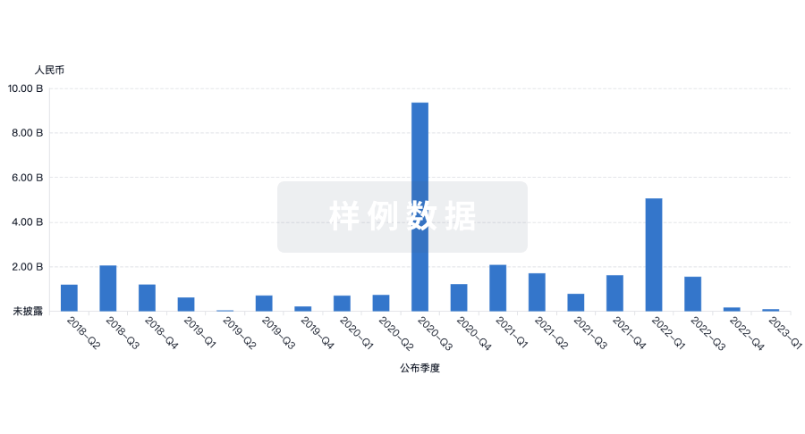
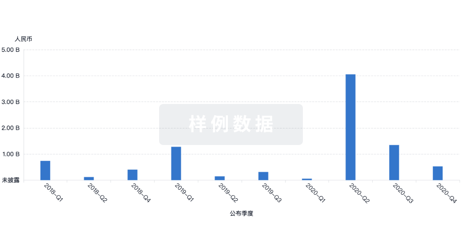
营收
使用 Synapse 探索超过 36 万个组织的财务状况。
登录
或
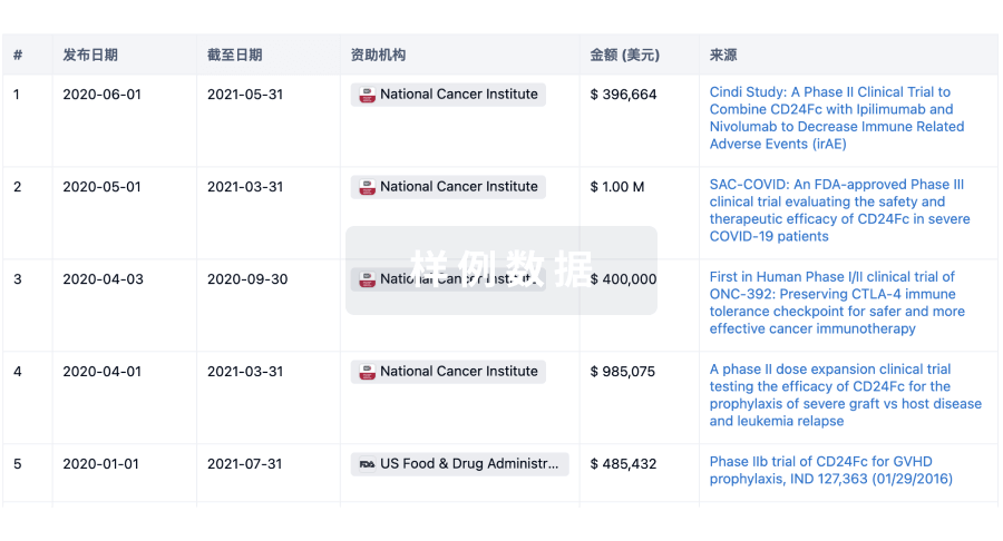
科研基金(NIH)
访问超过 200 万项资助和基金信息,以提升您的研究之旅。
登录
或
投资
深入了解从初创企业到成熟企业的最新公司投资动态。
登录
或
融资
发掘融资趋势以验证和推进您的投资机会。
登录
或
生物医药百科问答
全新生物医药AI Agent 覆盖科研全链路,让突破性发现快人一步
立即开始免费试用!
智慧芽新药情报库是智慧芽专为生命科学人士构建的基于AI的创新药情报平台,助您全方位提升您的研发与决策效率。
立即开始数据试用!
智慧芽新药库数据也通过智慧芽数据服务平台,以API或者数据包形式对外开放,助您更加充分利用智慧芽新药情报信息。
生物序列数据库
生物药研发创新
免费使用
化学结构数据库
小分子化药研发创新
免费使用