预约演示
更新于:2026-06-17
Anhui Normal University
更新于:2026-06-17
概览
标签
神经系统疾病
心血管疾病
肿瘤
小分子化药
化学药
多糖药物
疾病领域得分
一眼洞穿机构专注的疾病领域

技术平台
公司药物应用最多的技术
靶点
公司最常开发的靶点
关联
靶点 |
作用机制 |
在研机构 |
原研机构 |
在研适应症 |
非在研适应症 |
最高研发阶段 |
首次获批国家/地区 |
首次获批日期 |
靶点 |
作用机制 |
在研机构 |
原研机构 |
在研适应症 |
非在研适应症 |
最高研发阶段 |
首次获批国家/地区 |
首次获批日期 |
靶点 |
作用机制 |
在研机构 |
原研机构 |
在研适应症 |
非在研适应症 |
最高研发阶段 |
首次获批国家/地区 |
首次获批日期 |
NCT07452302
A 12-Week School-Based Mixed Physical Education Intervention Combining Cooperative Sports Games and Ball Sports to Improve Mental Health and Physical Fitness in Rural Left-Behind Children
100 项与 安徽师范大学 相关的临床结果
登录后查看更多信息
登录后查看更多信息
2026-12-31International Journal of Qualitative Studies on Health and Well-Being
Navigating stigma and support: a qualitative study of help-seeking for mental health challenges among Chinese university students
Article
作者: Fang, Shuanghu ; Zeb, Irum ; Khan, Aashiq
PURPOSE:
This study explores how students' help-seeking behaviors are shaped by their Psychological Capital (PsyCap) and perceived social support amidst the structural challenges they face.
METHODS:
Using a qualitative approach, we analyzed semi-structured interviews with 30 students from a Chinese university. Themes related to internal resources (e.g., resilience, hope, self-efficacy, optimism) and external resources (e.g., support from peers and family) were identified through thematic analysis.
RESULTS:
The findings suggest that students experiencing mental health difficulties often rely on their PsyCap and social support networks, such as peers and family, to cope. However, significant barriers such as stigma and a lack of mental health literacy hinder effective help-seeking. Moreover, the study highlights the influence of academic stress and environmental pressures on students' mental health and on their use of cognitive and emotional regulation strategies.
CONCLUSIONS:
These findings call for the development of mental health literacy programs that integrate PsyCap and social support elements to foster better help-seeking behaviors and improve mental health outcomes. Such programs can facilitate access to mental health services and contribute to a healthier academic environment.
2026-08-01JOURNAL OF AFFECTIVE DISORDERS
Machine learning models for detecting suicidal ideation in Chinese in-patients with major depressive disorder: A single-centre retrospective study
Article
作者: Gu, Changyan ; Xie, Yu ; Chen, Juan ; Wang, Anzhen ; Cheng, Xialong ; Zhang, Xulai ; Pan, Min ; Zheng, Hongxin
PURPOSE:
This study aimed to develop and internally validate machine-learning (ML) models that exploit routine electronic medical record (EMR) data to identify recent SI in Chinese in-patients with MDD.
PATIENTS AND METHODS:
A retrospective cohort of 721 in-patients with major depressive disorder (MDD), including 399 with suicidal ideation (SI-positive), was recruited from the Fourth People's Hospital of Hefei between January 2020 and August 2023. The dataset was stratified into training (70%) and test (30%) sets. All preprocessing steps (median imputation and Z-score normalization) and Boruta feature selection were performed exclusively on the training set using R software (version 4.4.2), with multicollinearity removed for variables with a variance inflation factor (VIF) > 5 or a pairwise Pearson correlation coefficient |r| > 0.75. Six machine learning algorithms-random forest (RF), logistic regression (LR), LightGBM, support vector machine (SVM), K-nearest neighbor (KNN), and XGBoost-were trained using GridSearchCV combined with 10-fold stratified cross-validation, with model fine-tuning via the class_weight = 'balanced' parameter. Model performance was evaluated on the independent test set using multiple metrics, including the area under the receiver operating characteristic curve (AUC), area under the precision-recall curve (PR-AUC), accuracy, sensitivity, specificity, positive predictive value (PPV), and negative predictive value (NPV). SHAP analysis, implemented in Python (version 3.12), was used to enhance model interpretability. Stratified subgroup analysis (stratified by sex and age) and sensitivity analysis (comparing the optimal RF model with the traditional LR baseline model via DeLong's test) were conducted to verify the robustness and superiority of the proposed model.
RESULTS:
Random Forest achieved the best discrimination (AUC 0.857) and maintained stable discrimination across sexes (AUC = 0.843 in females and 0.822 in males), demonstrating higher sensitivity in females (0.883) and higher specificity in males (0.900).Top risk features: Compared with SI-negative patients, SI-positive patients were predominantly male (74.4% vs. 53.7%), married (80.7% vs. 63.4%), and had a lower educational level (83.7% vs. 29.5% without higher education). Furthermore, both their current age and age at depression onset were significantly greater (all P < 0.001).
CONCLUSION:
ML models, especially Random Forest, can effectively identify recent SI risk in Chinese MDD patients using readily available clinical data.
2026-07-01BIOTECHNOLOGY ADVANCES
From molecules to field: Integrated insights into cuticle-mediated drought tolerance in plants
Review
作者: Iqbal, Babar ; Wang, Yong-Feng ; Khan, Amir Abdullah ; Du, Daolin ; Akbar, Rasheed
Drought stress exacerbates non-stomatal water loss, which hinders agricultural growth and global food security. The cuticle, a layer of cutin, waxes, and other polymers, protects plants from the negative impact of drought. One crucial adaptation that has enabled terrestrial plants to survive and flourish in drought-prone regions is the development of cuticle structures. In this review, we (1) provide an overview of the molecular and enzymatic pathways involved in the biosynthesis of cutin and wax; (2) analyze important regulatory networks that control the formation of cuticles, such as abscisic acid (ABA) mediated signaling and transcription factors (SHN1/WIN1, MYB, NAC); (3) synthesis functional evidence of cuticle contributions to drought resilience across species; (4) evaluate how cuticle-related mutants and transgenic lines of genes such as ECERIFERUM1, Fatty Acyl-CoA Reductase 1 and Lipid Transfer Protein GPI-Anchored 22 (e.g., CER1, FAR1, LTPG22) change the composition of lipids and impact drought phenotypes; and (5) present lipidomic as a targeted phenotyping method to measure changes in very-long-chain alkanes, primary alcohols, and cutin monomers under water deficit. In Arabidopsis, camelina, cotton, rice, wheat, and turfgrass, we employ gas chromatography-flame ionization detection (GC-FID) for accurate detection and quantification of fatty acid content, and gas chromatography-mass spectrometry (GC-MS) for the qualitative and quantitative analysis of lipid components to identify lipid-remodeling patterns that are consistent across all of these plants and are associated with drought tolerance and barrier integrity. Finally, we discuss how to create "drought-smart" crops by quantitative trait loci (QTL) mapping, marker-assisted selection, CRISPR-Cas9 editing of KCS (3-ketoacyl-CoA synthase) and ERF (ethylene-responsive factor) genes, and overexpression approaches. This establishes the foundation for integrating cuticle biology with novel omics methods.
100 项与 安徽师范大学 相关的药物交易
登录后查看更多信息
100 项与 安徽师范大学 相关的转化医学
登录后查看更多信息
组织架构
使用我们的机构树数据加速您的研究。
登录
或
管线布局
2026年07月12日管线快照
管线布局中药物为当前组织机构及其子机构作为药物机构进行统计,早期临床1期并入临床1期,临床1/2期并入临床2期,临床2/3期并入临床3期
药物发现
1
4
临床前
登录后查看更多信息
当前项目
登录后查看更多信息
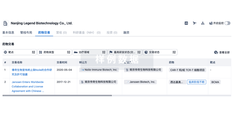
药物交易
使用我们的药物交易数据加速您的研究。
登录
或
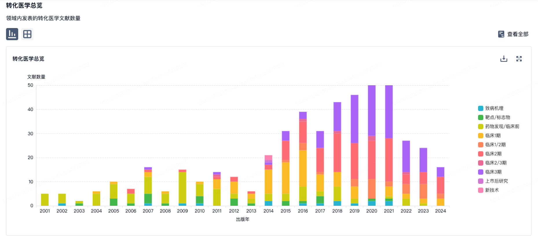
转化医学
使用我们的转化医学数据加速您的研究。
登录
或
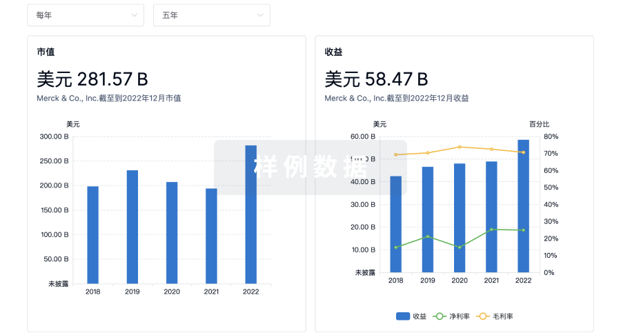
营收
使用 Synapse 探索超过 36 万个组织的财务状况。
登录
或
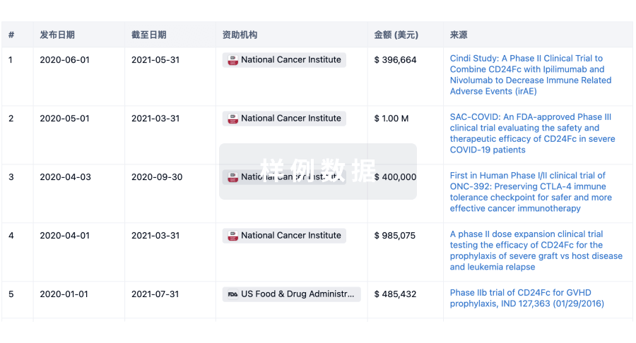
科研基金(NIH)
访问超过 200 万项资助和基金信息,以提升您的研究之旅。
登录
或
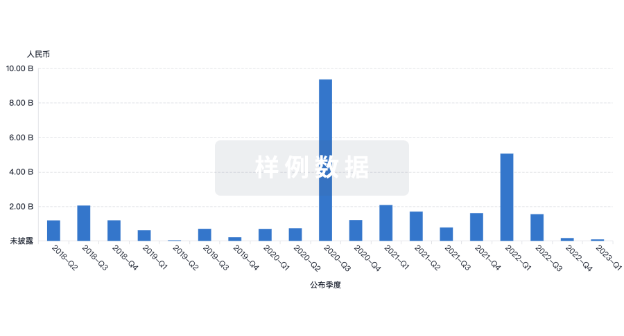
投资
深入了解从初创企业到成熟企业的最新公司投资动态。
登录
或
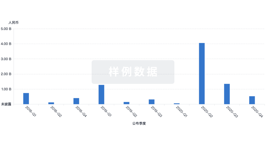
融资
发掘融资趋势以验证和推进您的投资机会。
登录
或
生物医药百科问答
全新生物医药AI Agent 覆盖科研全链路,让突破性发现快人一步
立即开始免费试用!
智慧芽新药情报库是智慧芽专为生命科学人士构建的基于AI的创新药情报平台,助您全方位提升您的研发与决策效率。
立即开始数据试用!
智慧芽新药库数据也通过智慧芽数据服务平台,以API或者数据包形式对外开放,助您更加充分利用智慧芽新药情报信息。
生物序列数据库
生物药研发创新
免费使用
化学结构数据库
小分子化药研发创新
免费使用