预约演示
更新于:2025-05-07
PD-L1 positive stomach adenocarcinoma
PD-L1阳性胃腺癌
更新于:2025-05-07
基本信息
别名- |
简介- |
关联
2
项与 PD-L1阳性胃腺癌 相关的药物靶点 |
作用机制 PDL1抑制剂 |
在研机构 |
原研机构 |
最高研发阶段批准上市 |
首次获批国家/地区 美国 |
首次获批日期2017-03-23 |
靶点- |
作用机制 细菌替代物 [+1] |
在研机构 |
原研机构 |
非在研适应症- |
最高研发阶段临床2期 |
首次获批国家/地区- |
首次获批日期1800-01-20 |
1
项与 PD-L1阳性胃腺癌 相关的临床试验NCT05419362
A Phase II Study to Evaluate the Safety and the Efficacy of GEN-001 in Combination With Avelumab for Patients With PD-L1 Positive Advanced Gastric or Gastroesophageal Junction Adenocarcinoma
This is a phase II, multicenter, open-label study to evaluate the antitumor activity, efficacy and safety of GEN-001 in combination with avelumab as a third line (3L) or greater line treatment which is not received the Standard of Care (SOC) for patients with PD-L1 positive advanced GC/Gastroesophageal Junction Adenocarcinoma who are not received cancer immunotherapy regimens as mono or combination therapy.
开始日期2022-04-07 |
申办/合作机构  Genome & Co. Genome & Co. [+1] |
100 项与 PD-L1阳性胃腺癌 相关的临床结果
登录后查看更多信息
100 项与 PD-L1阳性胃腺癌 相关的转化医学
登录后查看更多信息
0 项与 PD-L1阳性胃腺癌 相关的专利(医药)
登录后查看更多信息
9
项与 PD-L1阳性胃腺癌 相关的文献(医药)2017-11-01·Clinical Lung Cancer2区 · 医学
Computed Tomography Features of Lung Adenocarcinomas With Programmed Death Ligand 1 Expression
2区 · 医学
Article
作者: Katsura, Masakazu ; Akamine, Takaki ; Okamoto, Tatsuro ; Matsubara, Taichi ; Shoji, Fumihiro ; Shimokawa, Mototsugu ; Takada, Kazuki ; Takamori, Shinkichi ; Oda, Yoshinao ; Haratake, Naoki ; Kozuma, Yuka ; Toyokawa, Gouji ; Maehara, Yoshihiko
2017-05-01·Journal of Thoracic Oncology
Correlation between Classic Driver Oncogene Mutations in EGFR , ALK , or ROS1 and 22C3–PD-L1 ≥50% Expression in Lung Adenocarcinoma
Article
作者: Le, Xiuning ; Rangachari, Deepa ; Huberman, Mark S ; VanderLaan, Paul A ; Costa, Daniel B ; Kobayashi, Susumu S ; Shea, Meghan
1987-01-01·The Japanese Journal of Pharmacology
Effect of 16,16-dimethyl prostaglandin E2 on gastric surface epithelial cell damage induced by 20% ethanol in rats.
Article
作者: Takeuchi, Koji ; Okabe, Susumu ; Nakagawa, Momoyo ; Ohno, Tomochika ; Nishimura, Seiichiro
分析
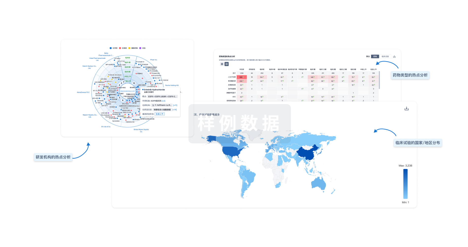
对领域进行一次全面的分析。
登录
或

生物医药百科问答
全新生物医药AI Agent 覆盖科研全链路,让突破性发现快人一步
立即开始免费试用!
智慧芽新药情报库是智慧芽专为生命科学人士构建的基于AI的创新药情报平台,助您全方位提升您的研发与决策效率。
立即开始数据试用!
智慧芽新药库数据也通过智慧芽数据服务平台,以API或者数据包形式对外开放,助您更加充分利用智慧芽新药情报信息。
生物序列数据库
生物药研发创新
免费使用
化学结构数据库
小分子化药研发创新
免费使用
