预约演示
更新于:2026-07-11
[68Ga] anti-PD-L1 nanobody(Suzhou SmartNuclide Biopharmaceutical)
镓[68Ga]纳坦重组PD-L1单域抗体(Suzhou SmartNuclide Biopharmaceutical)
更新于:2026-07-11
概要
基本信息
原研机构 |
非在研机构- |
权益机构- |
最高研发阶段临床1期 |
首次获批日期- |
最高研发阶段(中国)临床1期 |
特殊审评- |
登录后查看时间轴
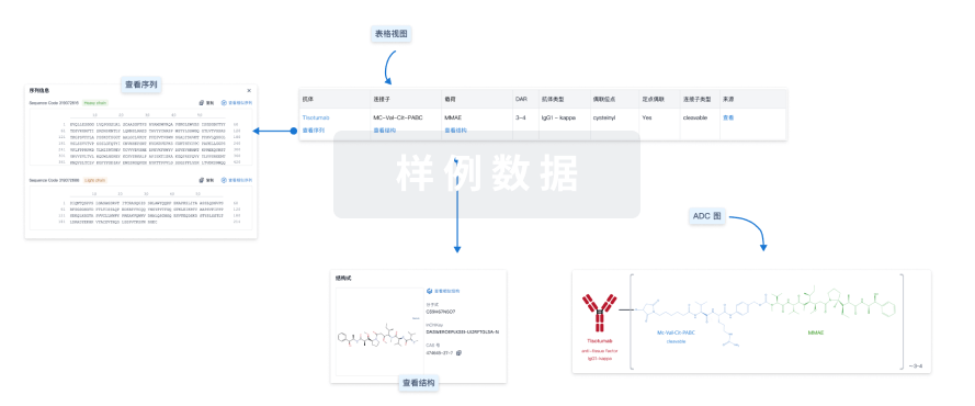
结构/序列
使用我们的ADC技术数据为新药研发加速。
登录
或
关联
3
项与 镓[68Ga]纳坦重组PD-L1单域抗体(Suzhou SmartNuclide Biopharmaceutical) 相关的临床试验CTR20232251
一项评价[68Ga]Ga-NOTA-SNA002在实体瘤患者中安全耐受性、辐射吸收剂量、分布特征的I期临床试验
NCT05989997
Phase I Clinical Trial of Evaluating 68Ga-NOTA-SNA002 for the Safety Tolerance, Radiation Absorbed Dose and Dosimetry in Patients With Solid Tumor
NCT05490264
[68Ga]Ga-NOTA-SNA002 (PD-L1 PET Tracer) for PET/CT in Patients With Solid Tumors
100 项与 镓[68Ga]纳坦重组PD-L1单域抗体(Suzhou SmartNuclide Biopharmaceutical) 相关的临床结果
登录后查看更多信息
100 项与 镓[68Ga]纳坦重组PD-L1单域抗体(Suzhou SmartNuclide Biopharmaceutical) 相关的转化医学
登录后查看更多信息
100 项与 镓[68Ga]纳坦重组PD-L1单域抗体(Suzhou SmartNuclide Biopharmaceutical) 相关的专利(医药)
登录后查看更多信息
1
项与 镓[68Ga]纳坦重组PD-L1单域抗体(Suzhou SmartNuclide Biopharmaceutical) 相关的文献(医药)EJNMMI Physics
A hybrid curve-fitting method for radiation dosimetry evaluation and simultaneous multi-organ biodistribution assessment in total-body dynamic PET/CT: application to a novel PD-L1-targeted tracer
Article
作者: Zhang, Lichao ; Xie, Yunze ; Liu, Guobing ; Shi, Hongcheng ; Tang, Wenxin
OBJECTIVE:
This study explored a hybrid curve-fitting method optimized for radiation dosimetry evaluation in total-body dynamic positron emission tomography/computed tomography (PET/CT) imaging, compared to conventional methods reliant on multi-time-point static acquisition protocols, and evaluated the radiation dosimetry results and organ biodistribution of [68Ga]Ga-NOTA-SNA002.
METHODS:
A total of 16 patients with solid tumors underwent a 60-min dynamic PET acquisition immediately after administration of [68Ga]Ga-NOTA-SNA002, followed by a 20-min static PET scan at about 120 min post-injection. Volumes of interest (VOIs) were manually delineated on the CT images for subsequent radiation dosimetry estimation and biodistribution analysis. Two datasets were created to generate organ time-activity curves (TACs): (1) two PET frames reconstructed at 15-35 min and 40-60 min from the dynamic PET dataset, and one frame from static PET acquisition, thereby simulating a conventional multi-time-point static protocol; (2) 55 frames from the dynamic PET dataset and a frame from static PET. To calculate the time-integrated activity coefficients (TIACs) for the tracer in source organs, two methods were used: a routine method (RM) that fitted Dataset 1 by using a conventional bi-exponential curve fitting approach, and a hybrid method (HM) that fitted Dataset 2 by combining rectangular integration for the early phase of TACs with exponential fitting for the later phase. Absorbed and effective radiation doses were subsequently estimated using OLINDA/EXM version 1.1.
RESULTS:
The [68Ga]Ga-NOTA-SNA002 primarily accumulated in the urinary system, with relatively low overall uptake levels in other source organs. Analysis of tumor-to-organ standardized uptake value (SUV) ratios revealed that all ratios reached their highest values at 120 min post-injection. The TIACs derived from the HM were consistently higher than those obtained using the RM method, while preserving a similar rank order of distribution across organs. In both methods, the kidneys, liver, and lungs exhibited the largest TIACs. The bladder wall, kidneys, and spleen received the highest absorbed doses. The total effective dosimetry calculated with RM and HM were 20.47 ± 3.08 µSv/MBq and 36.33 ± 6.18 µSv/MBq, respectively.
CONCLUSION:
The radiation dosimetry results obtained from both the RM and the HM consistently support the safety and feasibility of total-body PET/CT imaging using [68Ga]Ga-NOTA-SNA002. Meanwhile, the HM provides theoretically more accurate results compared to the RM, and is therefore recommended for radiation dosimetry evaluation of other novel tracers.
21
项与 镓[68Ga]纳坦重组PD-L1单域抗体(Suzhou SmartNuclide Biopharmaceutical) 相关的新闻(医药)2025-11-11
·谈市说市
放射疗法并购
2024-09-13
·同写意
蛋白降解靶向嵌合体引进/卖出临床申请
2024-09-13
蛋白降解靶向嵌合体引进/卖出临床申请放射疗法
100 项与 镓[68Ga]纳坦重组PD-L1单域抗体(Suzhou SmartNuclide Biopharmaceutical) 相关的药物交易
登录后查看更多信息
研发状态
10 条进展最快的记录, 后查看更多信息
登录
| 适应症 | 最高研发状态 | 国家/地区 | 公司 | 日期 |
|---|---|---|---|---|
| 乳腺癌 | 临床1期 | 中国 | 2023-11-01 | |
| 黑色素瘤 | 临床1期 | 中国 | 2023-11-01 | |
| 非小细胞肺癌 | 临床1期 | 中国 | 2023-11-01 | |
| 头颈部鳞状细胞癌 | 临床1期 | 中国 | 2023-11-01 | |
| 实体瘤 | 临床1期 | 中国 | 2022-01-30 |
登录后查看更多信息
临床结果
临床结果
适应症
分期
评价
查看全部结果
登录后查看更多信息
转化医学
使用我们的转化医学数据加速您的研究。
登录
或

药物交易
使用我们的药物交易数据加速您的研究。
登录
或

核心专利
使用我们的核心专利数据促进您的研究。
登录
或

临床分析
紧跟全球注册中心的最新临床试验。
登录
或

批准
利用最新的监管批准信息加速您的研究。
登录
或

生物类似药
生物类似药在不同国家/地区的竞争态势。请注意临床1/2期并入临床2期,临床2/3期并入临床3期
登录
或
特殊审评
只需点击几下即可了解关键药物信息。
登录
或

芽仔
全新生物医药AI Agent 覆盖科研全链路,让突破性发现快人一步
立即开始免费试用!
智慧芽新药情报库是智慧芽专为生命科学人士构建的基于AI的创新药情报平台,助您全方位提升您的研发与决策效率。
立即开始数据试用!
智慧芽新药库数据也通过智慧芽数据服务平台,以API或者数据包形式对外开放,助您更加充分利用智慧芽新药情报信息。
生物序列数据库
生物药研发创新
免费使用
化学结构数据库
小分子化药研发创新
免费使用